Last updated on January 10, 2022
Content By: Dr. Nikita Toshi BDS, Assistant Manager (Medical Review), Dr. Ritu Budania MBBS, MD (Pharmacology) Head, Medical Affairs
Last updated on January 10, 2022
This article discusses in detail the structure and function of heart and its role in the human body. When your hand is clenched into a fist, it is approximately the size of your heart. The most vital organ of our body is located in the centre of the chest and slightly to the left of the breastbone.
Since heart disease is very common and usually quiet until it strikes, it is important to be aware of the factors that put your heart in danger. Numerous risk factors for coronary heart disease are controllable. By implementing lifestyle changes, you can reduce your risk of cardiac diseases.

Written by
BDS, Assistant Manager (Medical Review)

Reviewed by
MBBS, MD (Pharmacology) Head, Medical Affairs
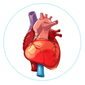
The heart is located in the middle of your chest, between the lungs and under the rib cage slightly to the left of the sternum (breastbone). The primary function of the heart is to pump blood and supply oxygen and nutrients to your cells. The heart consists of four chambers – two upper chambers called the right and left atria and two lower sections called the right and left ventricles. There are valves between the upper and lower chambers that ensure blood flows only in a single direction through your heart.
A thorough understanding of the shape and size of the heart is essential to understanding the structure and function of the heart. The size of your heart is almost the same as the size of your clenched fist. Men have larger hearts than women. Athletes engaged in high-intensity training tend to have larger hearts, too. The heart weighs in the range of 7 and 15 ounces (200 to 425 grams). By the end of a long life, an individual’s heart might have extended and contracted more than 3.5 multiple times, which is a sign of the normal working of the human heart.
The double-layered covering called the pericardium surrounds the heart like a sac. It consists of 2 layers. The external layer is fibrous in nature and is called the parietal layer and the internal layer is smooth and is called the visceral layer. The pericardium provides mechanical protection for the heart and the large blood vessels and helps in reducing the friction between the heart and the surrounding structures.

There are many supplements that can boost your heart health.
You can order them online at PharmEasy and get them home delivered!
Four chambers, layers of the heart wall and valves constitute the human heart and are responsible for the systematic functioning of the circulatory system. Read on to find out more about each of these parts of the heart.
Three layers of tissue form the heart wall namely:
Together, these three layers play a significant role in the normal working of the human heart.
The heart is divided into four chambers, namely:
The atria, which are thin-walled chambers, receive blood from the veins and pump it to the ventricles. The ventricles, which are thick-walled chambers, pump the blood out of the heart. The atria and ventricles are connected together by valves which ensure that blood flows only in one direction inside the heart.
The wall that separates the left and right sides of the heart is called the septum. The left side of the heart receives oxygen-rich blood from the pulmonary veins (the only veins that carry oxygenated blood from the lungs to the heart) and pumps it into the aorta (the largest artery), while the right part of the heart receives low-oxygen blood directly from the the largest vein or the vena cava and transports it to the lungs via the pulmonary veins.
The heart valves act like gates at the chamber openings, they open and close to allow blood to flow through the chambers. Their main function is to ensure that blood flows only in one direction through the heart.
The atrioventricular (AV) valves are located between your upper and lower heart chambers. They include:
Semilunar (SL) valves, which are located between the ventricles and the arteries that emerge from the heart, consist of:
For a healthy functioning of valves, it is important that:
Oxygen poor blood from the rest of the body flows into the right atrium of the heart through two large veins. Then, the blood flows into the right ventricle and is pumped into the pulmonary arteries present in the lungs. After collecting oxygen, the blood flows back to into the left atrium through the pulmonary veins and then to the left ventricle. The aorta, then onward, carries the fully oxygenated blood to the rest of the body.
The myocardium (middle layer of the heart wall) is a functioning muscle that needs a non-stop supply of oxygen and nutrients to work efficiently. Therefore, the cardiovascular muscle has a large network of arteries, veins and capillaries to carry oxygen to the contracting cells and eliminate byproducts.
Your heart’s electrical system controls the rhythm and pace of your heartbeat. It consists of:
Your heart also has a network of electrical bundles and fibres that help in the conduction of electrical impulses well throughout the muscular structure of the
Include the right nutritional supplements in your diet to keep your heart healthy!
Order them at PharmEasy now and get great discounts in just a few clicks!
The heart is a strong organ located in the chest behind and slightly towards the left of the breastbone. The heart constantly works, pumping blood through the network of pipes (blood vessels), consisting of arteries, veins and capillaries. The heart and the blood vessels form the cardiovascular system. The right atrium and right ventricle are referred to as the right heart and the left chamber comprises the left atrium and left ventricle.
The heart plays the following roles in the human body:
The human body is a network of blood vessels that is responsible for circulating blood to every cell of the body. Proper blood circulation helps organs function normally. The right side of the heart receives blood with lesser amounts of oxygen from the veins. It then pumps this blood to the lung and picks up oxygen. The left side of the heart receives oxygen-rich blood from the lungs and pumps it into the arteries, which then reaches other organs in the body. It is the function of arteries and veins to transport blood through this network.
Human blood carries oxygen, hormones and nutrients that are required by the organs to function well. Apart from this, the blood pumped by the heart also carries carbon dioxide to the lungs, which allows them to breathe it out. The function of the heart valves is to help the blood flow in the right direction.
Blood circulation takes place through two circuits in the heart. The first is called the pulmonary circuit (across the lungs) and the second is the systemic circuit (across the body). It is in the pulmonary circuit that the deoxygenated blood exits the right ventricle through the pulmonary artery and reaches the lungs. This blood returns as oxygenated blood to the heart through the pulmonary vein.
Blood pressure originates as a result of the pumping action of the heart. It is a measurable force exerted by blood against the walls of blood vessels. The vessels stretch in response to this force and the resultant contraction is pivotal in maintaining a normal blood flow through the vascular system.
Manage the symptoms of your heart disease with the right medicines!
Order online on PharmEasy and get it home delivered at your convenience!
Blood is a life-sustaining fluid that courses through the whole body. It transports the following to our body tissues:
![]() Nourishment
Nourishment
![]() Antibodies
Antibodies
![]() Hormones
Hormones
![]() Electrolytes
Electrolytes
![]() Heat
Heat
![]() Oxygen
Oxygen
Blood is also responsible for removing the following from body tissues:
![]() Waste matter
Waste matter
![]() Carbon dioxide
Carbon dioxide
The circulatory system transports nutrients and oxygen to all the parts of the body. It consists of the heart, as well as the arteries, veins and capillaries that run throughout the body. The veins return blood to the heart. The function of arteries and veins is organized in a tree-like pattern: The “storage compartment” – the primary course (aorta) – branches into massive pipes that lead to smaller vessels. Finally, the smallest course is a collection of minuscule vessels. These are the various components of the circulatory system.
Two pathways come from the heart – pulmonary circulation and systemic circulation.
In pulmonary circulation
The pulmonary artery is a major artery that emerges from the heart. It splits into two fundamental branches and carries blood from the heart to the lungs. In the lungs, the blood gets oxygen and drops off carbon dioxide.
In systemic circulation
Next, the blood returns to the heart having received loads of oxygen from the lungs. So, now the oxygenated blood can be circulated throughout the body. The aorta is a major conduit that carries oxygenated blood.
The signs of poor blood circulation in the body may present as:
![]() Tingling or numbness in hands and feet
Tingling or numbness in hands and feet
![]() Cold hands or feet
Cold hands or feet
![]() Swollen ankles and legs
Swollen ankles and legs
![]() Loss of memory and difficulty in concentrating
Loss of memory and difficulty in concentrating
![]() Issues with the digestion of food
Issues with the digestion of food
![]() Fatigue
Fatigue
![]() Muscle cramps
Muscle cramps
![]() Changes in skin color
Changes in skin color
![]() Occurrence of ulcers
Occurrence of ulcers
![]() Varicose veins
Varicose veins
If you are experiencing one or more of these symptoms, your blood circulation may need improvement. Consult a doctor to find out if you need to improve your blood circulation and the steps you can take in this direction. The best treatment for bad circulation depends upon the reason and issues in the function of arteries and veins.
Blood coagulation in simple terms means clotting. It is when the consistency of blood changes from liquid to a semi-solid state. Blood clotting is due to any accident or cuts in the human flesh. These clots are not harmful but helpful. Occasionally, a blood clot will form without a trigger (such as an injury or cut) which can be harmful and head to various complications.
You can undoubtedly forestall numerous heart illnesses by heart screening or making the previously mentioned lifestyle changes. Heart disease prevention is, in every situation, better compared to treatments!
Get the best medicines to maintain proper blood circulation
Order online on PharmEasy and get great discounts in just a few clicks!
Here are 5 interesting facts about the heart:
The human heart is located in the chest between the lungs under the protection of your rib cage and muscles of the chest region.
AB negative is the rarest blood group among the eight primary blood types.
Your health depends on a number of parameters – nutrition and lifestyle, genetics, psychosocial factors, environmental factors, etc. These parameters are common for all blood types. However, if you have Blood type O, you should be happy to know that studies have shown that this blood type has a lower risk of heart diseases and some cancers. However, there is no explanation for this observation.
Medical studies indicate that usually, blood vessels heal as a part of the wound healing process. The duration of wound healing depends on the severity of the injury, medical care and rest given. Diet chart for heart patients, hydration and supplements can also aid in the healing process.
Given below are the signs of poor blood circulation
Vitamin B complex and vitamin C are essential for the healthy functioning of the blood vessels. However, all essential nutrients play a role in the repair and maintenance of the blood vessels.
Leave a Comment