Article

Castor Plant Adaptation to Salinity Stress during Early Seedling Stage by Physiological and Transcriptomic Methods

1
College of Landscape Architecture, Northeast Forestry University, Harbin 150040, China
2
National and Local Joint Engineering Research Center of Ecological Treatment Technology for Urban Water Pollution, Zhejiang Provincial Key Laboratory for Water Environment and Marine Biological Resources Protection, College of Life and Environmental Science, Wenzhou University, Wenzhou 325035, China
*
Authors to whom correspondence should be addressed.
These authors contributed equally to this work.
Agronomy 2023, 13(3), 693; https://doi.org/10.3390/agronomy13030693
Submission received: 13 January 2023 / Revised: 13 February 2023 / Accepted: 24 February 2023 / Published: 27 February 2023
(This article belongs to the Topic Plant Responses and Tolerance to Salinity Stress)

Abstract

:
The early seedling stage is considered the most vulnerable period for plants, especially under salinity conditions. The castor plant (Ricinus communis) is a well-known oil and energy crop worldwide that can survive under stressful conditions. However, the specific mechanisms of this species during its early seedling stage under salt stress are still not clearly understood. Here, the physiological and transcriptome changes in the cotyledons and roots of the castor plant were evaluated. The results indicated that salt stress (150 mM NaCl, 6 d) increased malondialdehyde (MDA) and proline content, whereas it decreased dry weight (DW) and soluble sugar content. The Illumina Hiseq 2500 platform was used to analyze transcriptome profiles in the cotyledons and roots under salt stress conditions. The results showed that 1580 differentially expressed genes (DEGs) were found in the cotyledons (880 upregulated and 700 downregulated) and 1502 DEGs in the roots (732 upregulated and 770 downregulated). Furthermore, we found that salt stress significantly regulated 22 genes (e.g., 29520.t000005, 29633.t000030, and 29739.t000024) involved in protein processing in the endoplasmic reticulum of the cotyledons. However, salt stress induced the expression of 25 genes (e.g., 30068.t000101, 30076.t000022, 29970.t000022, and 29957.t000027) involved in phenylpropanoid biosynthesis in the roots. In addition, a large number of genes participating in plant hormone signal transduction, starch and sucrose metabolisms, and arginine and proline metabolisms were induced in both cotyledons and roots. In conclusion, this study demonstrated that the different expression patterns in cotyledons and roots as well as their synergic relationship contributed to enhancing the salt tolerance of castor plants.

1. Introduction

Soil salinization has become a serious threat to crop productivity worldwide; it is also extremely detrimental to ecological environments [1]. The Earth’s saline-alkali land area is, at present, approximately 1 × 10⁹ hm2, accounting for 7% of total land area [2]. In the northeastern region of China, the saline-alkali area is about 7 × 10⁶ hm2, which is increasing due to global climate change and poor irrigation and fertilization management [3]. Salt stress is an abiotic stress that mostly impacts plant growth and development. Salt stress induces osmotic and oxidative stresses, ionic toxicity, and changes in gene expression due to the high level of salt, thereby inhibiting plant growth and hindering normal physiological processes [4].
Plants have developed a series of physiological and molecular mechanisms to cope with the damage caused by salt stress, such as accumulation of osmotic regulators (e.g., soluble sugars and proline) and reactive oxygen species, maintenance of ion balance, and changes in membrane permeability and related transporters [5,6,7]. In general, salt tolerance is always a complex trait controlled by genetic factors. Several transcriptome sequencing studies have revealed genes related to salt tolerance in various plants, including Betula platyphylla [8], Lycium ruthenicum [9], Setaria viridis [10], Fagopyrum esculentum [11], and Ricinus communis [12]. These studies suggested a large number of salt-tolerance genes encoding hormone signaling, accumulation of specific osmolytes, membrane permeability, ion transportation, and redox reactions. They are all beneficial for deeply understanding the mechanisms of plants under salt stress. Therefore, transcriptomics and quantitative reverse transcription–polymerase chain reaction (qRT-PCR) verification are very useful tools for analyzing plant responses to salt stress. Existing studies are deficient in terms of exploring the mechanisms of the plant above and below ground during the young seedling stage (cotyledon vs. root) under such stress conditions.
The early seedling stage is considered the most vulnerable period for plants, especially under stress conditions. Cotyledons are not only the main components of the seed, but also the autotrophic organs before the appearance of the first real leaf; they play an extremely important role in subsequent seedling establishment [13,14]. Cotyledons can perform photosynthesis, thereby providing nutrients and enough energy for seedling growth [15]. Previous studies have revealed that shading cotyledons decreased the dry weight and seedling survival rate of Suaeda salsa and removing cotyledons reduced the growth and photosynthesis of castor seedlings under salt stress [16,17]. To date, a majority of studies have explored cotyledon functions such as nutrient storage and transportation as well as their photosynthesis ability [18], but little is known about their physiological and transcriptome changes under salt stress. Moreover, the roots of plants are directly exposed to the salinity in the soil, and seem more sensitive compared with the aboveground organ. Meanwhile, based on the close relationship between cotyledons and roots, how castor plants respond to salt stress (cotyledons vs. roots) during the early seedling stage using the transcription approach is still poorly understood.
Castor (Ricinus communis L.) belongs to the Euphorbiaceae family; it is a well-known oil and energy crop worldwide. It is widely distributed in temperate and tropical regions such as China, Brazil, and India [19]. This species has great commercial potential because the oil content of its seeds is as high as 45–55%, which is much higher than that of other oil crops [20]. The oil can be widely used as lubricating oil and medicine, as well as incorporated in paint. It is also a promising alternative fuel that can replace petroleum [21]. In addition, this plant can also survive under stressful conditions and has strong stress resistance and good adaptability to high temperature, drought, and salt stresses [22]. In recent years, the castor plant has been used in the process of salt-affected soil improvement, especially in northeast China [23]. However, its salt-tolerant mechanism is still not clear, especially the relationship between cotyledons and roots during the early seedling stage under such conditions.
Therefore, in this study physiological and transcriptomic analyses were carried out in the cotyledons and roots of castor plants under salt stress during the early seedling stage. We aimed to determine whether the cotyledons and roots of this species had different expression patterns as well as a synergic relationship between them under salt stress.

2. Materials and Methods

2.1. Plant Materials and Stress Treatments

Seeds of Ricinus communis (Fenbi 10) were provided by the Shanxi Academy of Agricultural Sciences of China. Full seeds that were consistent in size were selected; the seeds were soaked in 10% sodium hypochlorite (NaClO) for 10 min, then the surface of the seeds was rinsed with distilled water. Seeds were sown in plastic pots with 2 kg soil, 8 per pot, and then placed in a greenhouse (relative humidity: 60 ± 5%, ambient temperature: 25 ± 1 °C, photo period: 16 h light/8 h dark). After 16 days of seedling emergence, plants were randomly divided into control (0 mM NaCl) and salt stress (150 mM NaCl), with three replicates for each treatment. The cotyledons and roots of the castor plants were harvested after 6 days of salt stress and kept in a storage room at −80 °C for subsequent physiological assays and transcriptome analysis.

2.2. Determination of Plant Biomass

The fresh and dry weight of the cotyledons and roots of the castor plants were determined in three replicates and expressed as g plant−1. These were de-enzymed at 105 °C for 15 min and at 80 °C for 48 h in an oven to constant weight, at which point the dry weight was measured.

2.3. Determination of Malondialdehyde (MDA) Content

The MDA content in the cotyledons and roots was determined as described by [24]. A total of 0.5 g of cotyledons and roots was homogenized in 5 mL of 5% trichloroacetic acid (TCA). After centrifugation, 1 mL of supernatant was taken, then 1 mL 0.67% thiobarbituric acid (TBA) was added and boiled for 10 min. Absorbance of the supernatant at 450 nm, 532 nm, and 600 nm was recorded.

2.4. Determination of Soluble Sugar and Proline Content

The cotyledons and roots (0.5 g) were boiled with 5 mL of distilled water for 30 min, cooled, centrifuged, and the process repeated. Then, 0.2 mL of the supernatant was mixed with 0.8 mL of absolute ethanol and 5 mL of anthrone reagent, and placed in a water bath at 90 °C for 15 min. The absorbance at 630 nm was recorded.
The proline content was measured using sulfosalicylic acid [25]. Proline can react with an acidic ninhydrin solution and become red after heating, it is then extracted by toluene. The absorbance was recorded at 520 nm.

2.5. Transcriptome Analysis

2.5.1. RNA Extraction and Sequencing and Analysis of Sequence Data

Cotyledons and roots under the control and salt treatments (three biological replicates, 12 samples in total) were sent to OE biotech Co., Ltd. (Shanghai, China) for RNA-seq. Total RNA was isolated using a mirVana miRNA Isolation Kit (Ambion). RNA integrity was evaluated using the Agilent 2100 Bioanalyzer (Agilent Technologies, Santa Clara, CA, USA). Samples with an RNA integrity number (RIN) ≥ 7 were subjected to subsequent analysis. The libraries were constructed using a TruSeq Stranded mRNA LTSample Prep Kit (Illumina, San Diego, CA, USA); these libraries were then sequenced on the Illumina sequencing platform (HiSeqTM 2500 or Illumina HiSeq X Ten) and generated 125 bp/150 bp paired-end reads.
After using Trimmomatic to process the raw data (raw reads), removing low-quality reads and reads containing ploy-N to obtain the clean reads, the clean reads were then mapped to reference genomes using hisat2. The gene ontology (GO) annotation and classification were searched using blast2GO. Kyoto Encyclopedia of Genes and Genomes (KEGG) pathways were annotated using the KEGG Automatic Annotation Server. FDR ≤ 0.05, log2 FPKM ratio ≥ 1, and FPKM ≥ 2 were set as thresholds to identify differentially expressed genes. After selecting the differential genes, GO and KEGG enrichment analyses were performed to find DEGs and then locate the affected biological functions or pathways.

2.5.2. Quantitative Reverse Transcription–Polymerase Chain Reaction (qRT-PCR) Analysis

The expression of 6 genes selected randomly from the RNA-seq results were analyzed using qRT-PCR (Table 1). Reverse transcription of total RNA was carried out using the 5× Transcript All-in-one Super Mix for qPCR. qRT-PCR was performed on an ABI 7500 Real Time PCR System using 2 × perfectstartTM Green qPCR SuperMix. The thermal cycling conditions were 10 min at 90 °C, 40 cycles of 5 s at 95 °C, 30 s at 60 °C, and then 72 °C for 30 s.

2.6. Statistical Analysis

All data were from three biological replicates, which were analyzed using SPSS software v.26.0 and represented as mean ± SE (standard error). One-way analysis of variance (ANOVA) was used to test for significance and Tukey’s test was used to determine the differences among each treatment (p < 0.05). Figures were plotted using SigmaPlot v.13.0.

3. Results

3.1. Growth and Content of Malondialdehyde, Soluble Sugars, and Proline under Salt Stress

Salt stress significantly decreased the dry weight (DW) compared with the control (p < 0.05). The DW of cotyledons and roots decreased significantly under salt stress compared with that in the control group (p < 0.05). The DW of the roots decreased more noticeably than that of the cotyledons, by 29.17% and 66.67%, respectively (Figure 1a).
The malondialdehyde (MDA) content in both cotyledons and roots under salt stress showed an increasing trend compared with that in the control group, and the MDA content in the cotyledons and roots significantly increased by 2.23- and 1.54-fold, respectively (p < 0.05) (Figure 1b). The soluble sugar content in the roots did not change significantly under such conditions, while that in the cotyledons significantly decreased by 18.62% (p < 0.05) (Figure 1c). The proline content in the cotyledons and roots significantly increased, by 7.34- and 1.82-fold, respectively (p < 0.05) (Figure 1d).

3.2. Assembly and Transcriptome Quality Assessment

A transcriptome analysis was performed in this study to evaluate the specific mechanisms of gene expression in the cotyledons and roots of castor plants. This result indicated that 85.43 G clean reads were obtained, with an average Q30 value between 95.19% and 96.15% (Table 2). The clean reads were then mapped to the genome database, and the results indicated that 94.71–96.42% of total reads were mapped to the genome. Therefore, the utilization rate of the transcriptome was high, which could ensure the effectiveness of sequence assembly and post-analysis. Principal component analysis (PCA) of 12 samples was performed to evaluate consistency among the biological replicates (Figure 2a). The results indicated a statistically significant difference between cotyledons and roots in the castor plants. Moreover, the repeatability of transcriptome sequencing samples of the correlation coefficient from the three biological replicates under the same treatment was close to 1, indicating that the repeatability between samples was highly consistent.

3.3. Gene Expression and Differentially Expressed Gene Analysis

DEGs were determined at a threshold of p < 0.05 with |log2 (fold change)| ≥ 2. We obtained the following results by comparing and analyzing the transcriptome data of castor cotyledons and roots between the control and salinity. Further, 1580 DEGs (880 upregulated and 700 downregulated) were identified in the CKC (control and cotyledons) and 150C (salt stress and cotyledons) comparison, 1502 DEGs (732 upregulated and 770 downregulated) in the CKR (control and roots) and 150R (salt stress and roots) comparison (Figure 2b). A total of 283 DEGs co-varied in both cotyledons (178 upregulated and 105 downregulated) and roots (176 upregulated and 107 downregulated) (Figure 2c; Tables S1 and S2). By comparing and analyzing the common pathways of cotyledons and roots, it was found that 283 genes were significantly involved in plant hormone signal transduction, starch and sucrose metabolism, and arginine and proline metabolism (Tables S1 and S2). More DEGs were upregulated in the cotyledons under salt stress, while more DEGs were downregulated in the roots, highlighting the different expression patterns of DEGs in the two tissues.

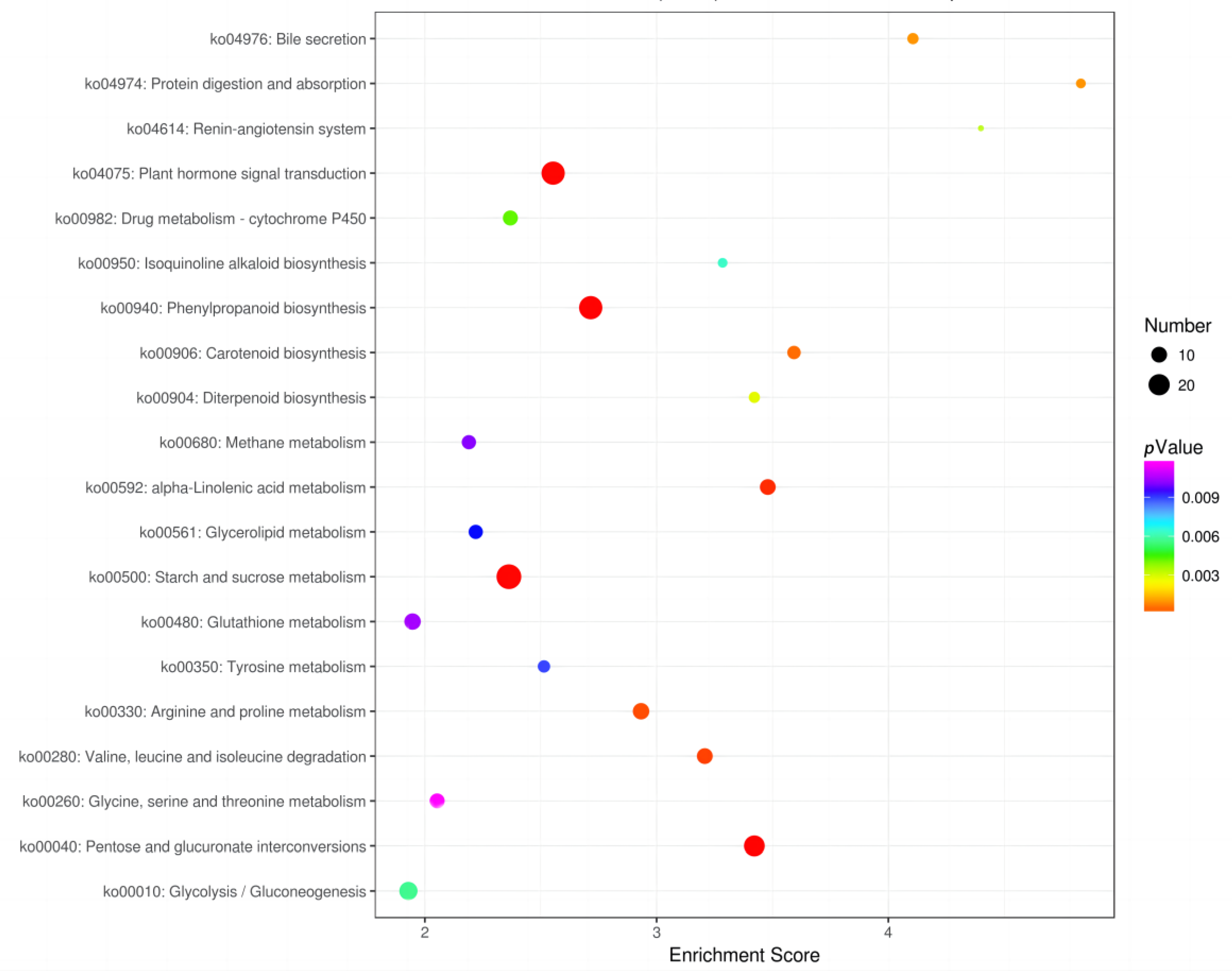
3.4. Functional Annotation and Enrichment Analysis

A GO enrichment analysis of the DEGs was performed for the CKC and 150C comparisons and the CKR and 150R comparisons in order to understand the potential mechanisms of castor plants under salt stress. The results showed that differentially expressed genes were more concentrated in the cotyledons than in the roots under salt stress. Moreover, 1580 DEGs were annotated into 3 categories of biological process, cellular components, and molecular functions in the cotyledons (Figure 3a). In terms of relation to biological process (BP), the abscisic acid metabolic process, ammonium transportation, and response to chitin formed the largest group under salt stress. In terms of cellular components (CC), the integral component of the plasma membrane, plasmodesmatal endoplasmic reticulum, and extracellular matrix were dramatically enriched. In terms of molecular function (MF), DNA binding transcription factor (TF) activity, sequence-specific DNA binding, and sulfate transmembrane transporter activity were dominant terms under salt stress. Hence, 1502 DEGs were classified into 30 terms in the roots, including BP (cellular response to sucrose starvation, pectin catabolic process, and secondary metabolite biosynthetic process), CC (extracellular region, apoplast, and cell wall), and MF (heme binding, pectinesterase inhibitor activity, and iron binding) (Figure 3b).
Furthermore, we performed a KEGG pathway analysis to conduct comprehensive, active biological pathways in DEGs between salt stress and control of cotyledons and roots.
Firstly, 726 DEGs were annotated into 192 KEGG pathways in the cotyledons, and 20 pathways were selected that were most significantly enriched in DEGs (Figure 4). KEGG enrichment analysis of DEGs in the cotyledons was conducted under salt stress; they were mainly distributed in ko04075 (plant hormone signal transduction, 29 genes), ko04141 (protein processing in endoplasmic reticulum, 22 genes), ko00500 (starch and sucrose metabolism, 22 genes), ko00052 (galactose metabolism, 9 genes), ko04213 (longevity regulating pathway, 9 genes), ko04612 (antigen processing and presentation, 8 genes), ko00460 (cyanoamino acid metabolism, 8 genes), ko00561 (glycerolipid metabolism, 8 genes), and ko00330 (arginine and proline metabolism, 22 genes).
Secondly, 602 DEGs in the roots were annotated into 162 KEGG pathways (Figure 5). The DEGs were significantly enriched in ko00500 (starch and sucrose metabolism, 29 genes), ko04075 (plant hormone signal transduction, 25 genes), ko00940 (phenylpropanoid biosynthesis, 25 genes), ko00040 (pentose and glucuronate interconversions, 19 genes), ko00010 (glycolysis/gluconeogenesis, 14 genes), ko00330 (arginine and proline metabolism, 11 genes), ko00480 (glutathione metabolism, 11 genes), ko00592 (alpha-linolenic acid metabolism, 10 genes), and ko00280 (valine, leucine, and isoleucine degradation, 10 genes).

3.5. TF Responses to Salt Stress

TFs are a class of protein molecules with special structures that can regulate gene expression. They control key downstream reactions by regulating the transcription of target genes, thereby causing a series of physiological and biochemical changes. Therefore, in this study, DEGs were further analyzed and annotated into 58 TF families. Both the numbers and classifications of TFs differed in the cotyledons and roots under salt stress. ERF (ethylene responsive factor), MYB-related (v-myb avian myeloblastosis viral oncogene homolog-related), NAC (NAM, ATAF1, ATAF2, and CUC2), bHLH (basic helix-loop-helix), C2H2 (Cys2/His2), and WRKY were the main enrichment families for DEGs in the cotyledons (Figure 6a), whereas MYB-related, bHLH, ERF, MYB, LBD (lateral organ boundaries domain), and NAC were the main enrichment families in the roots (Figure 6b).

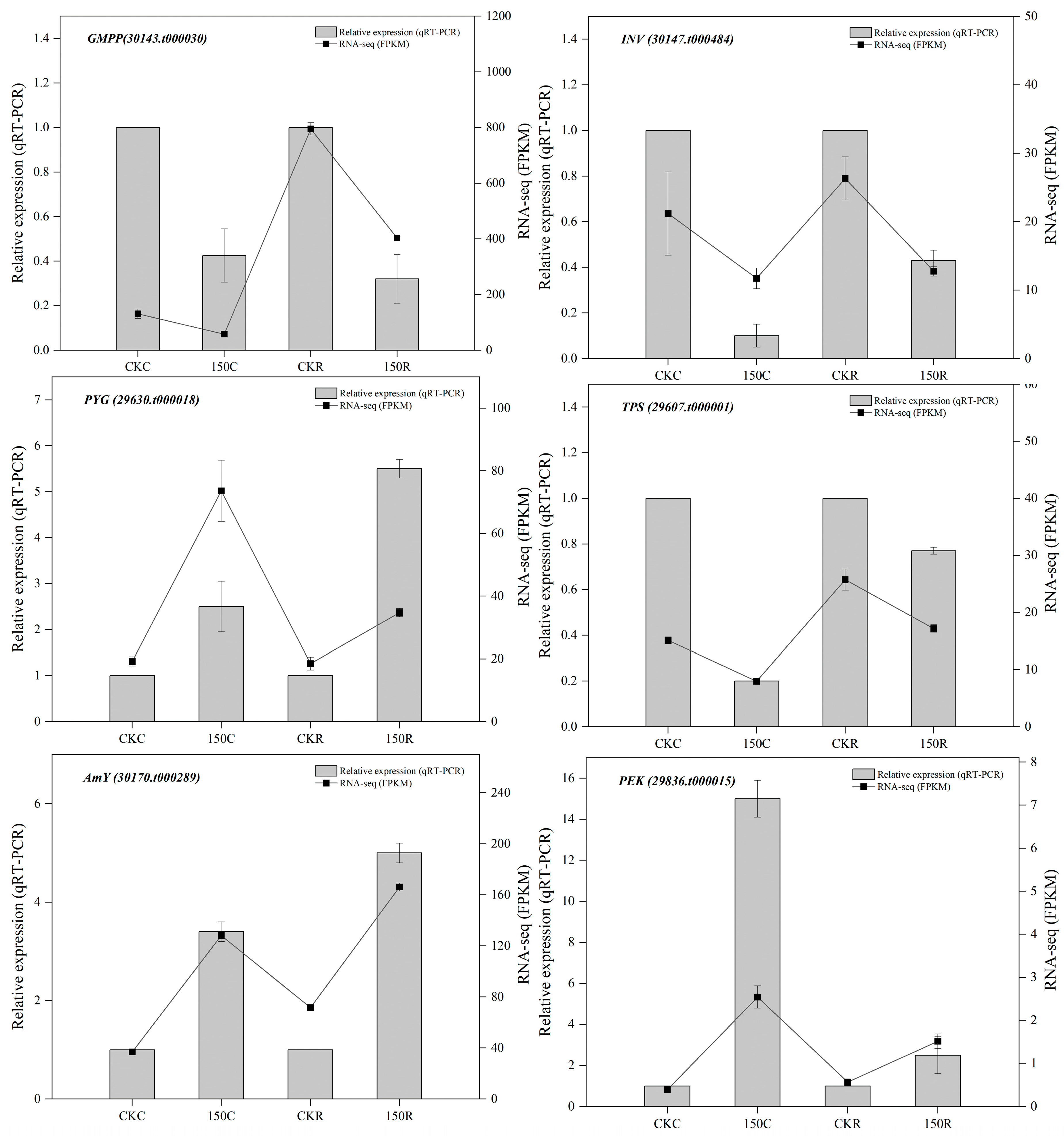
3.6. Validation of RNA-Seq by qRT-PCR
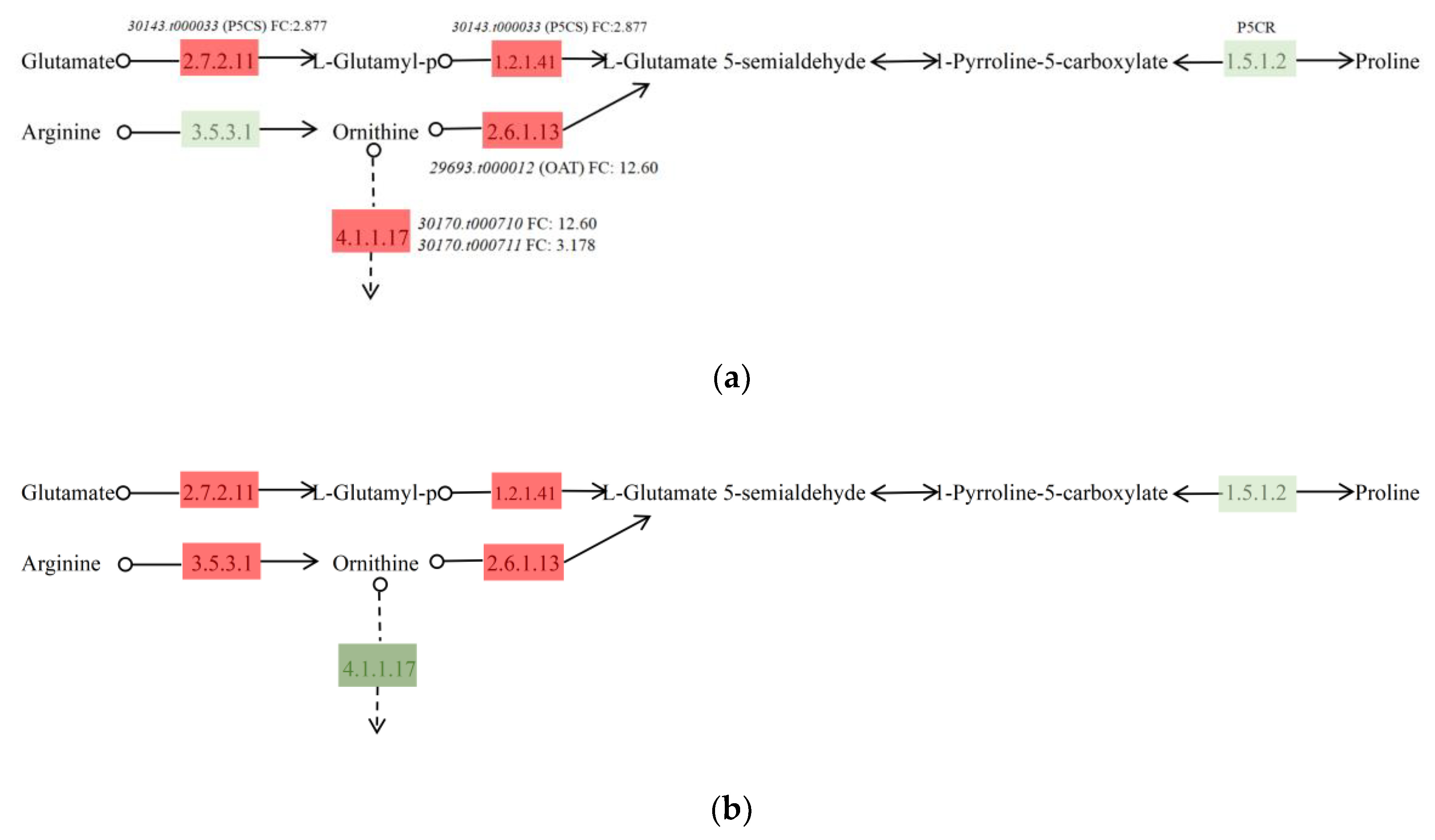
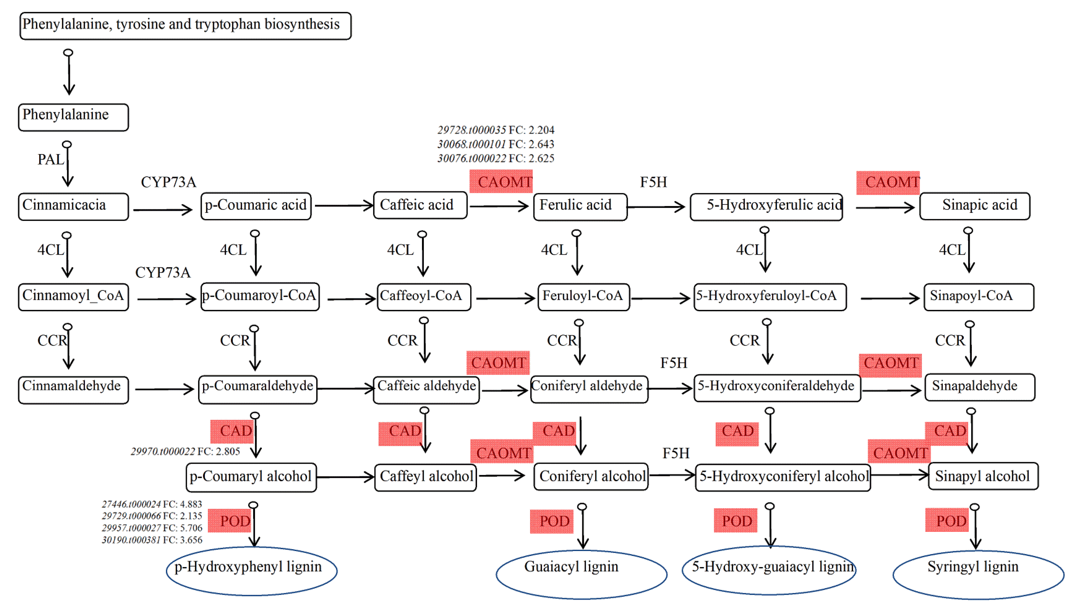
The relative expression of six genes was evaluated using qRT-PCR to validate the reliability of the data. Six genes were randomly selected: 30143.t000030 (GMPP), 30147.t000484 (INV), 29630.t000018 (PYG), 29607.t000001 (TPS), 30170.t000289 (AmY), and 29836.t000015 (PEK). The product of GMPP has the function of transporting mannose; INV is a key enzyme in sucrose metabolism, which irreversibly hydrolyzes sucrose into glucose and fructose and plays an important role in response to stress; PYG catalyzes the breakdown of glycogen; TPS is involved in the synthesis of trehalose, which has been observed to be associated with tolerance to various types of stress, including salt stress; AmY mediates starch degradation and plays a central role in the accumulation of soluble sugars; and PEK is a key enzyme in the glycolysis pathway [26,27,28,29,30]. The expression patterns of the six genes analyzed using qRT-PCR were consistent with those in RNA-seq data (Figure 7). Therefore, these results suggested that the RNA-seq data were reliable.

4. Discussion

Morpho-physiology, transcriptional regulation, and biochemical metabolism all play critical roles in plant responses to environmental stresses. Salt stress is always considered a major abiotic stress. Plants respond to salt stress via morphological, physiological, and biochemical changes, as well as the regulation of numerous genes. Castor plants have an ability to adapt to salt stress [31]. In this study, 16-day young seedlings were treated with 150 mM NaCl for 6 days, and their physiological activities and RNA-seq were examined. The results clearly indicated physiological and molecular responses with a tissue-specific pattern. Studies focusing on different expression patterns and the synergic relationship between cotyledons and roots in the adaptation to salt stress in the early seedling stage of plants are rare.
Plants undergo membrane lipid peroxidation to produce MDA under stress conditions [32]. MDA content can be used to assess the integrity of plant cell membranes under abiotic stress. The results indicated that salt stress could largely affect the status of the cell membrane in the seedlings of castor plants. The MDA content in both cotyledons and roots significantly increased under salt stress compared with that in the control group and was much higher in the cotyledons (Figure 1b). This indicated that the cotyledon membrane system was substantially damaged under salt stress, which was consistent with the fact that the DEGs of GO functional annotation classification in the cotyledons were mainly enriched in integral components of the plasma membrane. Improving osmotic regulation ability through osmotic regulation accumulation is also an important mechanism for plants in adapting to stress. Plants can reduce intracellular osmotic potential, improve cellular water retention, and maintain water balance by accumulating organic solutes such as soluble sugars, soluble proteins, and proline. The proline content in the cotyledons and roots of castor increased under salt stress, and the increase was greater in the cotyledons, which was consistent with the 22 DEGs and 11 DEGs participating in arginine and proline metabolism in the transcriptome analysis, respectively.
Plants always respond to salt stress via complex physiological and molecular mechanisms. Transcriptomes have been widely used to explore the effects of salt stress on plants and their regulatory mechanisms under such conditions. TFs are very important in modulating abiotic stress tolerance in plants. Many TFs, such as MYB, NAC, WRKY, ERF, bZIP (basic leucine zipper), and bHLH are closely associated with defense against abiotic stresses, including salt stress [33,34,35,36]. In this study, we found 58 groups of TF genes differentially regulated between cotyledons and roots under salt stress. Some TF genes were unique to the cotyledons and roots. These findings revealed that a large group of TF genes was involved in transcription regulation between cotyledons and roots in a specific manner under salt stress. Previous studies showed that the over-expression of OsMYB3R-2 resulted in an increase in tolerance to cold, drought, and salt stresses in transgenic Arabidopsis [37]. HcERF4 was found to play a positive role in regulating salt and drought stresses in kenaf [38]. Furthermore, studies showed that NAC was found to enhance resistance to salt stress in Populus [16]. Therefore, identifying some unique salt response genes in castor plants was possible. Further analysis of the functions and expressions controlling the mechanism of these genes would not only provide the opportunity of isolating and identifying some novel genes but also enhance our understanding of specific mechanisms of salt tolerance in cotyledons and roots during the early seedling stage of this species. Furthermore, previous studies showed that DEGs were mainly enriched in plant hormone signal transduction, phenylpropanoid biosynthesis, and starch, sucrose, and amino acid metabolism, in Musa acuminata [39]. In this study, DEGs in different tissues of castor plants under salt stress were analyzed using transcriptomics technology, and the significantly enriched KEGG pathways were determined. DEGs of cotyledons and roots were mainly co-enriched in plant hormone signal transduction, starch and sucrose metabolism, and arginine and proline metabolism. However, the two tissues also had differences in responding to salt stress. The DEGs of cotyledons were highly enriched in the protein processing of the endoplasmic reticulum, but in the roots were significantly enriched in phenylpropanoid biosynthesis.
Phytohormones are the most important endogenous substances and play a vital role in modulating abiotic stress tolerance in plants [40]. The results of this study indicated that plant hormone signal transduction (namely, ko04075) was significantly enriched in both cotyledons and roots (Figure 8). Auxin is a hormone that regulates the speed and direction of plant growth and is produced by cell regions with division and enlargement activity; it plays an indispensable role during the early seedling stage. In cotyledons, ARF (30170.t000573) was upregulated, promoting the downregulation of AUX/IAA (29598.t000019, 29598.t000020, 29844.t000021, 29883.t000047, and 29833.t000048) and the upregulation of SAUR (30147.t000725 and 30190.t000227), while ARF (29742.t000033), AUX/IAA (29844.t000020, 29844.t000021, and 29883.t000047), and SAUR (30169.t000414) were upregulated in the roots. These might further inhibit auxin accumulation in the roots. This result was not consistent with the fact that the genes encoding auxin were downregulated in the leaves of Podocarpus macrophyllus under salt stress [41]. This result probably differed from previous findings due to different salt concentrations and different tissues. ABA (abscisic acid) is a key regulator in plants and plays a crucial role in coordinating complex signal transduction mechanisms [42]. In this study, ABA signal transduction was significantly affected under salt stress, such as the downregulated genes encoding PYR/PYC (29729.t000023 and 29820.t000023) and upregulated genes encoding PP2C (29751.t000085 and 30169.t000292) in both cotyledons and roots, but ABF (29801.t000121) was only upregulated in the roots. Previous studies also indicated that the upregulation of PP2C, SnPK2, and ABF could enhance salt tolerance in Arabidopsis thaliana and Solanum tuberosum [43,44]. JA (jasmonic acid) is a major signaling molecule that controls plant growth and development [45]. The results showed that JAZ (29727.t000033) was significantly upregulated in the cotyledons, which directly controlled the transcription of MYC2 (27964.t000011) and negatively regulated the entire JA response, while the JA-related regulatory genes in the roots did not change significantly. This indicated that the cotyledons could resist salt stress.
Osmotic stress always occurs under salt stress in plants. Increasing evidence indicates that the accumulation of soluble sugars and proline could control membrane stability to alleviate salt stress [46]. In this study, we found that DEGs in the cotyledons and roots were significantly enriched in starch and sucrose metabolism, and also in arginine and proline metabolism. A gene (30143.t000033) encoding P5CS (pyrroline-5-carboxylate synthase) promoted proline synthesis in the glutamate pathway. In addition, ornithine generated P5C (pyrroline-5-carboxylate) under the catalysis of OAT (29693.t000012) and subsequently generated proline under P5CR (pyrroline-5- carboxylate reductase) activity (Figure 9a). In roots, arginine generated ornithine under arginase activity, which promoted the ornithine pathway (Figure 9b). Consistent with the RNA-seq analysis, the proline content in the roots was significantly higher than that in the cotyledons. As the roots are influenced by salt stress first, they may accumulate more osmotic substances to cope with the adverse effects. Previous studies found that P5CS could increase salt tolerance in Sorghum bicolor and Saccharum officinarum [47,48]. Furthermore, plants increased their concentration of enchylema and reduced its osmotic potential by breaking down starch into soluble sugars, including sucrose, glucose, fructose and trehalose [49]. Several genes were involved in soluble sugar metabolism (Tables S3–S6; Figures S1 and S2). We hypothesized that salt stress might alter gene expression and the enzyme activities related to sugar metabolism in castor plants to enhance plant tolerance.
Cotyledons and roots had a synergistic relationship to enhance salt tolerance, but they also had different expression patterns. The DEGs in cotyledons were highly enriched in protein processing of the endoplasmic reticulum in KEGG analysis (Table S7). Salt stress can cause protein misfolding and assembly, and Hsp70 has an important role in endoplasmic reticulum stress. In this study, multiple genes encoding Hsp70, Hsp90, Hsp40, and sHsf were upregulated (Figure S3). Consistent with our findings, previous studies suggested that Hsp70 was involved in abiotic stress responses in Arabidopsis, and the over-expression of Hsp70 could increase the heat and salt tolerance abilities of this species [50]. Therefore, we hypothesized that the upregulated expression of Hsp70 affected protein folding and assembly, thereby playing an important role in the response of cotyledons to salt stress. Phenylpropanoid biosynthesis involved lignin metabolism (Figure 10). Lignin is the main component of plant cell walls, and the degree of lignification increases under salt stress, which can improve the ability of plants to resist abiotic stresses. Salt stress alters the enzyme activity related to lignin biosynthesis, including PAL (phenylalanine ammonia-lyase), 4CL (4-coumarate–CoA ligase), CAD (cinnamyl alcohol dehydrogenase), and CAOMT (caffeic acid -O-methyltransferase), by upregulating these expression patterns and then improving the ability to adapt to salt stress [51]. For example, CmCAD2 was highly upregulated in melon under salt stress [52]. A previous study showed that TsCAD1 gene expression in the roots was significantly higher than that in stems and leaves [53]. In this study, we obtained one DEG encoding CAD, namely 29970.t000022, which was significantly upregulated, and we hypothesized that it might be the main gene in lignin synthesis responding to salt stress. In addition to CAD, we identified four upregulated genes responsible for encoding a POD (peroxidase) precursor, namely 7446.t00024, 29729.t000066, 29957.t000027, and 30190.t000381. Previous studies also found some upregulated DEGs encoding POD, which changed physiological and biochemical processes by accumulating POD to eliminate reactive oxygen species and then reduced the damage to the membrane [30]. Studies reported that PAL, CAOMT, 4CL, and CCR (cinnamoyl-CoA reductase) were major enzymes in plant lignification [51]. Currently, NAC, MYB, bHLH, and WRKY TFs also have been verified to be involved in regulating the phenylpropanoid biosynthesis pathway [54,55]. These results suggest that TFs regulated genes related to lignin synthesis and increased lignin content, which then enhanced salt tolerance in castor roots in the seedling stage.

5. Conclusions

This study provided an overview of the change in cotyledons and roots under salt stress. The results indicated that castor seedlings could accumulate osmotic regulators (e.g., soluble sugars and proline) and phytohormones in their cotyledons and roots to improve their salt tolerance. Furthermore, specific changes in terms of protein processing of the endoplasmic reticulum were observed in the cotyledons, and the roots could specifically increase the synthesis of lignin to reduce damage to the membrane of this species under salt stress. Hence, this study provides novel insights into the transcriptomic responses of castor, which were distinct in cotyledons and roots, as well as their synergic relationship under salt stress during the early seedling stage. Moreover, the functional genes were found to have the potential for enhancing the ability of castor plants to adapt to salt stress.

Supplementary Materials

The following are available online at https://www.mdpi.com/article/10.3390/agronomy13030693/s1, Figure S1: Ko00500 (arginine and proline metabolism) in the cotyledons of castor under salt stress. Red boxes represent upregulated genes, dark green boxes represent downregulated genes, and light green and light purple are castor-specific genes; Figure S2: Ko00500 (arginine and proline metabolism) in the roots of castor under salt stress. Red boxes represent upregulated genes, dark green boxes represent downregulated genes, and light green and light purple are castor-specific genes; Figure S3: Ko04141 (protein processing in endoplasmic reticulum) in the cotyledons of castor under salt stress. Red boxes represent upregulated genes, dark green boxes represent downregulated genes, and light green and light purple are castor-specific genes; Table S1. 283 DEGs (178 upregulated and 105 downregulated) involved in cotyledons of castor under salt stress; Table S2. 283 DEGs (176 upregulated and 107 downregulated) involved in roots of castor under salt stress; Table S3: Upregulated DEGs involved in starch and sucrose metabolism pathways in the cotyledons of castor under salt stress; Table S4: Downregulated DEGs involved in starch and sucrose metabolism pathways in the cotyledons of castor under salt stress; Table S5: Upregulated DEGs involved in starch and sucrose metabolism pathways in the roots of castor under salt stress; Table S6: Downregulated DEGs involved in starch and sucrose metabolism pathways in the roots of castor under salt stress; Table S7: DEGs involved in protein processing in the endoplasmic reticulum pathway in the cotyledons of castor under salt stress.

Author Contributions

Writing—original draft preparation, X.D.; experimentation, X.D., Y.M., S.C., and J.L. (Junyu Liu); data, C.S. and Z.J.; writing—review and editing, X.Y.; conceptualization and direction, J.L. (Jixiang Lin). All authors have read and agreed to the published version of the manuscript.

Funding

This research was funded by the Natural Science Foundation of Heilongjiang Province (YQ2019C005) and the Postdoctoral Foundation of Heilongjiang Province (LBH-Q19063).

Data Availability Statement

Not applicable.

Conflicts of Interest

The authors declare that there are no conflicts of interest.

References

  1. Wang, W.; Vinocur, B.; Altman, A. Plant responses to drought, salinity and extreme temperatures: Towards genetic engineering for stress tolerance. Planta 2003, 218, 1–14. [Google Scholar] [CrossRef]
  2. Munns, R.; Gilliham, M. Salinity tolerance of crops–what is the cost? New Phytol. 2015, 208, 668–673. [Google Scholar] [CrossRef] [Green Version]
  3. Fang, S.; Tu, W.; Mu, L.; Sun, Z.; Hu, Q.; Yang, Y. Saline alkali water desalination project in Southern Xinjiang of China: A review of desalination planning, desalination schemes and economic analysis. Renew. Sustain. Energy Rev. 2019, 113, 109268. [Google Scholar] [CrossRef]
  4. Zhao, S.; Zhang, Q.; Liu, M.; Zhou, H.; Ma, C.; Wang, P. Regulation of plant responses to salt stress. Int. J. Mol. Sci. 2021, 22, 4609. [Google Scholar] [CrossRef] [PubMed]
  5. Liang, W.; Ma, X.; Wan, P.; Liu, L. Plant salt-tolerance mechanism: A review. Biochem. Biophys. Res. Commun. 2018, 495, 286–291. [Google Scholar] [CrossRef] [PubMed]
  6. Yang, Y.; Guo, Y. Elucidating the molecular mechanisms mediating plant salt-stress responses. New Phytol. 2018, 217, 523–539. [Google Scholar] [CrossRef] [Green Version]
  7. Zhao, C.; Zhang, H.; Song, C.; Zhu, J.K.; Shabala, S. Mechanisms of plant responses and adaptation to soil salinity. Innov. 2020, 1, 100017. [Google Scholar] [CrossRef] [PubMed]
  8. Mijiti, M.; Zhang, Y.; Zhang, C.; Wang, Y. Physiological and molecular responses of Betula platyphylla Suk to salt stress. Trees 2017, 31, 1653–1665. [Google Scholar] [CrossRef]
  9. Mo, S.; Biao, A.; Wang, Z.; Lin, S.; Yang, T.; Pan, L.; Zeng, S. Spatio transcriptome uncover novel insight into the Lycium ruthenicum seedling tolerant to salt stress. Ind. Crops Prod. 2022, 177, 114502. [Google Scholar] [CrossRef]
  10. Zhang, D.; He, S.; Fu, Y.; Yu, R.; Gao, X.; Wang, Z.; Chen, M. Transcriptome analysis reveals key genes in response to salinity stress during seed germination in Setaria italica. Environ. Exp. Bot. 2021, 191, 104604. [Google Scholar] [CrossRef]
  11. Li, H.; Lv, Q.; Deng, J.; Huang, J.; Cai, F.; Liang, C.; Chen, Q. Transcriptome analysis reveals key seed-development genes in common buckwheat (Fagopyrum esculentum). Int. J. Mol. Sci. 2019, 20, 4303. [Google Scholar] [CrossRef] [PubMed] [Green Version]
  12. Han, P.L.; Li, S.X.; Yao, K.S.; Geng, H.Y.; Liu, J.Y.; Wang, Y.N.; Lin, J.X. Integrated metabolomic and transcriptomic strategies to reveal adaptive mechanisms in castor plant during germination stage under alkali stress. Environ. Exp. Bot. 2022, 203, 105031. [Google Scholar] [CrossRef]
  13. Hanley, M.E. Seedling herbivory, community composition and plant life history traits. Perspect. Plant Ecol. 1998, 1, 191–205. [Google Scholar] [CrossRef]
  14. Xu, X.; Zhang, L.; Cao, X.; Liu, L.; Jiang, B.; Zhang, C.; Han, T. Cotyledons facilitate the adaptation of early-maturing soybean varieties to high-latitude long-day environments. Plant. Cell Environ. 2021, 44, 2551–2564. [Google Scholar] [CrossRef]
  15. Shi, S.; An, L.; Mao, J.; Aluko, O.O.; Ullah, Z.; Xu, F. The CBL interacting protein kinase NtCIPK23 positively regulates seed germination and early seedling development in tobacco (Nicotiana tabacum L.). Plants 2021, 10, 323. [Google Scholar] [CrossRef]
  16. Wang, Y.; Xu, Y.; Peng, X.; Yan, J.; Yan, X.; Zhou, Z.; Lin, J. Cotyledon removal decreases salt tolerance during seedling establishment of Ricinus communis, an oilseed energy crop species. Ind. Crops Prod. 2019, 142, 111857. [Google Scholar] [CrossRef]
  17. Zhou, J.C.; Zhao, W.W.; Yin, C.H.; Song, J.; Wang, B.S.; Fan, J.L.; Feng, G. The role of cotyledons in the establishment of Suaeda physophora seedlings. Plant Biosyst. -Int. J. Deal. All Asp. Plant Biol. 2014, 148, 584–590. [Google Scholar]
  18. Wang, Y.N.; Peng, X.Y.; Hua, X.Y.; Zang, W.; Yang, X.F.; Lin, J.X. Research progress on the physiological function of plant cotyledons. Pratacultural. Sci. 2018, 35, 2988–2997. [Google Scholar]
  19. Ribeiro, P.R.; Fernandez, L.G.; Castro, R.D.; Ligterink, W.; Hilhorst, H.W. Physiological and biochemical responses of Ricinus communis seedlings to different temperatures: A metabolomics approach. BMC Plant Biol 2014, 14, 223–237. [Google Scholar] [CrossRef] [Green Version]
  20. Huang, X.Y.; Lu, Y. Research advance in preparation and application of castor seed oil and its derivatives. China Oils Fats 2011, 36, 52–56. [Google Scholar]
  21. Baldwin, B.S.; Cossar, R.D. Castor yield in response to planting date at four locations in the south-central United States. Ind. Crops Prod. 2009, 29, 316–319. [Google Scholar] [CrossRef]
  22. El-Nashar, Y.I.; Hassan, B.A.; Aboelsaadat, E.M. Response of Nemesia (Nemesia×hybridus) plants to different irrigation water sources and arbuscular mycorrhizal fungi inoculation. Agric. Water Manag. 2021, 243, 106416. [Google Scholar] [CrossRef]
  23. Sarula, L.P. Research progress on adaptation of castor (Ricinus communis L.) to Abiotic Stress. J. Inn. Mong. Univ. Natl. 2021, 36, 135–139. [Google Scholar]
  24. Heath, R.L.; Packer, L. Photoperoxidation in isolated chloroplasts: I. Kinetics and stoichiometry of fatty acid peroxidation. Arch. Biochem. Biophys. 1968, 125, 189–198. [Google Scholar] [CrossRef] [PubMed]
  25. Bates, L.S.; Waldren, R.P.; Teare, I.D. Rapid determination of free proline for water-stress studies. Plant Soil 1973, 39, 205–207. [Google Scholar] [CrossRef]
  26. Han, R.C.; Liu, L.L.; Liu, J.L.; Xie, D.M.; Peng, D.Y.; Yu, N.J. Cloning and quantitative expression analysis of GMPP gene from Dendrobium huoshanense. China J. Chin. Mater. Med. 2019, 44, 1552–1557. [Google Scholar]
  27. Tie, Y.Y.; Tian, J. Cloning of sativum inwertase gene AsINV and its expression analysis in response to low temperature and drought stress. Plant Physiol. J. 2021, 57, 2258–2270. [Google Scholar]
  28. Zhang, Y.Y.; Lv, Y.; Xuan, W.Y.; Zhang, X.Q.; Xu, N.J.; Sun, X. Response of trehalose—6-phosphate synthetase (TPS) to abiotic stress in Gracilariopsis lemaneiformis. Oceanol. Et Limnol. Sin. 2021, 52, 777–785. [Google Scholar]
  29. Yue, C.; Cao, H.L.; Lin, H.Z.; Hu, J.; Ye, L.J.; Li, J.M.; Hao, Z.L.; Hao, X.Y.; Sun, Y.; Yang, Y.J.; et al. Expression patterns of alpha-amylase and beta-amylase genes provide insights into the molecular mechanisms underlying the responses of tea plants (Camellia sinensis) to stress and postharvest processing treatments. Planta 2019, 250, 281–298. [Google Scholar] [CrossRef]
  30. Wang, C.; Zhang, J.; Xie, Y. Cloning and hpRNAi Vector Construction of Brassica napus Pyrophosphate-dependent Phosphofructokinase (PFK). Mol. Plant Breed. 2006, 14, 199–204. [Google Scholar]
  31. Huang, F.L.; Jiang, Y.X.; Zhang, S.B.; Liu, S.; Eh, T.J.; Meng, F.J.; Lei, P. A Comparative Analysis on Morphological and Physiological Characteristics between Castor Varieties (Ricinus communis L.) under Salt Stress. Sustainability 2022, 14, 10032. [Google Scholar] [CrossRef]
  32. Diao, H.Y.; Wang, A.Z.; Yuan, F.H.; Guan, D.X.; Sun, Y.; Wu, J.B. Applications of compound-specific isotope analysis in tree non-structural carbohydrates research: A review. Ying Yong Sheng Tai Xue Bao= J. Appl. Ecol. 2020, 31, 4291–4300. [Google Scholar]
  33. Zhu, J.K. Salt and drought stress signal transduction in plants. Annu. Rev. Plant Biol 2002, 53, 247–273. [Google Scholar] [CrossRef] [Green Version]
  34. Liu, L.; Tang, X.F.; Wu, Y.M.; Huang, Y.B.; Tang, Y.X. The roles of MYB transcription factors on plant defense responses and its molecular mechanism. Yi Chuan/Hered. 2008, 30, 1265–1271. [Google Scholar] [CrossRef] [PubMed]
  35. Lang, Y.; Liu, Z. Basic Helix-Loop-Helix (bHLH) transcription factor family in Yellow horn (Xanthoceras sorbifolia Bunge): Genome-wide characterization, chromosome location, phylogeny, structures and expression patterns. Int. J. Biol. Macromol. 2020, 160, 711–723. [Google Scholar] [CrossRef] [PubMed]
  36. Lei, P.; Liu, Z.; Hu, Y.; Kim, H.C.; Liu, S.; Liu, J.Q.; Xu, L.P.; Li, J.X.; Zhao, Y.; Yu, Z.L.; et al. Transcriptome analysis of salt stress responsiveness in the seedlings of wild and cultivated Ricinus communis L. J. Biotechnol. 2021, 327, 106–116. [Google Scholar] [CrossRef]
  37. Dai, X.; Xu, Y.; Ma, Q.; Xu, W.; Wang, T.; Xue, Y.; Chong, K. Overexpression of an R1R2R3 MYB gene, OsMYB3R-2, increases tolerance to freezing, drought, and salt stress in transgenic Arabidopsis. Plant Physiol. 2007, 143, 1739–1751. [Google Scholar] [CrossRef] [Green Version]
  38. Yue, J.; Tang, M.; Zhang, H.; Luo, D.; Cao, S.; Hu, Y.; Chen, P. The transcription factor HcERF4 confers salt and drought tolerance in kenaf (Hibiscus cannabinus L.). Plant Cell 2022, 150, 207–221. [Google Scholar] [CrossRef]
  39. Wei, J.; Liu, D.; Liu, Y.; Wei, S. Physiological analysis and transcriptome sequencing reveal the effects of salt stress on banana (Musa acuminata cv. BD) Leaf. Front. Plant Sci. 2022, 13, 822838. [Google Scholar] [CrossRef]
  40. Zhu, T.; Lin, J.; Zhang, M.; Li, L.; Zhao, C.; Chen, M. Phytohormone involved in salt tolerance regulation of Elaeagnus angustifolia L. seedlings. J. For. Res. 2019, 24, 235–242. [Google Scholar] [CrossRef]
  41. Zou, L.; Li, T.; Li, B.; He, J.; Liao, C.; Wang, L.; Wu, Q. De novo transcriptome analysis provides insights into the salt tolerance of Podocarpus macrophyllus under salinity stress. BMC Plant Biol. 2021, 21, 1–17. [Google Scholar] [CrossRef] [PubMed]
  42. Sicilia, A.; Testa, G.; Santoro, D.F.; Cosentino, S.L.; Lo Piero, A.R. RNA Seq analysis of giant cane reveals the leaf transcriptome dynamics under long-term salt stress. BMC Plant Biol. 2019, 19, 1–24. [Google Scholar] [CrossRef] [PubMed] [Green Version]
  43. Muñiz García, M.N.; Cortelezzi, J.I.; Fumagalli, M.; Capiati, D.A. Expression of the Arabidopsis ABF4 gene in potato increases tuber yield, improves tuber quality and enhances salt and drought tolerance. Plant Mol. Biol. 2018, 98, 137–152. [Google Scholar] [CrossRef] [PubMed]
  44. Ma, X.; Liu, J.N.; Yan, L.; Liang, Q.; Fang, H.; Wang, C.; Wu, D. Comparative transcriptome analysis unravels defense pathways of Fraxinus velutina Torr against salt stress. Front. Plant Sci. 2022, 13, 842726. [Google Scholar] [CrossRef]
  45. Moons, A.; Prinsen, E.; Bauw, G.; Van Montagu, M. Antagonistic effects of abscisic acid and jasmonates on salt stress-inducible transcripts in rice roots. Plant Cell 1997, 9, 2243–2259. [Google Scholar]
  46. Zhu, J.K. Abiotic stress signaling and responses in plants. Cell 2016, 167, 313–324. [Google Scholar] [CrossRef] [Green Version]
  47. Su, M.; Li, X.F.; Ma, X.Y.; Peng, X.J.; Zhao, A.G.; Cheng, L.Q.; Liu, G.S. Cloning two P5CS genes from bioenergy sorghum and their expression profiles under abiotic stresses and MeJA treatment. Plant Sci. 2011, 181, 652–659. [Google Scholar] [CrossRef]
  48. Iskandar, H.M.; Widyaningrum, D.; Suhandono, S. Cloning and characterization of P5CS1 and P5CS2 genes from Saccharum officinarum L. under drought stress. J. Trop. Crop Sci. 2014, 1, 23–30. [Google Scholar] [CrossRef]
  49. Geng, G.; Lv, C.; Stevanato, P.; Li, R.; Liu, H.; Yu, L.; Wang, Y. Transcriptome analysis of salt-sensitive and tolerant genotypes reveals salt-tolerance metabolic pathways in sugar beet. Int. J. Mol. Sci. 2019, 20, 5910. [Google Scholar] [CrossRef] [Green Version]
  50. Montero-Barrientos, M.; Hermosa, R.; Cardoza, R.E.; Gutierrez, S.; Nicolas, C.; Monte, E. Transgenic expression of the Trichoderma harzianum hsp70 gene increases Arabidopsis resistance to heat and other abiotic stresses. J. Plant Physiol. 2010, 167, 659–665. [Google Scholar] [CrossRef]
  51. Shafi, A.; Chauhan, R.; Gill, T.; Swarnkar, M.K.; Sreenivasulu, Y.; Kumar, S.; Singh, A.K. Expression of SOD and APX genes positively regulates secondary cell wall biosynthesis and promotes plant growth and yield in Arabidopsis under salt stress. Plant Mol. Biol. 2015, 87, 615–631. [Google Scholar] [CrossRef] [PubMed]
  52. Liu, W.; Jin, Y.; Li, M.; Dong, L.; Guo, D.; Lu, C.; Qi, H. Analysis of CmCADs and three lignifying enzymes in oriental melon (‘CaiHong7’) seedlings in response to three abiotic stresses. Sci. Hortic. 2018, 237, 257–268. [Google Scholar] [CrossRef]
  53. Sui, J.; Deng, H.; Yang, J.; Sun, J.; Qu, C. Cloning and expression characteristics of TsCAD1 gene under abiotic stress condition in Toona sinensis. Genom. Appl. Biol. 2019, 38, 4617–4625. [Google Scholar]
  54. Zhao, W.; Ding, L.; Liu, J.; Zhang, X.; Li, S.; Zhao, K.; Chen, F. Regulation of lignin biosynthesis by an atypical bHLH protein CmHLB in Chrysanthemum. J. Exp. Bot. 2022, 73, 2403–2419. [Google Scholar] [CrossRef]
  55. Guo, G.Y.; Bai, F.; Liu, W.; Mi, C.L. Advances in research of the regulation of transcription factors of lignin biosynthesis. Sci. Agric. Sin. 2015, 48, 1277–1287. [Google Scholar]
Figure 1. Dry weight (a) and content of malondialdehyde (b), soluble sugar (c) and proline (d) in the cotyledons and roots of castor plants under salt stress. CKC: (control, cotyledons), 150C: (salt stress, cotyledons), CKR: (control, roots), 150R: (salt stress, roots). Bars represent mean ± SD (n = 3). Values marked with different letters differ significantly from each other (p < 0.05).
Agronomy 13 00693 g001
Figure 2. Principal component analysis of castor seedling expression levels under salt stress (a), the number of differentially expressed genes (DEGs) upregulated and downregulated in castor seedlings under salt stress (b), and Venn diagram analysis of DEGs annotated in the NR database (c). CKC: (control, cotyledons), 150C: (salt stress, cotyledons), CKR: (control, roots), 150R: (salt stress, roots).
Agronomy 13 00693 g002
Figure 3. Classification of unigenes in GO annotation of castor cotyledons and roots under salt stress: (a) CKC (control, cotyledons) vs. 150C (salt stress, cotyledons) and (b) CKR (control, roots) vs. 150R (salt stress, roots).
Agronomy 13 00693 g003
Figure 4. The 20 most significantly enriched KEGG pathways of DEGs in the cotyledons under salt stress (CKC vs. 150C). CKC: control, cotyledons; 150C: salt stress, cotyledons. The dot size and color indicate gene number and p-value, respectively.
Agronomy 13 00693 g004
Figure 5. The 20 most significantly enriched KEGG pathways of DEGs in the roots under salt stress (CKR vs. 150R). CKR: control, roots; 150R: salt stress, roots. The dot size and color indicate gene number and p-value, respectively.
Agronomy 13 00693 g005
Figure 6. Transcription factor family analysis of castor cotyledons and roots under salt stress: (a) CKC (control, cotyledons) vs. 150C (salt stress, cotyledons) and (b) CKR (control, roots) vs. 150R (salt stress, roots).
Agronomy 13 00693 g006
Figure 7. Expression pattern validation of six randomly selected genes in the cotyledons and roots of castor under salt stress by qRT-PCR. CKC: (control, cotyledons), 150C: (salt stress, cotyledons), CKR: (control, roots), 150R: (salt stress, roots). The normalized expression level (FPKM) of RNA-Seq is indicated on the y-axis. Bars represent mean ± SD (n = 3).
Agronomy 13 00693 g007
Figure 8. Analysis of plant hormone signal transduction in terms of DEGs: (a) ko04075 (plant hormone signal transduction) in the cotyledons of castor under salt stress. (b) ko04075 (plant hormone signal transduction) in the roots of castor under salt stress. Red boxes represent upregulated genes, dark green boxes represent downregulated genes, yellow has both upregulated and downregulated genes, and light green and light purple are castor-specific genes.
Agronomy 13 00693 g008
Figure 9. Analysis of arginine and proline metabolism in terms of DEGs: (a) Ko00300 (arginine and proline metabolism) in the cotyledons of castor under salt stress. (b) ko00300 (arginine and proline metabolism) in the roots of castor under salt stress. Red boxes represent upregulated genes, dark green boxes represent downregulated genes, yellow has both upregulated and downregulated genes, and light green and light purple are castor-specific genes.
Agronomy 13 00693 g009
Figure 10. Overview of lignin biosynthesis in the roots of castor under salt stress. Red boxes represent upregulated genes. PAL, phenylalanine ammonia-lyase; 4CL, 4-coumarate–CoA ligase; CCR, cinnamoyl-CoA reductase; CYP73A, trans-cinnamate 4-hydroxylase; CAOMT, caffeic acid -O-methyltransferase; CAD, cinnamyl alcohol dehydrogenase; POD, peroxidase; F5H, ferulate 5-hydroxylase.
Agronomy 13 00693 g010
Table 1. Primer sequences of real-time quantitative PCR of castor.
Gene IDForward PrimerReverse Primer
AMY (30170.t000289)CTTCAAGCTGCTGTGCAAGGGTTCCTTCAGCCCCCAATCA
PFK (29836.t000015)GGTATTGGCCTGGTGAAGCTAACAACAAGAACCGCATGGC
PYG (29630.T000018)GCACTGGTTCGCATTTGTGTAAGACTCCGGTTACTTGGCG
TPS (29607.t000001)GGGTGATGTTGTTTGGTGCCCACTGACAAAATGCCGTGCA
GMPP (30143.t000030)GGATGGACATTGGACAGCCAACACAGCCTGGACCTATTGC
INV (30147.t000484)AGGTCCATTTGGGCTTCTGGCGCAGCTTTTCATCGTCGAG
Table 2. Sequencing data quality and mapping results of castor cotyledons and roots under salt stress.
SampleRaw ReadsRaw BasesClean ReadsClean BasesValid BasesQ30GCMapped Reads
CKC150.78M7.62G50.31M7.32G96.14%95.45%43.26%95.61%
CKC250.19M7.53G49.77M7.21G95.72%95.78%43.33%96.32%
CKC348.08M7.21G47.66M6.89G95.54%95.74%43.51%96.52%
CKR147.67M7.15G47.32M6.83G95.47%96.15%43.19%95.60%
CKR251.55M7.73G51.16M7.36G95.25%96.02%43.18%96.25%
CKR348.77M7.31G48.38M6.94G94.87%96.11%43.18%95.27%
S150C148.04M7.21G47.60M6.85G95.03%95.86%43.25%95.39%
S150C250.49M7.57G50.06M7.24G95.58%95.96%43.28%95.28%
S150C348.91M7.34G48.55M7.04G96.00%95.83%43.48%95.52%
S150R150.95M7.64G50.55M7.31G95.63%95.69%43.29%95.58%
S150R249.55M7.43G49.18M7.13G95.89%95.78%43.36%95.16%
S150R350.56M7.58G50.10M7.31G96.40%95.17%43.43%94.71%
Disclaimer/Publisher’s Note: The statements, opinions and data contained in all publications are solely those of the individual author(s) and contributor(s) and not of MDPI and/or the editor(s). MDPI and/or the editor(s) disclaim responsibility for any injury to people or property resulting from any ideas, methods, instructions or products referred to in the content.

