Article

Shell Deformities in the Green-Lipped Mussel Perna viridis: Occurrence and Potential Environmental Stresses on the West Coast of Peninsular Malaysia

1
Department of Biology, Faculty of Science, Universiti Putra Malaysia, Serdang 43400, Selangor, Malaysia
2
School of Biology, Universiti Teknologi MARA, Negeri Sembilan Branch, Kuala Pilah Campus, Kuala Pilah 72000, Negeri Sembilan, Malaysia
3
Graduate School of Maritime Sciences, Faculty of Maritime Sciences, Kobe University, Kobe 658-0022, Japan
4
Faculty of Science and Marine Environment, Universiti Malaysia Terengganu, Kuala Nerus 21030, Terengganu, Malaysia
5
Ocean Pollution and Ecotoxicology (OPEC) Research Group, Universiti Malaysia Terengganu, Kuala Nerus 21030, Terengganu, Malaysia
6
Fisheries Research Institute, Batu Maung 11960, Pulau Pinang, Malaysia
7
Department of Environmental Science, Faculty of Mathematics and Natural Sciences, Universitas Sebelas Maret, Jl. Ir. Sutami 36A, Surakarta 57126, Central Java, Indonesia
8
Biodiversity Research Group, Universitas Sebelas Maret, Jl. Ir. Sutami 36A, Surakarta 57126, Central Java, Indonesia
9
Faculty of Health and Life Sciences, INTI International University, Persiaran Perdana BBN, Nilai 71800, Negeri Sembilan, Malaysia
10
Physics Department, Faculty of Science, Al-Azhar University, Asyut 71524, Egypt
11
Institute of Physics and Technology, Ural Federal University, 620002 Yekaterinburg, Russia
*
Author to whom correspondence should be addressed.
Pollutants 2023, 3(3), 406-418; https://doi.org/10.3390/pollutants3030028
Submission received: 3 June 2023 / Revised: 20 July 2023 / Accepted: 25 July 2023 / Published: 4 September 2023

Abstract

:
The green-lipped mussel Perna viridis’ sensitive nature and characteristic as a benthos organism that filters the sediment in its environment make it one of the possible bioindicators for pollution in the aquatic ecosystem. The present study aimed to determine the percentages of total shell deformities in comparison to the past data in the coastal waters of Peninsular Malaysia. It was found that several types of discontinuous, continuous, and unexplained shell abnormalities contributed to the overall range of shell deformities of 15.8–87.5%, which was greater in comparison to that (0.0–36.8%). The present study showed that the highest overall proportion of shell abnormalities occurred in Teluk Jawa, whereas the lowest percentages were found in Kampung (Kg.) Pasir Puteh. The regulative mechanisms at the well-known polluted sites at Kg. Pasir Puteh could be the explanation. Further research should be conducted to determine the degree of heavy metal that may be the source of these malformations in the mussel shells.

1. Introduction

Mariculture of bivalves has a long history dating back to 2000 years ago in China for oysters [1]. It is known for being feasible, as it requires no artificial feeding processes or an expensive farming structure. Bivalve mollusks such as oysters, mussels, and clams have been an important food source for many coastal residences and the general population ever since. In 2020, bivalves were estimated to hold about a 2.8% value of the global aquatic products. A total of 17.7 million tonnes of mollusks, primarily bivalves, were farmed and exported globally. The total value of export was USD 4.3 billion [2].
Other than consumption uses, bivalve mollusks have also been studied as an indicator species of the ocean. Indicator species are responsive to environmental changes but also tolerant, so they are used to estimate the ecosystem’s health and environmental conditions. One of the bivalve mollusks species found commonly on the west coast of Peninsular Malaysia is Perna viridis (Linnaeus, 1758), known as the green-lipped mussels. P. viridis is suitable for biomonitoring on the west coast of Peninsular Malaysia due to eight criteria, which are wide geographical distribution, sedentary lifestyle, easy sampling, the simple correlation between the metal levels in mussels and the environment, species with low morphological variations for easier identification, ability to accumulate pollutants within the tissues of the mussel, sensitive to environmental changes but tolerant, and potential health risk as they are consumed commercially. One of the many methods of P. viridis’ indication of environmental change is the presence of shell deformity. There are multiple reasonings to the cause of shell deformity, which include environmental conditions, such as temperature, pH levels or pollutants, nutrient deficiencies [3], infections and parasitism from barnacles, bacteria, or algae, lack of substratum, genetic factors, and physical trauma [4].
The presence of shell abnormalities in P. viridis collected from Peninsular Malaysia was believed to be caused by exposure to high concentrations of heavy metals cadmium (Cd), copper (Cu), lead (Pb), and zinc (Zn) in the substrate [5]. The flow of heavy metals into the estuaries and oceans, where mussels reside, is likely due to human activities. Heavy metals such as Cd, Cu, and Zn are often used as feed additives in pig farms as growth promoters and antibiotic alternatives [6]. In a study by Yap et al. [7], the heavy metal content found in the Sepang Besar River was assessed and compared over a 12-year period. After the 1998 piggery ban, the heavy metal content had drastically diminished. For example, the concentration of Zn was 9.40 times lower, and Cu was 36.9 times lower by 2010. However, the heavy metal content found in the manure and waste of the piggery farm was still present and persistent after 12 years. Heavy metals can also be unleashed through mining, smelting, or metal-based activities within the factories [8]. The unregulated disposal of chemical wastes and sewage from agricultural farms and industrial factories can also cause heavy metal pollution. Fertilizers used for palm oil plantations and other open farms can also runoff into the sewer or rivers after the incidence of flash flooding and heavy rains during the monsoon seasons of Malaysia [9].
For this study, only the shell of mussels was sampled and sorted into different deformity types. The soft tissue analysis, ocean water quality, and environmental information of the sample sites were not recorded. Therefore, the heavy metal content or pollutants within the water were not measured. However, shell deformity can still be used as a bioindicator. In a study conducted by Yap et al. [10], heavy metal sensitivity was compared between the periostracum (the outermost layer of the mollusks shell) and the soft tissue of P. viridis. The result showed that periostracum was a better biomonitoring material for Pb but not Cd.
Studies of shell deformations in P. viridis, thought to be effects of environmental conditions, can be used for biological monitoring. Based on a comprehensive review of previous studies through 2021, Yap et al. [11] concluded that bivalves’ shell has the potential for biomonitoring metal pollution, and it is believed that they can be reconstructed to reflect the history of metal pollution. The idea is to use the bivalves’ shells over the use of soft tissues to monitor metal pollution, which is acceptable and practical. Shells can be collected and easily stored for an extended period without freezing them. Data on the bioaccumulation of metals in mussel shells can be collected and compared to past pollution. Early in larval development, shell formation is initiated in mollusks. Sequential carbonate deposition continues and contributes to shell growth throughout life after metamorphosis. Metals incorporated into the shell and soft tissues of mollusks were absorbed through their food or directly from seawater [12]. The research focused mainly on the accumulation of metals in soft tissues. However, using shells to monitor metal contamination has much less variability. It offers a number of advantages over tissues [13,14]: serial incorporation of elements throughout the period of shell formation, higher preservation potential even after the organism’s death, and relatively cheap and easy storage [15]. Trace metals incorporated into shell components during growth must have been assimilated by the organism. Elliptio complanata is an example of a species often used as a biomonitor; the component not exposed to water or erosion is the nacre. Incorporation of trace metals into the shell accompanies shell formation and is a slow process compared to accumulation and release from soft tissue. The relationship between trace metal concentrations in the nacre and the environment is less variable and more consistent than for tissues. Because of its higher sensitivity, shell nacre is a more sensitive indicator and can potentially be a good biomonitor of metal concentrations in the environment [14].
The objective of the present study was to determine the percentages of total shell deformities compared to the past data in the coastal waters of Peninsular Malaysia. New speculation could be derived from the previous heavy metal data and the present shell deformity data.

2. Materials and Methods

2.1. Sampling and Shell Measurements

Live samples of P. viridis were purchased from fishermen at six different sites (Table 1, Figure S1) located along the west coast of Peninsular Malaysia (Figure 1) with no pre-order. Furthermore, samples were bought in weight units instead of counting to save time from counting exactly 100 samples for every purchase. Moreover, the fishermen might not have had enough harvest of P. viridis on the date of purchase. Therefore, some sites will have more mussels than others. This means that the samples obtained were random and not biased.
Live P. viridis was cooked to open the shell and cleaned by removing barnacles attached to the shells. Then, shell weight (g ± SE), shell length (mm ± SE), shell height (mm ± SE), and shell width (mm ± SE) were measured using a vernier caliper and electronic scale (Figure 2). All recorded morphological data were calculated for mean, median, standard error, minimum, and maximum values.

2.2. Statistical Analysis
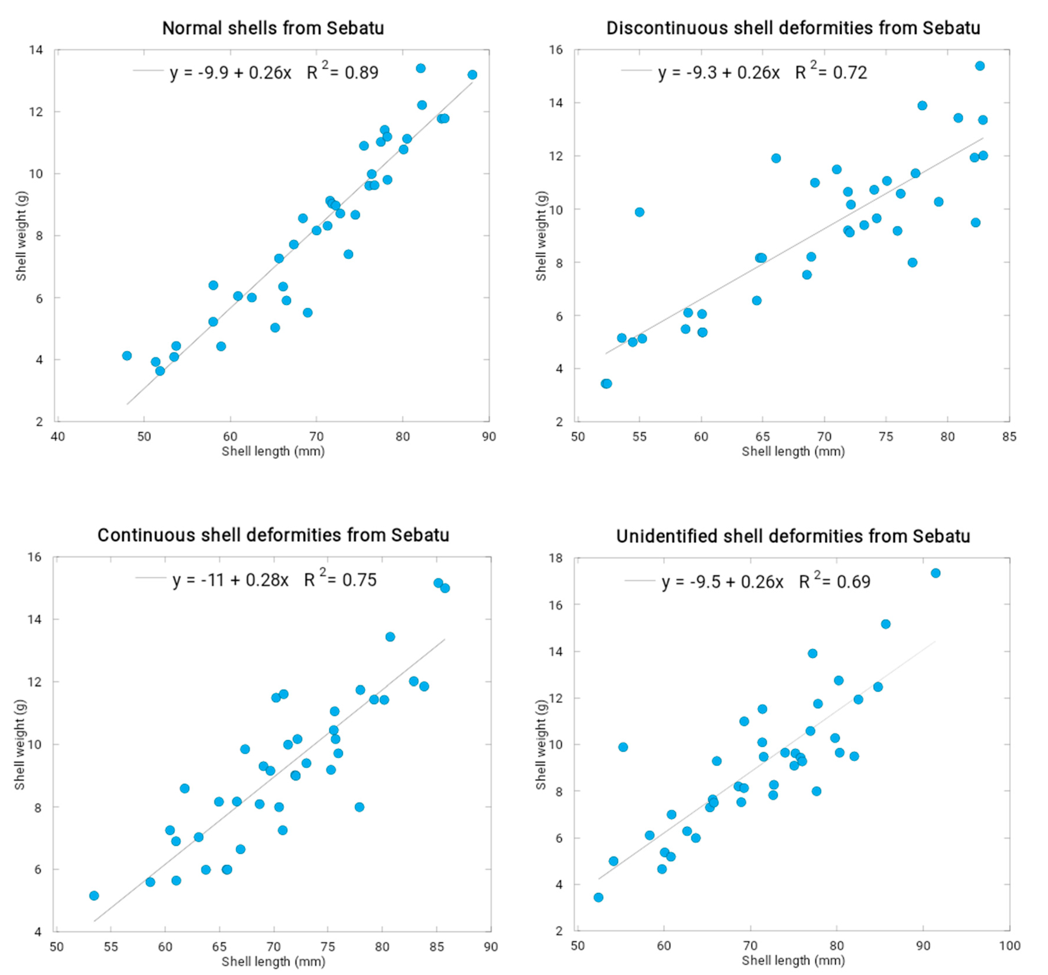
A KaleidaGraph (Version 3.08, Synergy Software, Eden Prairie, MN, USA) was used for statistical analysis and to create the graphical bar charts in this study. The relationships between the shell length and other shell’s allometric parameters (shell width, shell height, and shell weight) were investigated using multiple linear regression analysis in STATISTICA (Version 10; StatSoft. Inc., Tulsa, OK, USA, 1984–2011).

3. Results

Table S1 shows all recorded parameters, which include the mean, median, standard error, minimum, and maximum of each shell’s characteristics (length, width, height, and weight). Also, the total shell count, percentage of discontinuous shell deformity from the site, percentage of continuous shell deformity from the site, and the percentages of total shell deformity were recorded. The result can be summarized into shell deformity count and the percentage of total shell deformity.
Shell deformities of different types, their count, and the percentage of total shell deformity are listed in Table 2. The P. viridis shell deformity classification was obtained based on the study by Sunila and Lindstrom [5] for the blue mussel Mytilus edulis, where shell deformity could be classified into continuous or discontinuous deformities. The classification of deformity types by Sunila and Lindstrom [5] is described as follows: posterior score (growth ring curved towards the apex and formed a deep hollow); ventral/dorsal score (groove around the byssus cleft); flattening of posterior/anterior parts (direction of growth changes); and streak (brown or yellow streak, formed a collar bending posteriorly). Every other deformity that does not fit these criteria will be listed under unidentified deformity. In this study, we found discontinuous shell deformities such as dorsal streak, posterior streak, and flattening of the anterior part. On the contrary, we found continuous shell deformities such as posterior score, dorsal score, and ventral score (Figure 3). The listed deformity can be severe or slight, but it was not described within this study. Teluk Jawa sites recorded a high percentage of shell deformities (87.5%), while Kampung (Kg.) Pasir Puteh showed the least percentage of deformities (15.8%) (Table 2).
In the present study, 35.3% of the 201 mussels collected in Parit Jawa had shell deformities. About 28.9% were found as discontinuous, and 6.47% were continuous deformities. The mean for shell length, width, height, and weight in discontinuous and continuous deformities is 75.46 mm ± 1.75; 24.86 mm ± 0.62; 33.07 mm ± 0.56; 10.22 g ± 0.43 and 68.61 mm ± 2.15; 25.04 mm ± 1.2; 31.84 mm ± 0.98; 9.35 g ± 0.88, respectively. A previous study for heavy metal contamination in Parit Jawa by Edward et al. [16] reported that bivalve species, Polymesoda erosa, have high concentrations of Pb, Ni, Zn, and Fe, which were 12.2, 7.70, 222, and 1307 mg/kg, respectively.
A total of 122 mussels were retrieved from the Parit Karang Tangkak, where 53.3% of the mussels were found to have shell deformities. Discontinuous and continuous deformities were observed. A total of 39.3% of the deformities were discontinuous, which included flattening of the shells’ anterior part, dorsal streak, and posterior streak. The mean values of length, width, height, and weight of deformed mussels were 74.67 mm ± 1.63, 27.19 mm ± 0.4, 135.93 mm ± 0.5, and 9.66 g ± 0.43, respectively. Meanwhile, continuous deformities such as posterior, dorsal, and ventral scores were observed. They constituted 13.9% of the deformities, with means of length, width, height, and weight of 72.11 mm ± 1.77, 25.59 mm ± 0.73, 32.04 mm ± 0.71, and 9.24 g ± 0.75, respectively. Mussels with no shell deformities have the mean length, width, height, and weight of 73.85 mm ± 0.91, 26.07 mm ± 00.55, 31.46 mm ± 0.35, and 9.64 g ± 0.95, respectively.
A total of 164 mussels were collected from Kuala Sebatu, Johore. It has one of the highest shell deformity percentages of 75.6%. Also, many of its shells had multiple deformities present in one shell. Of all the shells observed, 45.1% were discontinuous, 13.4% were continuous, and 17.1% were unidentified deformities. The mean length, width, height, and width of each shell deformity type of mussel shells are listed in Table 3.
A total of 37.6% of mussels collected from Kuala Masai in Johore Straits were found to possess shell deformities. About 25.5% were found as discontinuous, 10.07% were continuous, and 2.01% were unidentified deformities. The mean values for shell length, width, height, shell weight in discontinuous, continuous, and unidentified deformities are 66.01 mm ± 2.00; 23.70 mm ± 0.52; 30.65 mm ± 0.65; 7.84 g ± 0.52, 68.71 mm ± 3.00; 24.04 mm ± 0.72; 31.03 mm ± 1.23; 8.38 g ± 1.00, and 62.01 mm ± 5.71; 21.39 mm ± 2.08; 26.27 mm ± 1.49; 6.44 g ± 1.67, respectively. In recent studies in Sg Masai, Zn and Cu (38.9 and 7.77 µg/g) accumulated at a higher concentration than Pb and Cd (0.41 and 0.14 µg/g) in P. viridis. Sg Masai is adjacent to urban areas, with relatively few nearby industrial activities. The findings suggested that the deformities in the shell could be caused by heavy metal contamination in the collected area. Over time, industrial activities and urban development around the Straits of Johore have increased, potentially causing heavy metal pollution in the aquatic environment [17].
In total, only 48 shells were collected from Teluk Jawa. This site was found to have the mean highest shell deformity percentage of 87.5%. Among the deformed shells, 56.3% were discontinuous, 4.17% were continuous, and 27.1% were unidentified deformities. The mean length, width, height, and width of each shell deformity type of mussel shells are listed in Table 4.
From Kg Pasir Puteh, a total of 139 mussels were retrieved. Only 15.8% showed shell deformities, where 13.7% had discontinuous shell deformities, and only 2.16% had continuous shell deformities. Normal shells (84.2% of the total) have means of 58.49 mm ± 1.74, 24.10 mm ± 0.24, 24.76 mm ± 0.26, and 5.38 g ± 0.24 for shell length, width, height, and weight, respectively. Discontinuous shell deformities, such as posterior streak and dorsal streak, have the means of 50.66 mm ± 1.81, 21.84 mm ± 0.95, 26.05 mm ± 1.22, and 2.07 g ± 0.24 in length, width, height, and weight, respectively. As for continuous shell deformities, only posterior and dorsal scores were observed, with means of 56.07 mm ± 8.47, 18.83 mm ± 0.65, 20,73 mm ± 0.80, and 3.80 g ± 0.45 in length, width, height, and weight, respectively.
From Figure 4, the coefficient of determinations (R2) between the shell lengths and shell weights of the normal shells is higher (R2 = 0.89) than those of the discontinuous shell deformities (R2 = 0.72), continuous shell deformities (R2 = 0.75), and unidentified shell deformities (R2 = 0.69). This indicated that shell deformities of all types had caused more deviations and anomalies of P. viridis shell growths. Multiple linear regression analyses show the highest R2 value (0.90) for the normal shell length when correlated to shell width, shell height, and total shell weight. It is higher than the R2 values for the deformed shell lengths for discontinuous type (R2 = 0.78), continuous type (R2 = 0.84), and unidentified shell deformity type (R2 = 0.74) (Table S2).

4. Discussion

Due to the complexities of the environment, no single factor can be easily isolated as a specific cause of shell deformities. Shell abnormalities should be regarded as a bioindicator of unfavorable environmental conditions, even though their potential for metal pollution bioindication is unproven based on the few data available in the literature [18].
Shell morphology is a remarkably consistent characteristic that serves as the foundation for bivalve classification. As a result, changes in shell size, shape, thickness, weight, bilateral asymmetry, growth of carbonate structures on the internal shell surface, and the development of abnormalities may indicate a variety of events in the organism or the surrounding environment, such as diseases, attack by epibionts, parasites, substrate type, water current, oxygen concentration, and aquatic pollution [19,20,21,22,23,24].
Shell deformities in green mussels were suggested to be influenced by many factors, such as the presence of heavy metal contamination (Cd and Cu) that were discharged by the nearby industrial areas, hydrocarbons contamination [25], as well as microplastic contamination from [26]. Yap et al. [27] previously found that 9.52% of the green mussels in Kg. Pasir Puteh suffered from shell deformities, which were relatively lower than the present finding, which recorded a total of 15.83% shell deformities. This indicates a potential increase in heavy metal effluent discharge from industrial areas over the decade. The study from Zahid et al. [26] on the microplastic accumulation in green mussels at the same site found that the smaller green mussels accumulated more microplastic inside the soft tissue than the larger-sized mussel, suggesting that microplastic accumulation might also be the potential factor that affected shell deformities. Other possible factors, such as polonium (Po) contamination, were higher in wild green mussels than the cultured and market green mussels due to direct contamination from industrial and domestic discharges [28].
The high shell deformities recorded in the populations of Teluk Jawa (87.5%) and Kuala Sebatu (75.6%) could be due to several reasons. As mentioned earlier, there are multiple reasons why P. viridis may contribute to the occurrence of shell deformities. Firstly, it could be the change in environmental conditions, such as the temperature of the water, pH levels of the water, salinity of the water, conductivity of the water, and the amount of oxygen dissolved in the water [29]. Kuala Sebatu is known as a site with no human disturbance [29] and relatively fewer heavy metal pollutants than Muar. Heavy metal pollutants have been speculated as the key important environmental factor that causes shell deformity. Previous heavy metal analysis on the mussel shells retrieved from Sebatu [10] reported around 26 mg/g dry weight of Pb in the shell and around 19 mg/g dry weight of Pb in the soft tissue. Furthermore, the average shell length of samples collected in two Sebatu sites was about 63.04 mm~85.4 mm, which gives an average of 74.22 mm. Compared to the data presented in Table S1, the average shell length of green-lipped mussels has decreased slightly (4 mm). Previously, Yap et al. [29] reported that byssus had the highest heavy metal concentration among other body parts of P. viridis collected from Sebatu. However, the high attachment of biofouling barnacles on the mussel shells (Figure 5) could also cause a high percentage of shell deformity in the Sebatu population.
Numerous species and habitats have had their development and structure, as well as the extent to which they fluctuate in relation to environmental conditions [30,31]. In fact, the degree of shape variation could be utilized as a useful predictor of habitat change [32], and Mytilus shell alterations may represent reactions to circumstances that were selected for particular features [33]. According to Telesca et al. [32], Mytilus exhibits comparable shell-shape responses to unfavorable environmental circumstances across various geographical scales. They discovered that the flexibility of shell form serves as a potent signal for understanding how blue mussel communities change in rapidly changing surroundings. According to Strayer [22], freshwater mussels’ malformed shells are only seen in streams with significant effluents of domestic wastes and agricultural pesticides. In a conceptual model put forth by Zuykov et al. [34], the influence of the alga’s photosynthesis on the parameters of the carbonate system at the site of calcification could be the key to explaining how wild mytilid mussels Mytilus spp. develop L-shaped shell deformities.
Blue mussel shell abnormalities or deformities were reportedly seen in Norsminde Fjord Harbour, according to Yaqin et al. [35]. The results are crucial for analyzing the environmental concerns posed by genotoxic chemicals (tributyltin), which are employed as antifouling agents in marine paints. The mean shell thickness index of blue mussels from the contaminated location was higher than those from predicted clear waters. According to Zuykov et al. [36], wild mytilid mussels infected with photosynthetic Coccomyxa-like algae are the only ones known to have an L-shaped shell deformity (LSSD) on the posterior shell edge. The LSSD developed when excess shell material was visible, and it only happened when the mussel was infected with the alga.
In the case of the Teluk Jawa population, the high-water turbidity in the Straits of Johore, where the mussels were collected, could contribute to such a high percentage of shell deformity. In a news blog by Shah [37], it was reported that a waterfront development project by the Straits of Johore was invading the regions of Teluk Jawa. A local fisherman (personal communication with Mr. Del Jatun) stated that the water had turned muddy and marine life had dwindled. This could be a sign of high turbidity, which is generally not favorable for many marine organisms. Aside from this information, no data on shell deformity or water parameters were recorded in the area.
The concentration of heavy metal is relative on the lower end compared to the data of Muar. Heavy metal concentration may have increased over the past 18 years due to an increase in pollutant disposal and human activities. Since we lack information on the Teluk Jawa site, there may have been an increase in human activity and pollution disposal in the past 11 years. The next reason why there is a presence of shell deformity could be due to a lack of nutrition in the water. In a study done by Epifanio [31,38], bivalve Argopecten irradians (bay scallops) were fed with four species of cultured algae. During this study period of 25 weeks, the growth rate of bivalves was slower than those found in the natural areas. Furthermore, shell deformity was present after 18 weeks of culture. Mariculture of P. viridis requires no artificial feeding as they are a filter feeder that obtains nutrition from the natural seawater. Also, when collecting the samples from Sebatu for this study, the fisherman mentioned that the mussels are all grown naturally and left alone for a year. The absence of water nutrient assessment and human intervention in feeding may cause an inadequate diet. The same could be said for Teluk Jawa, as it lacks any information on the site.
All mussels collected from each site had a presence of barnacles, some infested with many while others had none or just one. Zuykoy et al. [34] examined Mytilus spp. mussels infected with Coccomyxa algae affected the calcification site of mussels. Therefore, the mussel shells were deformed. Moreover, removing barnacles from the mussel shell may have caused a physical injury, which may have led to shell deformity. Next, a study about biofouling relating to shell deformity in bivalves using Clinocardium nuttallii as a model where there is an accumulation of living organisms on the surface of the bivalve shell was conducted by Dunham and Marshall [39]. Two methods were tested: (1) a short-term increase in bivalve density during peak plankton concentrations and biofouling settlement periods and (2) adding an artificial growth medium (clay) to culture enclosures. Both treatments resulted in reduced shell deformity found in C. nuttallii, and the biofouling rate of the barnacle was reduced by 67% and 83%, respectively, to the methods used. Finally, it could be genetic factors that may have also caused shell deformity. The extremely high deformity percentage of 87.50% found in the Teluk Jawa population could be due to the sample size. Only 48 samples out of the total of 823 samples were assessed. The deformity percentage may decrease with a bigger sample size.
Lastly, the low percentage of shell deformities observed at the well-known polluted site at Kg. Pasir Puteh [23] could be due to several reasons. First, the regulative mechanism of metals such as Zn in the soft tissues of P. viridis could be the factor [40]. Second, the mussel could have developed tolerance and resistance to the ever-changing and stressful due to the higher polymorphic loci in Kg. Pasir Puteh population [41]. Third, the sampling site of the mussel population was conducted far enough (300 m) from the riverine and near-shore surroundings. These well-cultured mussels from the long-line technique made the mussels well suspended, thus avoiding the polluted sedimented particles. Consequently, the growth of the mussel shells has less contact with the polluted sediments. Of course, these assumptions should be verified by conducting further ecotoxicological investigations, although the direct correlation to heavy metal pollution could not be ruled out [42].

5. Conclusions

Shell deformities in P. viridis are one of the potential bioindicators for pollution within the aquatic ecosystem due to its sensitivity and characteristic as a benthos organism that filters the sediment within its habitat. We found several types of shell deformities, including discontinuous, continuous, and unidentified deformities. Teluk Jawa recorded the highest total percentage of shell deformities among the study sites, while Kg. Pasir Puteh has the lowest percentage of shell deformities. The regulative mechanisms at the well-known polluted sites at Kg. Pasir Puteh could be the explanation. The current results provide an up-to-date insight into the shell deformities of P. viridis in Malaysian coastal waters, as described in a previous study [27]. Further studies should be conducted to measure the level of heavy metals, which might be the cause of these deformities.

Supplementary Materials

The following supporting information can be downloaded at: https://www.mdpi.com/article/10.3390/pollutants3030028/s1, Table S1. Compilation of allometric data in the green-lipped mussels obtained from the coastal waters of Peninsular Malaysia; Table S2. Multiple linear regression analysis between shell lengths (SL) (as dependent variable), and the shell widths, shell heights, and total shell weights in the normal shells and deformed shells of all types (discontinuous, continuous, and unidentified deformities) (as independent variables), of Perna viridis collected from Sebatu (N = 40); Figure S1. Sampling sites in the Straits of Johore.

Author Contributions

Conceptualization, C.K.Y.; methodology, C.K.Y., S.A.W., J.M.C. and J.S.; software, W.M.S.; validation, C.K.Y., N.A.A.A. and M.M.; formal analysis, W.M.S., N.A.A.A., M.M., H.O. and Y.H.; investigation, S.A.W., J.M.C. and J.S.; resources, R.N., H.O. and Y.H.; data curation, S.A.W., J.M.C., M.S.I., H.M.H.Z. and J.S.; writing—original draft preparation, S.A.W., J.M.C. and J.S.; writing—review and editing, C.K.Y., M.C.O., M.S.I., A.D.S., K.K., H.M.H.Z. and W.H.C.; visualization, K.K. and M.C.O.; supervision, C.K.Y.; project administration, C.K.Y., R.N. and W.H.C.; funding acquisition, C.K.Y. All authors have read and agreed to the published version of the manuscript.

Funding

This research received no external funding.

Data Availability Statement

Not applicable.

Acknowledgments

We appreciate the undergraduate students who helped in the collection of mussels from the present study.

Conflicts of Interest

The authors declare no conflict of interest.

References

  1. Mao, Y.; Lin, F.; Fang, J.; Fang, J.; Li, J.; Du, M. Bivalve production in China. In Goods Services of Marine Bivalves; Springer: Cham, Switzerland, 2018; pp. 51–72. [Google Scholar] [CrossRef]
  2. FAO. The State of World Fisheries and Aquaculture 2022; FAO: Rome, Italy, 2022. [Google Scholar] [CrossRef]
  3. Karakulak, F.S.; Yilmaz, I.; Yilmaz, C. The effects of diet, water quality and season on the occurrence of shell deformities in mussels (Mytilus galloprovincialis) in Izmir Bay (Aegean Sea, Turkey). J. Mar. Biol. Assoc. UK 2013, 93, 1609–1616. [Google Scholar]
  4. Chen, C.C.; Chen, J.S. Shell deformity in the pacific oyster, Crassostrea gigas, associated with environmental factors and disease. Aquaculture 2014, 437, 238–242. [Google Scholar]
  5. Sunila, I.; Lindstrom, R. Survival, growth and shell deformities of copper- and cadmium-exposed mussels (Mytilus edulis L.) in brackish water. Estuar. Coast. Shelf Sci. 1985, 21, 555–565. [Google Scholar]
  6. Li, N.; Li, H.; Su, G.; Chen, J. Heavy metal distribution profiles in soil and groundwater near pig farms in China. Chemosphere 2022, 294, 133721. [Google Scholar] [CrossRef] [PubMed]
  7. Yap, C.K.; Al-Mutairi, K.A. Ecological-health risks of potentially toxic metals in mangrove sediments near estuaries after years of piggery farming bans in Peninsular Malaysia. Sustainability 2022, 14, 1525. [Google Scholar]
  8. Briffa, J.; Sinagra, E.; Blundell, R. Heavy metal pollution in the environment and their toxicological effects on humans. Heliyon 2020, 6, e04691. [Google Scholar] [CrossRef]
  9. Srivastava, V.; Sarkar, A.; Singh, S.; Singh, P.; de Araujo, A.S.F.; Singh, R.P. Agroecological responses of heavy metal pollution with special emphasis on soil health and plant performances. Front. Environ. Sci. 2017, 5, 64. [Google Scholar] [CrossRef]
  10. Yap, C.K.; Tan, S.G. A study on the potential of the periostracum of Perna viridis as a biomonitoring material for Pb in tropical coastal waters based on correlation analysis. Sains Malays. 2010, 40, 809–819. [Google Scholar]
  11. Yap, C.K.; Sharifinia, M.; Cheng WHAbdo Al-Shami, S.; Wong, K.W.; Al-Mutairi, K.A. A commentary on the use of bivalve mollusks in monitoring metal pollution levels. Int. J. Environ. Res. Public Health 2021, 18, 3386. [Google Scholar] [CrossRef]
  12. Brown, M.T.; Depledge, M.H. Determinants of trace metal concentrations in marine organisms. In Metal Metabolism in Aquatic Environments; Langston, W.J., Bebianno, M.J., Eds.; Springer: New York, NY, USA, 1998; pp. 185–217. Available online: http://link.springer.com/chapter/10.1007/978-1-4757-2761-6_7 (accessed on 25 April 2016).
  13. Bourgoin, B. Mytilus edulis shell as a bioindicator of lead pollution: Considerations on bioavailability and variability. Mar. Ecol. Prog. Ser. 1990, 61, 253–262. [Google Scholar] [CrossRef]
  14. Lingard, S.M.; Evans, R.D.; Bourgoin, B.P. Method for the estimation of organic-bound and crystal-bound metal concentrations in bivalve shells. Bull. Environ. Contam. Toxicol. 1992, 48, 179–184. [Google Scholar] [CrossRef] [PubMed]
  15. Protasowicki, M.; Dural, M.; Jaremek, J. Trace metals in the shells of blue mussels (Mytilus edulis) from the Poland coast of Baltic sea. Environ. Monit. Assess. 2007, 141, 329–337. [Google Scholar] [CrossRef] [PubMed]
  16. Edward, F.B.; Yap, C.K.; Ismail, A.; Tan, S.G. Interspecific variation of heavy metal concentrations in the different parts of tropical intertidal bivalves. Water Air Soil Pollut. 2008, 196, 297–309. [Google Scholar]
  17. Sheng, T.L.; Han, P.J.; Tan, E.; Hing, Y.T.; Ramli, M.H.; Kassim, Z.; Ong, M.C. Concentration of heavy metals in Perna viridis collected from Straits of Johor, Malaysia. Malays. J. Anal. Sci. 2021, 25, 930–939. [Google Scholar]
  18. Zuykov, M.; Pelletier, E.; Harper, D.A.T. Bivalve mollusks in metal pollution studies: Bioaccumulation to biomonitoring. Chemosphere 2013, 93, 201–208. [Google Scholar]
  19. Mortensen, S.; Harkestad, L.S.; Stene, R.O.; Renault, T. Picoeucaryot alga infecting blue mussel Mytilus edulis in southern Norway. Dis. Aquat. Org. 2005, 63, 25–32. [Google Scholar] [CrossRef]
  20. Voets, J.; Talloen, W.; de Tender, T.; van Dongen, S.; Covaci, A.; Blust, R.; Bervoets, L. Microcontaminant accumulation, physiological condition and bilateral asymmetry in zebra mussels (Dreissena polymorpha) from clean and contaminated surface waters. Aquat. Toxicol. 2006, 79, 213–225. [Google Scholar]
  21. Sokolowski, A.; Pawlikowski, K.; Wolowicz, M.; Garcia, P.; Namies’nik, J. Shell deformations in the Baltic clam Macoma balthica from southern Baltic Sea (the Gulf of Gdansk): Hypotheses on environmental effects. AMBIO 2008, 37, 93–100. [Google Scholar]
  22. Strayer, D.L. A new widespread morphological deformity in freshwater mussels from New Jersey. Notes Northeast. Nat. 2008, 15, 149–151. [Google Scholar]
  23. Smolarz, K.; Bradtke, K. Bioindicative potential of shell abnormalities occurring in the clam Macoma balthica (L.) from the Baltic Sea. Mar. Pollut. Bull. 2011, 62, 1421–1426. [Google Scholar]
  24. Minguez, L.; Lang, A.S.; Beisel, J.N.; Giambérini, L. Is there a link between shell morphology and parasites of zebra mussels? J. Invertebr. Pathol. 2012, 109, 229–234. [Google Scholar] [CrossRef]
  25. Zakaria, M.P.; Horinouchi, A.I.; Tsutsumi, S.; Takada, H.; Tanabe, S.; Ismail, A. Oil pollution in the Straits of Malacca, Malaysia: Application of molecular markers for source identification. Environ. Sci. Technol. 2000, 34, 1189–1196. [Google Scholar] [CrossRef]
  26. Zahid, A.Z.M.; Kamaruddin, S.F.; Saifullizam, N.Z.; Chik, W.S.W.; Rani, N.H.A. Microplastics uptake in wild Asian green mussels sampled from Pasir Putih estuary in Johor, Malaysia. In IOP Conference Series: Earth and Environmental Science; IOP Publishing: Bristol, UK, 2022; Volume 1121, p. 012008. [Google Scholar]
  27. Yap, C.K.; Ismail, A.; Tan, S.G.; Omar, H. Occurrence of shell deformities in green-lipped mussel Perna viridis (Linnaeus) collected from Malaysian coastal waters. Bull. Environ. Contam. Toxicol. 2002, 69, 877–884. [Google Scholar] [CrossRef] [PubMed]
  28. Lim, M.P.; Mohamed, C.A.R. Radioactivity of 210Po in Green Mussels (Perna viridis) at the West Coast of Johore Straits, Malaysia. Malays. J. Anal. Sci. 2019, 23, 980–990. [Google Scholar]
  29. Yap, C.K.; Al-Barwani, S.M. A comparative study of condition indices and heavy metals in Perna viridis populations at Sebatu and Muar, Peninsular Malaysia. Sains Malays. 2012, 41, 1063–1069. [Google Scholar]
  30. Kroeker, K.J.; Sanford, E.; Rose, J.M.; Blanchette, C.A.; Chan, F.; Chavez, F.P.; Gaylord, B.; Helmuth, B.; Hill, T.M.; Hofmann, G.E.; et al. Interacting environmental mosaics drive geographic variation in mussel performance and predation vulnerability. Ecol. Lett. 2016, 19, 771–779. [Google Scholar] [CrossRef]
  31. Bergström, P.; Lindegarth, M. Environmental influence on mussel (Mytilus edulis) growth—A quantile regression approach. Estuar. Coast. Shelf Sci. 2016, 171, 123–132. [Google Scholar] [CrossRef]
  32. Telesca, L.; Michalek, K.; Sanders, T.; Peck, L.S.; Thyrring, J.; Harper, E.M. Blue mussel shell shape plasticity and natural environments: A quantitative approach. Sci. Rep. 2018, 8, 2865. [Google Scholar] [CrossRef] [PubMed]
  33. Fitzer, S.C.; Vittert, L.; Bowman, A.; Kamenos, N.A.; Phoenix, V.R.; Cusack, M. Ocean acidification and temperature increase impact mussel shell shape and thickness: Problematic for protection? Ecol. Evol. 2015, 5, 4875–4884. [Google Scholar] [CrossRef]
  34. Zuykov, M.; Anderson, J.; Pelletier, E. Does photosynthesis provoke formation of shell deformity in wild mytilid mussels infested with green microalgae Coccomyxa?—A conceptual model and research agenda. J. Exp. Mar. Biol. Ecol. 2018, 505, 9–11. [Google Scholar] [CrossRef]
  35. Yaqin, K. DNA damage and shell malformation in Blue Mussel, Mytilus edulis. Akuatikisle J. Akuakultur Pesisir Dan Pulau-Pulau Kecil 2022, 6, 65–74. [Google Scholar] [CrossRef]
  36. Zuykov, M.; Galina, K.; Philippe, A.; Michel, G.; Julia, A.; Christopher, W.M.; Graeme, S.; Michael, S. Shell deformity as a marker for retrospective detection of a pathogenic unicellular alga, Coccomyxa sp., in mytilid mussels: A first case study and research agenda. J. Invert. Pathol. 2020, 169, 107311. [Google Scholar] [CrossRef]
  37. Shah, M.F. Malaysia: Johor Waterfront Project Destroys Mangroves, Encroaches on Orang Asli Village. 2011. Available online: https://wildsingaporenews.blogspot.com/2011/10/malaysia-johor-waterfront-project.html (accessed on 1 April 2023).
  38. Epifanio, C.E. Shell deformity among scallops (Argopecten irradians Lamark) cultured in a recirculating seawater system. Aquaculture 1976, 9, 81–85. [Google Scholar] [CrossRef]
  39. Dunham, A.; Marshall, R.D. Using stocking density modifications and novel growth medium to control shell deformities andbiofouling in suspended culture of bivalves. Aquaculture 2012, 324–325, 234–241. [Google Scholar] [CrossRef]
  40. Yap, C.K.; Ismail, A.; Tan, S.G.; Omar, H. Correlations between speciation of Cd, Cu, Pb and Zn in sediment and their concentrations in total soft tissue of green-lipped mussel Perna viridis from the west coast of Peninsular Malaysia. Environ. Int. 2002, 28, 117–126. [Google Scholar] [CrossRef] [PubMed]
  41. Yap, C.K.; Tan, S.G.; Ismail, A.; Omar, H. Allozyme polymorphisms and heavy metal levels in the green-lipped mussel Perna viridis (Linnaeus) collected from contaminated and uncontaminated sites in Malaysia. Environ. Int. 2004, 30, 39–46. [Google Scholar] [CrossRef]
  42. Yap, C.K.; Ismail, A.; Tan, S.G.; Rahim Ismail, A. Could the occurrence of shell deformities in the green-lipped mussel Perna viridis (Linnaeus), collected from the west coast of Peninsular Malaysia, be related to heavy metal contamination? Malays. Appl. Biol. 2006, 35, 67–70. [Google Scholar]
Figure 1. Google map showing sampling stations of Perna viridis from the west coast of Peninsular Malaysia. The numbers follow the sampling sites in Table 1.
Pollutants 03 00028 g001
Figure 2. Measurement of morphological traits for Perna viridis. (SL: shell length; SW: shell width; SH: shell height).
Pollutants 03 00028 g002
Figure 3. Types of shell deformities. Discontinuous (red arrow) (A) dorsal streak; (B) posterior streak), continuous (red arrow); (C) ventral score; (D) posterior score; (E) dorsal score, and unidentified deformities (F). Black arrow indicates a normal specimen for each deformity.
Pollutants 03 00028 g003
Figure 4. Relationships between shell lengths (mm) and shell weights (g) in the normal shells and deformed shells (discontinuous, continuous, and unidentified types) in the green-lipped mussel Perna viridis from Sebatu (N = 40).
Pollutants 03 00028 g004
Figure 5. Attachments of barnacles on the shells of the Perna viridis population collected from Sebatu. The arrow shows the magnification of the samples in a bigger picture.
Pollutants 03 00028 g005
Table 1. Sampling site information.
Site NameLatitudeLongitude
1. Kuala Sebatu2°11′46.5″ N 102°46′41.0″ E
2. Parit Karang Tangkak2°08′00.0″ N 102°52′38.4″ E
3. Parit Jawa1°95′19.80″ N 102°63′39.1″ E
4. Teluk Jawa1°47′82.0″ N103°84′59.0″ E
5. Kg. Kuala Masai1°47′61.0″ N 103°87′18.0″ E
6. Kg. Pasir Puteh1°26′30.0″ N103°55′50.0″ E
Table 2. Occurrences of shell deformities of all types in the green-lipped mussel Perna viridis populations from the coastal waters of Peninsular Malaysia.
SitesDeformity TypesShell CountsTotal Shell Deformities of All Types
Parit JawaNormal13035.3%
Discontinuous58
Continuous13
Unidentified0
Sub-total201
Parit Karang TangkakNormal5753.3%
Discontinuous48
Continuous17
Unidentified0
Sub-total122
Kuala SebatuNormal4075.6%
Discontinuous126
Continuous17
Unidentified64
Sub-total164
Kuala MasaiNormal9337.6%
Discontinuous38
Continuous15
Unidentified3
Sub-total149
Teluk JawaNormal687.5%
Discontinuous38
Continuous2
Unidentified13
Sub-total48
Kg. Pasir PutehNormal11715.8%
Discontinuous19
Continuous3
Unidentified0
Sub-total 139
Total823
Table 3. Allometric data of shell characteristics obtained from the Kuala Sebatu population.
Types of Shell DeformitiesMean Length (mm)Mean Width (mm)Mean Height (mm)Mean Weight (g)
Normal70.03 ± 1.6131.77 ± 0.7622.50 ± 0.728.28 ± 0.44
Discontinuous70.06 ± 0.7232.62 ± 0.2922.71 ± 0.288.98 ± 0.23
Continuous70.99 ± 1.1032.85 ± 0.4422.53 ± 0.329.37 ± 0.36
Unidentified70.10 ± 0.9932.43 ± 0.3822.41 ± 0.318.95 ± 0.32
Table 4. Allometric data of shell characteristics in obtained from the Teluk Jawa population.
Shell Deformity TypeMean Length (mm)Mean Width (mm)Mean Height (mm)Mean Weight (g)
Normal90.87 ± 2.4841.54 ± 1.1529.82 ± 0.7819.19 ± 1.82
Discontinuous94.36 ± 1.8841.74 ± 0.6330.52 ± 0.6422.04 ± 1.10
Continuous98.92 ± 2.6244.74 ± 1.3731.90 ± 0.6925.12 ± 1.85
Unidentified94.45 ± 2.5641.16 ± 0.9730.39 ± 0.7122.33 ± 1.40
Disclaimer/Publisher’s Note: The statements, opinions and data contained in all publications are solely those of the individual author(s) and contributor(s) and not of MDPI and/or the editor(s). MDPI and/or the editor(s) disclaim responsibility for any injury to people or property resulting from any ideas, methods, instructions or products referred to in the content.

