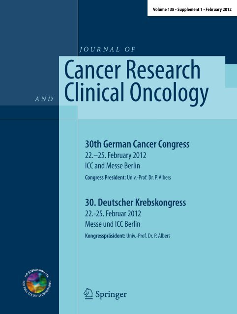
30th German Cancer Congress - February 2012, Berlin
30th German Cancer Congress - February 2012, Berlin
30th German Cancer Congress - February 2012, Berlin

Create successful ePaper yourself
Turn your PDF publications into a flip-book with our unique Google optimized e-Paper software.
A N D<br />
J O U R N A L O F<br />
HIJ<br />
Volume 138 • Supplement 1 • <strong>February</strong> <strong>2012</strong><br />
<strong>Cancer</strong> Research<br />
Clinical Oncology<br />
<strong>30th</strong> <strong>German</strong> <strong>Cancer</strong> <strong>Congress</strong><br />
22.–25. <strong>February</strong> <strong>2012</strong><br />
ICC and Messe <strong>Berlin</strong><br />
<strong>Congress</strong> President: Univ.-Prof. Dr. P. Albers<br />
30. Deutscher Krebskongress<br />
22.-25. Februar <strong>2012</strong><br />
Messe und ICC <strong>Berlin</strong><br />
Kongresspräsident: Univ.-Prof. Dr. P. Albers
Inhaltsverzeichnis<br />
This supplement was not sponsored<br />
by outside commercial interests . It was<br />
funded entirely by the publisher .<br />
Inhaltsverzeichnis<br />
Best of Poster<br />
Best of Poster – GI-Tumoren . . . . . . . . . . . . . . . . . . . . . . . . . . . . . . . . . . . . . . . . . . . . . . . . . . . . . . . . . . 3<br />
Best of Poster – Hauttumoren . . . . . . . . . . . . . . . . . . . . . . . . . . . . . . . . . . . . . . . . . . . . . . . . . . . . . . . . 4<br />
Best of Poster – Lungentumoren . . . . . . . . . . . . . . . . . . . . . . . . . . . . . . . . . . . . . . . . . . . . . . . . . . . . . 6<br />
Best of Poster – Lymphome/Leukämien/kindliche Tumoren . . . . . . . . . . . . . . . . . . . . . . . . . . . 8<br />
Best of Poster – Mammakarzinom/gynäkologische Tumoren . . . . . . . . . . . . . . . . . . . . . . . . . 10<br />
Best of Poster – Molekulare Onkologie . . . . . . . . . . . . . . . . . . . . . . . . . . . . . . . . . . . . . . . . . . . . . . . 12<br />
Best of Poster – Palliativmedizin/Supportivtherapie . . . . . . . . . . . . . . . . . . . . . . . . . . . . . . . . . . 13<br />
Best of Poster – Seltene Tumoren . . . . . . . . . . . . . . . . . . . . . . . . . . . . . . . . . . . . . . . . . . . . . . . . . . . . 15<br />
Best of Poster – Versorgungsstrukturen/Qualitätssicherung . . . . . . . . . . . . . . . . . . . . . . . . . . 17<br />
Best of Poster – Urologische Tumoren . . . . . . . . . . . . . . . . . . . . . . . . . . . . . . . . . . . . . . . . . . . . . . . 19<br />
Discussed Poster<br />
Discussed Poster – GI-Tumoren . . . . . . . . . . . . . . . . . . . . . . . . . . . . . . . . . . . . . . . . . . . . . . . . . . . . . . 20<br />
Discussed Poster – Mammakarzinom/gynäkologische Tumoren . . . . . . . . . . . . . . . . . . . . . . 23<br />
Discussed Poster – Molekulare Onkologie . . . . . . . . . . . . . . . . . . . . . . . . . . . . . . . . . . . . . . . . . . . . 26<br />
Discussed Poster – Seltene Tumoren . . . . . . . . . . . . . . . . . . . . . . . . . . . . . . . . . . . . . . . . . . . . . . . . . 28<br />
Discussed Poster – Supportivtherapie/Palliativmedizin . . . . . . . . . . . . . . . . . . . . . . . . . . . . . . . 30<br />
Discussed Poster – Versorgungsstrukturen/Qualitätssicherung . . . . . . . . . . . . . . . . . . . . . . . 33<br />
General Poster<br />
GI-Tumoren . . . . . . . . . . . . . . . . . . . . . . . . . . . . . . . . . . . . . . . . . . . . . . . . . . . . . . . . . . . . . . . . . . . . . . . . . 35<br />
Hauttumoren . . . . . . . . . . . . . . . . . . . . . . . . . . . . . . . . . . . . . . . . . . . . . . . . . . . . . . . . . . . . . . . . . . . . . . . 53<br />
Lungentumoren . . . . . . . . . . . . . . . . . . . . . . . . . . . . . . . . . . . . . . . . . . . . . . . . . . . . . . . . . . . . . . . . . . . . 55<br />
Lymphome/Leukämien/pädiatrische Tumoren . . . . . . . . . . . . . . . . . . . . . . . . . . . . . . . . . . . . . . . 57<br />
Mammakarzinom/gynäkologische Tumoren . . . . . . . . . . . . . . . . . . . . . . . . . . . . . . . . . . . . . . . . . 61<br />
Molekulare Onkologie . . . . . . . . . . . . . . . . . . . . . . . . . . . . . . . . . . . . . . . . . . . . . . . . . . . . . . . . . . . . . . .90<br />
Seltene Tumoren . . . . . . . . . . . . . . . . . . . . . . . . . . . . . . . . . . . . . . . . . . . . . . . . . . . . . . . . . . . . . . . . . . . 106<br />
Supportivmedizin/Palliativtherapie . . . . . . . . . . . . . . . . . . . . . . . . . . . . . . . . . . . . . . . . . . . . . . . . 119<br />
Urologische Tumoren . . . . . . . . . . . . . . . . . . . . . . . . . . . . . . . . . . . . . . . . . . . . . . . . . . . . . . . . . . . . . . 134<br />
Versorgungsstrukturen/Qualitätssicherung . . . . . . . . . . . . . . . . . . . . . . . . . . . . . . . . . . . . . . . . . 144<br />
KOK<br />
KOK . . . . . . . . . . . . . . . . . . . . . . . . . . . . . . . . . . . . . . . . . . . . . . . . . . . . . . . . . . . . . . . . . . . . . . . . . . . . . . . .151<br />
Autorenverzeichnis . . . . . . . . . . . . . . . . . . . . . . . . . . . . . . . . . . . . . . . . . . . . . . . . . . . . . . . . . . . . . . . . 153<br />
Journal of <strong>Cancer</strong> Research and Clinical Oncology Suppl 1 · 2011 |<br />
1
Abstracts<br />
J <strong>Cancer</strong> Res Clin Oncol (<strong>2012</strong>)<br />
[Suppl 1] · 138:2–162<br />
10 .1007/s00432-011-1144-4<br />
© Springer-Verlag <strong>2012</strong><br />
Programmkomitee DKK <strong>2012</strong><br />
Adam G (Hamburg), Albers P (Düsseldorf),<br />
Bartsch HH (Freiburg), Beckmann MW (Erlangen),<br />
Bokemeyer C (Hamburg), Brümmendorf<br />
T (Aachen), Bruns J (<strong>Berlin</strong>), Creutzig<br />
U (Hannover), Domagk K (Hamburg),<br />
Ehninger G (Dresden), Engers R (Düsseldorf),<br />
Enghofer E (Leverkusen), Feyer P (<strong>Berlin</strong>),<br />
Gabbert HE (Düsseldorf), Graeven U<br />
(Mönchengladbach), Gschwend J (München),<br />
Hallek M (Köln), Hauschild A (Kiel), Hegewisch-Becker<br />
(Hamburg), Helbig U (<strong>Berlin</strong>),<br />
Helou A (Bonn), Hofstädter F (Regensburg),<br />
Hohenberger W (Erlangen), Hölscher A<br />
(Köln), Hübner J (Frankfurt/M), Iro H (Erlangen),<br />
Kastenholz H (Bonn), Kerschgens C<br />
(<strong>Berlin</strong>), Kleeberg U (Hamburg), Klingebiel<br />
T (Frankfurt), Klinkhammer-Schalke M<br />
(Regensburg), Kohlhuber F (Bonn), Kortmann<br />
R-D (Leipzig), Kotzerke J (Dresden),<br />
Lang H (Mainz), Meier K (Soltau), Nettekoven<br />
G (Bonn), Ortmann, O (Regensburg),<br />
Paradies K (Hamburg), Propping P (Bonn),<br />
Riemann JF (Ludwigshafen), Schadendorf<br />
D (Essen), Schirren J (Wiesbaden), Schmidberger<br />
H (Mainz), Schmiegel W (Bochum),<br />
Schmutzler R (Köln), Singer S (Leipzig),<br />
Stummer W (Münster), Thomas M (Heidelberg),<br />
Wallwiener D (Tübingen), Weis J (Freiburg),<br />
Wesselmann S (<strong>Berlin</strong>), Wiedenmann<br />
B (<strong>Berlin</strong>), Wiegel T (Ulm), Wiestler O (Heidelberg),<br />
Wittekind C (Leipzig), Wolff K-D<br />
(München), Wylegalla C (Freiburg), Zeeb H<br />
(Bremen)<br />
Steering Comitee<br />
2 | Journal of <strong>Cancer</strong> Research and Clinical Oncology Suppl 1 · 2011<br />
30. Deutscher Krebskongress<br />
22.–25. Februar <strong>2012</strong><br />
Messe und ICC <strong>Berlin</strong><br />
Kongresspräsident<br />
Univ.-Prof. Dr. P. Albers<br />
Albers P (Düsseldorf), Beckmann MW (Erlangen),<br />
Bokemeyer C (Hamburg), Brümmendorf<br />
T (Aachen), Bruns J (<strong>Berlin</strong>), Graeven<br />
U (Mönchengladbach), Hallek M (Köln),<br />
Hauschild A (Kiel), Ortmann, O (Regensburg),<br />
Schmiegel W (Bochum), Zeeb H (Bremen)<br />
Gutachter <strong>2012</strong><br />
Al-Batran S (Frankfurt/M.), Albers P (Düsseldorf),<br />
Alberti W (Wuppertal), Bamberg<br />
M (Tübingen), Barth J (Gießen), Bartsch H<br />
(Freiburg), Beckmann M (Erlangen), Belka<br />
C (München), Berdel W (Münster), Berking<br />
C (München), Biersack H (Bonn), Bokemeyer<br />
C (Hamburg), Bosslet K (Penzberg),<br />
Branscheid D (Bielefeld), Britzen-Laurent<br />
N (Erlangen), Brossart P (Bonn), Budach<br />
W (Düsseldorf), Büttner R (Köln), Dartsch<br />
D (Hamburg), Debatin K (Ulm), Dunst J<br />
(Lübeck), Ebert M (Mannheim), Einsele H<br />
(Würzburg), Emons G (Göttingen), Engenhart-Cabillic<br />
R (Marburg), Engers R (Neuss),<br />
Engert A (Köln), Fehm T (Tübingen), Feyer<br />
P (<strong>Berlin</strong>), Folprecht G (Dresden), Freidank<br />
A (Fulda), Friedel G (Gerlingen), Gabbert H<br />
(Düsseldorf), Ganser A (Hannover), Geißler<br />
M (Esslingen a.N.), Glimm H (Heidelberg),<br />
Graeven U (Mönchengladbach), Gutzmer R<br />
(Hannover), Hallek M (Köln), Hartenstein<br />
R (München), Hartmann J (Kiel), Heike M<br />
(Dortmund), Henne-Bruns D (Ulm), Hense<br />
H (Münster), Hertenstein B (Bremen), Hillemanns<br />
P (Hannover), Hochhaus A (Jena),<br />
Hofheinz R (Mannheim), Hohenberger W<br />
(Erlangen), Hölscher A (Köln), Howaldt H<br />
(Gießen), Hübner J (Frankfurt/M.), Jaehde U<br />
(Bonn), Jocham D (Lübeck), Jonat W (Kiel),<br />
Jordan K (Halle/S.), Kaaks R (Heidelberg),<br />
Keller M (Heidelberg), Kiechle M (München),<br />
Kimmig R (Essen), Klug S (Dresden), Knüchel-Clarke<br />
R (Aachen), Koch U (Hamburg),<br />
Köhne C (Oldenburg), Kortmann R (Leipzig),<br />
Kreienberg R (Ulm), Krug B (Köln), Kubicka<br />
S (Reutlingen), Lang H (Mainz), Lehnert T<br />
(Bremen), Lipp M (<strong>Berlin</strong>), Loquai C (Mainz),<br />
Lordick F (Braunschweig), Lorenzen S (München),<br />
Lutz M (Saarbrücken), Mackensen A<br />
(Erlangen), Marmé D (Freiburg), Meier K<br />
(Soltau), Meyer H (Solingen), Meyer T (Ansbach),<br />
Miller K (<strong>Berlin</strong>), Möhler M (Mainz),<br />
Molls M (München), Müller R (Köln), Neuhaus<br />
P (<strong>Berlin</strong>), Niederle N (Leverkusen),<br />
Oettle H (Friedrichshafen), Ortmann O<br />
(Regensburg), Paradies K (Hamburg), Petersen<br />
C (Hamburg), Possinger K (<strong>Berlin</strong>),<br />
Propping P (Bonn), Reinacher-Schick A (Bochum),<br />
Riemann J (Ludwigshafen), Rödel C<br />
(Frankfurt/M.), Schäfer R (<strong>Berlin</strong>), Schirren<br />
J (Wiesbaden), Schlag P (<strong>Berlin</strong>), Schlegel U<br />
(Bochum), Schmidt E (Bremen), Schmidt-<br />
Wolf I (Bonn), Schmieder K (Mannheim),<br />
Schmiegel W (Bochum), Schöning T (Heidelberg),<br />
Schuler M (Essen), Schütte W Halle/S.,<br />
Schwarz M (Tübingen), Seufferlein T Halle/S.,<br />
Singer S (Leipzig), Stahl M (Essen), Stenzl A<br />
(Tübingen), Stuschke M (Essen), Tannapfel<br />
A (Bochum), Thomas M (Heidelberg), Trarbach<br />
T (Essen), Trefzer U (<strong>Berlin</strong>), Trepel M<br />
(Hamburg), Trümper L (Göttingen), Ukena<br />
D (Bremen), Unger C (Freiburg), Vanhoefer<br />
U (Hamburg), Wecht D (Marburg), Wiegel<br />
T (Ulm), Wirth M (Dresden), Wittekind C<br />
(Leipzig), Wylegalla C (Freiburg)
GI-Tumoren<br />
Best of Poster – GI-Tumoren<br />
B1 – 0077<br />
Protein profiling identifies HDAC2 and TXNL1 as aneuploidy-associated<br />
markers in colorectal cancer<br />
*T . Gemoll 1,2,3 , U . Roblick 1,2,3 , S . Szymczak 4 , T . Braunschweig 5 , S . Becker 3 ,<br />
B .-W . Igl 4 , H .-P . Bruch 1 , A . Ziegler 4 , U . Hellman 6 , M . Difilippantonio 7 , T . Ried 7 ,<br />
H . Jörnvall 2 , G . Auer 3 , J . Habermann 1,2,3,7<br />
1 University of Lübeck, Department of Surgery, Lübeck, Deutschland,<br />
2 Karolinska Institut, Stockholm, Schweden, 3 Karolinska Biomic Center,<br />
Stockholm, Schweden, 4 University of Lübeck, Institute for Medical Biometry<br />
and Statistics, Lübeck, Deutschland, 5 University Clinic RWTH Aachen,<br />
Institute of Pathology, Aachen, Deutschland, 6 Ludwig Institute for <strong>Cancer</strong><br />
Research, Uppsala, Schweden, 7 NCI/NIH, Genetics Department, Bethesda,<br />
Deutschland<br />
Background. DNA aneuploidy has been identified as prognostic factor<br />
for epithelial malignancies. Further understanding of the translation of<br />
DNA aneuploidy into protein expression will help to define therapies,<br />
prognosis and prevention. We therefore aimed at identifying aneuploidy-associated<br />
protein expression.<br />
Methods. DNA ploidy assessment by image cytometry identified three<br />
diploid and four aneuploid colorectal cancer cell lines. All cell lines were<br />
subjected to protein expression profiling by two-dimensional gel electrophoresis.<br />
Proteins were identified by mass spectrometry, subjected to<br />
Ingenuity Pathway Analysis (IPA), and target proteins were validated by<br />
Western Blot. Validated proteins were clinically evaluated by immunohistochemistry<br />
using a tissue microarray (TMA). The TMA comprised<br />
47 aneuploid and 31 diploid primary colorectal carcinomas, as well as 19<br />
adjacent normal mucosa specimens.<br />
Results. Two independent statistical analyses revealed 64 proteins that<br />
were significantly differentially expressed between the diploid and<br />
aneuploid cell lines. Of these, 26 proteins could be identified by mass<br />
spectrometry and were subjected to IPA. The majority of these proteins<br />
interacted in two overlapping high-ranked IPA networks maintaining<br />
Cellular Assembly and Organization, Cellular Function and Maintenance,<br />
Infection Mechanism, Cell Cycle, and Cellular Growth and<br />
Proliferation. Network proteins showed cancer-associated functions of<br />
Cellular Assembly and Organization, Cell-To-Cell Signalling, and Cell<br />
Death (p
Abstracts<br />
their therapeutical role for human gastrointestinal cancers has not yet<br />
been clarified.<br />
To define the inhibitory and pro-apoptotic effects of two PI3K inhibitors<br />
BEZ235 and BKM120, three human colon cancer (HT-29, HCT-116,<br />
DLD-1) and three gastric cancer cell lines (NCIn87, AGS, MKN-45) with<br />
different PIK3CA mutation status were analysed. First, viability, apoptosis<br />
and caspase assays were performed during incubation with these<br />
inhibitors alone or combined with classical cytotoxic agents. Second,<br />
molecular consequences for cell cycle and the signalling pathways were<br />
analysed by defining the protein levels by FACS and Western blot.<br />
Both, BKM120 and the dual PI3K/mTOR inhibitor BEZ235 induced a<br />
significant concentration-dependent reduction in cell viability and an<br />
increase of apoptotic cell death, while mutated cells reacted more sensitive<br />
to treatment. Here, BKM120 was more effective than BEZ235. It caused<br />
a G1 arrest in tumor cells. In contrast, BKM120 induced a G2 shift<br />
in all gastrointestinal cancer cell lines. BKM120 clearly down-regulated<br />
the AKT pathway and BEZ235 additionally inhibited the mTOR (via<br />
p70S6K) pathway. In colon cancer cells, BEZ235 caused a synergistic<br />
induction of apoptosis combined with irinotecan. Combinations with<br />
5-fluoruracil and both substances induced additive apoptotic effects.<br />
Human gastric cancer cells were less sensitive to BEZ235 and BKM120.<br />
In sum, BEZ235 and BKM120 had high pro-apoptotic effects in all human<br />
cell lines and in special cases a better response in the PIK3CA mutated<br />
cells. Our in vitro data further support the clinical development of<br />
these PI3K inhibitors BEZ235 and BKM120 as potential targeting agents<br />
for patients with different wild type or mutated gastrointestinal cancer<br />
cells.<br />
Best of Poster – GI-Tumoren<br />
B4 – 0499<br />
STAT3 is a potential molecular target to sensitize colorectal<br />
cancer cells to chemoradiotherapy<br />
*M . Grade1 , M . Spitzner1 , G . Emons1 , E . Kendziorra1 , M . Rave-Fränk2 , J . Gaedcke1<br />
, H . Becker1 , T . Beißbarth3 , M . Ghadimi1 1Universitätsmedizin Göttingen, Klinik für Allgemein- und Viszeralchirurgie,<br />
Göttingen, Deutschland, 2Universitätsmedizin Göttingen, Klinik für<br />
Strahlentherapie und Radioonkologie, Göttingen, Deutschland, 3Universi tätsmedizin Göttingen, Medizinische Statistik, Göttingen, Deutschland<br />
Background. Resistance to preoperative chemoradiotherapy represents<br />
a major clinical problem in the treatment of patients with locally advanced<br />
rectal cancer. Therefore, the identification of molecular biomarkers<br />
that differentiate responsive and resistant tumors is exceedingly important,<br />
because this may lead to the identification of novel molecular<br />
targets whose modification could be harnessed to sensitize a priori resistant<br />
tumors to multimodal treatment.<br />
Materials and methods. We recently established an in vitro model for<br />
5-FU based chemoradiotherapy, and correlated differences in treatment<br />
sensitivity of 12 colorectal cancer cell lines with pretherapeutic gene expression<br />
profiles. One gene the expression of which correlated positively<br />
with treatment resistance was the signal transducer and activator of<br />
transcription 3, STAT3. To test the functional relevance of this observation,<br />
we first determined STAT3 mRNA and protein expression levels in<br />
all cell lines. Next, we established doxycycline-inducible stable shRNA<br />
single-cell clone (SCC) populations. Successful silencing of STAT3 was<br />
detected by Western blot analysis. The induced SCCs were treated with<br />
3 µM 5-FU, and subsequently exposed to 0, 1, 2, 4, 6, and 8 Gy of Xrays.<br />
In addition, STAT3 was inhibited using two different siRNAs, and<br />
a small-molecular inhibitor (STATTIC).<br />
Results. STAT3 was significantly overexpressed in resistant cells. In<br />
SW480 and SW837 cells, both shRNA- and siRNA-mediated silencing<br />
as well as STATTIC-induced inhibition of STAT3-phosphorylation re-<br />
4 | Journal of <strong>Cancer</strong> Research and Clinical Oncology Suppl 1 · 2011<br />
sulted in a significantly increased chemoradiosensitivity, with dose-reduction<br />
factors of 1.8 to 2.<br />
Conclusion. These results highlight the potential relevance of STAT3 as a<br />
novel molecular target to sensitize a priori resistant tumor cells to chemoradiotherapy.<br />
Hauttumoren<br />
Best of Poster – Hauttumoren<br />
B5 – 0178<br />
Efficient induction of apoptosis in melanoma cells and reduced<br />
melanoma growth in nude mice by a gene therapy approach<br />
based on oncolytic adenoviral vectors with inducible expression<br />
of TRAIL<br />
*L .F . Fecker1 , H . Fechner2 , J . Eberle1 1Charité – Universitätsmedizin <strong>Berlin</strong> Klinik für Dermatologie, Venerologie<br />
und Allergologie, Hauttumorzentrum, AG Apoptoseregulation in Hauttumoren,<br />
<strong>Berlin</strong>, Deutschland, 2Technische Universität, Institut für Biotechnologie,<br />
<strong>Berlin</strong>, Deutschland<br />
Background. High mortality and therapy resistance of melanoma demands<br />
the development of new strategies, and overcoming of apoptosis<br />
deficiency appears as particularly promising. TNF-related apoptosis inducing<br />
ligand (TRAIL) has been shown previously as highly effective<br />
for apoptosis induction in melanoma cells and may apply for gene therapy<br />
due to its selective impact on tumor cells.<br />
Experimental procedures. Established adenoviral vectors AdV-TRAIL<br />
and AdV-CD95L encode for the death ligands TRAIL and CD95L, respectively.<br />
They are characterized by a variant adenoviral E1A protein<br />
and by deletion of E1B aiming at the restriction of viral replication to<br />
tumor cells. For melanoma selectivity, the viral replication gene E1A is<br />
controlled by a tyrosinase promoter. The tetracycline-responsive transactivator<br />
rtTA and the death ligand are further controlled by a bidirectional<br />
tetracycline-inducible promoter. Apoptosis was monitored by a<br />
DNA fragmentation ELISA and cell killing by crystal violet staining.<br />
For proliferation, real time cell analysis (RTCA) was used. Growth of<br />
tumors established in nude mice was monitored after intratumoral injections<br />
of AdV-TRAIL.<br />
Results. AdV-TRAIL mediated strong expression of E1A and doxycycline<br />
-dependent induction of TRAIL selectively in melanoma cells, which<br />
resulted in melanoma cell lysis and induction of apoptosis. In contrast,<br />
non-melanoma cells and normal human melanocytes appeared as protected.<br />
Comparison of AdV-TRAIL with AdV-CD95L revealed largely<br />
similar efficacies of both death ligands in melanoma cells. In melanoma<br />
xenotransplantation models, AdV-TRAIL demonstrated its efficacy by<br />
significant growth reduction of established melanomas after intratumoral<br />
application. Melanoma cell killing by AdV-TRAIL could be further<br />
improved in vitro by combinations with chemotherapeutics.<br />
Conclusions. We demonstrate that melanoma cells can be efficiently targeted<br />
by death ligand-based gene therapy. Possible resistance may be<br />
overcome by combined chemotherapy.<br />
The study was supported by the <strong>German</strong> <strong>Cancer</strong> Aid (Deutsche Krebshilfe,<br />
grants 107398 and 108008) .
Best of Poster – Hauttumoren<br />
B6 – 0275<br />
Cytotoxicity of new Duplex Drugs linking 3’-C-Ethynylcytidine<br />
and 5-Fluor-2’-deoxyuridine against human Melanoma cells<br />
*S . Schott1 , H . Niessner2 , T . Sinnberg2 , S . Venturelli3 , A . Berger3 , F . Meier2 ,<br />
C . Garbe2 , C . Busch2 1Universitätsklinikum Heidelberg, Universitätsfrauenklinik, Nationales<br />
Centrum für Tumorerkrankungen, Heidelberg, Deutschland, 2University of<br />
Tübingen, Section of Dermato-Oncology, Department of Dermatology and<br />
Allergology, Tübingen, Deutschland, 3University of Tübingen, Department<br />
of Internal Medicine I, Tübingen, Deutschland<br />
Introduction. Melanoma is an increasingly common and potentially fatal<br />
malignancy of the skin and some mucous membranes. Since no cure<br />
exists for metastatic disease (stage IV), there is an urgent need for novel<br />
drugs to overcome melanoma therapy resistance. 2’-Deoxy-5-fluorouridylyl-(3’-5’)-3’-C-ethynylcytidine<br />
[5-FdU(3’-5’)ECyd] and 3’-C-ethynylcytidinylyl-(5’->1-O)-2-O-octadecyl-sn-glycerylyl-(3-Oà5')-2'-deoxy-<br />
5-fluorouridine [ECyd-lipid-5-FdU] represent cytostatic active duplex<br />
drugs, which can be metabolized into various active antimetabolites.<br />
Methods. The cytotoxicity of these heterodinucleoside phosphate analogues,<br />
their corresponding monomers ECyd and 5-FdU and combinations<br />
thereof were evaluated on five metastatic melanoma cell lines and<br />
six ex-vivo patient-derived melanoma cells in comparison to current<br />
standard cytostatic agents and the BRAF V600E inhibitor Vemurafenib.<br />
Further, in vitro studies were performed for cell proliferation and<br />
cell-cycle analysis. Embyrotoxicity was assessed with the chicken embryo<br />
assay. In vivo efficacious in the LOX IMVI solid tumor xenograft<br />
mouse model was tested in the Developmental Therapeutic Program<br />
(DTP) of the National <strong>Cancer</strong> Institute (NCI, US).<br />
Results. In vitro (real-time) proliferation assays demonstrated that<br />
5-FdU(3’-5’)ECyd and ECyd-lipid-5-FdU had a high cytotoxic efficacy<br />
causing 75% melanoma cell death at concentrations in the nano- and<br />
micromolar range. Cytotoxicity was conducted by induction of DNA<br />
cleavage/apoptosis. Chicken embryotoxicity demonstrated that the duplex<br />
drugs were less toxic than their parental monomers. In vivo the<br />
duplex drug 5-FdU(3’-5’)ECyd was efficacious in the murine LOX IMVI<br />
solid tumor xenograph model upon administration of 11.2 mg/kg/injection<br />
every fourth day.<br />
Conclusion. Both duplex drugs are promising novel cytostatic agents for<br />
the treatment of malignant melanoma meriting clinical evaluation.<br />
Best of Poster – Hauttumoren<br />
B7 – 0332<br />
Inhibition of the PI3K-AKT signalling pathway to overcome therapy<br />
resistance in melanoma-derived brain metastasis<br />
*H . Niessner1 , A . Adam2 , A . Bornemann3 , J . Honegger4 , L . Quintanilla-Fend2 ,<br />
C . Busch1 , J . Bauer1 , L . Just5 , K . Hoek6 , C . Garbe1 , F . Meier1 1Universität Tübingen, Dermatologische Onkologie, Tübingen, Deutschland,<br />
2Universität Tübingen, Pathologie, Tübingen, Deutschland,<br />
3 4 Universität Tübingen, Hirnforschung, Tübingen, Deutschland, Universität<br />
Tübingen, Neurochirurgie, Tübingen, Deutschland, 5Universität Tübingen,<br />
Anatomie, Tübingen, Deutschland, 6Universität Zürich, Dermatologie,<br />
Zürich, Schweiz<br />
Introduction. Brain metastases occur in over 70% of patients with metastatic<br />
melanoma and are the most common cause of death. Current<br />
therapy options are neurosurgery, radiosurgery, whole brain radiation,<br />
chemotherapy and supportive care. The median survival time for mela-<br />
noma patients with brain metastasis ranges from 0.7 to 5 months depending<br />
on age and performance status. Therefore, new therapy strategies<br />
are mandatory.<br />
Methods. In melanoma, activation of the RAF-MEK-ERK and PI3K-<br />
AKT-mTOR signal transduction pathways makes a decisive contribution<br />
to tumor progression and treatment resistance. Ongoing clinical<br />
studies suggest a transient effect of BRAF inhibitors in melanoma brain<br />
metastases. We posed the question as to whether blockade of the RAF-<br />
MEK-ERK or/and PI3K-AKT-mTOR signalling pathways would be a<br />
promising strategy for the treatment of melanoma brain metastases. We<br />
blocked RAF-MEK-ERK or/and PI3K-AKT-mTOR signalling pathways<br />
at different levels using classical and new pharmacological inhibitors<br />
and investigated the effects on viability/proliferation and survival/<br />
apoptosis of >10 newly isolated cell lines derived from melanoma brain<br />
metastases. Furthermore, immunohistochemical analyses of brain metastases<br />
from >10 melanoma patients including matched extracerebral<br />
metastases from lung and liver in the same patients for p-ERK, ERK,<br />
AKT, p-AKT and PTEN were performed.<br />
Results. Growth inhibition was most pronounced with PI3K inhibitors<br />
achieving growth inhibition rates of up to 80%. Moreover, PI3K inhibition<br />
potently induced apoptosis in cerebral metastatic melanoma cells.<br />
Histochemical analysis showed that both cerebral and extracerebral<br />
metastases were highly positive for ERK and AKT. p-ERK was seen<br />
exclusively at the tumor periphery of both cerebral and extracerebral<br />
metastases. Interestingly, most melanoma brain metastases were highly<br />
positive for activated AKT, whereas matched extracerebral metastases<br />
from lung and liver in the same patients were weakly positive or negative<br />
for activated AKT. Moreover, PTEN appeared to be downregulated<br />
in brain metastases.<br />
Conclusion. Together, these findings suggest that activation of AKT is<br />
relevant for the survival and growth of melanoma cells in the brain parenchyma<br />
and that inhibition of PI3K-AKT signalling may be a suitable<br />
strategy to enhance and/or prolong the antitumor effect of BRAF inhibitors<br />
in melanoma brain metastases.<br />
Best of Poster – Hauttumoren<br />
B8 – 0489<br />
Validation of a fresh-tissue based prognostic gene signature in<br />
formalin-fixed, paraffin-embedded melanomas<br />
*G . Brunner1 , L . Suter1 , H .-J . Schulze2 , C . Berking3 , A . Heinecke4 , J . Atzpodien5 1Hauttumorzentrum Hornheide-Münster, Tumorforschung, Münster,<br />
Deutschland, 2Hauttumorzentrum Hornheide-Münster, Dermatologie,<br />
Münster, Deutschland, 3LMU München, Dermatologie und Allergologie,<br />
München, Deutschland, 4WWU Münster, Medizinische Informatik und Bioinformatik,<br />
Münster, Deutschland, 5Hauttumorzentrum Hornheide-Münster,<br />
Onkologie, Münster, Deutschland<br />
Melanoma incidence is rapidly increasing – with a doubling rate of 10–<br />
20 years. Precision and reliability of conventional histopathological and<br />
clinical staging, however, remain limited in predicting clinical outcome.<br />
On the other hand, complementary molecular prognostic markers<br />
are not yet available.<br />
We have recently identified, for the first time, a prognostic gene signature<br />
expressed in fresh-frozen primary melanomas (n=135), which is<br />
associated with overall survival (multivariate Cox regression analysis:<br />
p=0.0004, hazard ratio 3.83). The clinical value of a signature-based risk<br />
score is its ability to identify patients at low risk, not identified by conventional<br />
AJCC staging, and to define risk patients in need of adjuvant<br />
therapies. The purpose of this study was to establish analysis of the signature<br />
genes in formalin-fixed, paraffin-embedded (FFPE) melanoma<br />
tissue and to validate prognostic significance.<br />
Journal of <strong>Cancer</strong> Research and Clinical Oncology Suppl 1 · 2011 |<br />
5
Abstracts<br />
We developed a sensitive and robust methodology to analyze and normalize<br />
gene expression in FFPE tissue samples (some of them more<br />
than 20 years old): Total RNA was prepared from FFPE sections matching<br />
the above fresh-frozen primary melanomas (131 out of 135), quality-controlled,<br />
and transcribed into cDNA. Human reference RNA was<br />
included as an internal standard. Following pre-amplification of the<br />
cDNA, expression of the nine signature genes (KRT9, KBTBD10, DCD,<br />
ECG2/SPINK7, PIP, SCGB1D2, SCGB2A2, COL6A6, HES6) and of four<br />
house-keeping genes (18S rRNA, GAPDH, GUSB, BPNT1) was quantified<br />
by real-time PCR using TaqMan assays specific for short amplicons.<br />
Gene expression data were normalized, in two steps, to correct for interassay<br />
technical variability (based on the reference RNA data) as well as<br />
for inter-sample variability of RNA quality (based on the data for the<br />
house-keeping genes). Significance of correlation of FFPE gene expression<br />
data (CT values or estimated mRNA copy numbers) with data from<br />
matched fresh-frozen tissue samples (two-sided t-test) or with patient<br />
overall survival (univariate Cox regression analysis; clinical follow-up<br />
data up to 273 months) was evaluated.<br />
The majority of FFPE primary melanomas (125 out of 131) yielded<br />
mRNA of sufficient quality. In matched pairs of FFPE and fresh-frozen<br />
tissue samples, there was significant correlation of expression of all<br />
nine signature genes. Significance of correlation was higher with CT<br />
values (r=0.19–0.58; p=0.001–0.05) than with estimated copy numbers.<br />
Expression of 7 out of the 9 genes in FFPE melanomas (CT values or<br />
estimated copy numbers) was significantly associated with overall survival<br />
(p=0.0001–0.07).<br />
Thus, our prognostic melanoma gene signature was successfully transferred<br />
from fresh-frozen onto FFPE tissue samples. This greatly increases<br />
clinical applicability of a gene-signature based prognostic risk score<br />
and, in addition, allows the retrospective prognostic analysis of primary<br />
melanomas.<br />
Lungentumoren<br />
Best of Poster – Lungentumoren<br />
B9 – 0198<br />
Hypofractionated image-guided breath-hold radiotherapy of<br />
pulmonary tumors and metastases – clinical results<br />
A . Frauenfeld1 , *J . Boda-Heggemann1 , C . Weiss2 , U . Attenberger3 ,<br />
K . Siebenlist1 , F . Schneider1 , F . Wenz1 , F . Lohr1 1University Medical Center Mannheim, University of Heidelberg, Department<br />
of Radiation Oncology, Mannheim, Deutschland, 2University Medical<br />
Center Mannheim, University of Heidelberg, Abteilung für Medizinische<br />
Statistik, Biomathematik und Informationsverarbeitung, Mannheim,<br />
Deutschland, 3University Medical Center Mannheim, University of Heidelberg,<br />
Institute of Clinical Radiology and Nuclear Medicine, Mannheim,<br />
Deutschland<br />
Purpose. Stereotactic Ablative RadioTherapy (SABR) of stage I–II tumors<br />
has been shown to be a highly effective treatment modality with<br />
low toxicity. It is also a non-invasive therapy option for lung metastases.<br />
Outcome and toxicity were retrospectively evaluated in a single-institution<br />
patient cohort who had undergone hypofractionated image-guided<br />
breath-hold lung SBRT.<br />
Patients and methods. 50 lesions of 43 patients with NSCLC (n=27, St. I–<br />
IV including patients with controlled brain metastases or local relapse)<br />
and lung metastases of various primary tumors (n=16) were consecutively<br />
treated with image-guided breath-hold SBRT. After an initial dose<br />
finding phase, patients were irradiated with a regimen of 5×12 Gy. Dose<br />
calculation was performed with collapsed cone algorithm in 60% of<br />
the lesions. Breath hold was performed with Active Breathing Control<br />
6 | Journal of <strong>Cancer</strong> Research and Clinical Oncology Suppl 1 · 2011<br />
(ABC®, Elekta), daily imaging with EPIDs and repeat breath hold cone-beam<br />
CT. The data were evaluated retrospectively regarding overall<br />
survival (OS), progression-free-survival (PFS), progression pattern and<br />
local control (LC). Radiation parameters and follow-up CT images were<br />
analysed.<br />
Results. BED2 (Biologically Effective Dose) was 87±20 Gy (median<br />
83 Gy). 30 lesions were treated with a BED2 of 90 Gy. Median follow-up was 15 months. Median OS<br />
was 20 months, without significant difference in patients with primary<br />
lung tumor or metastases. Actuarial 1-year OS-rate was 67%; 2-years OS<br />
rate 43%, respectively. 27% of the patients died on non-disease related<br />
reason (cardiovascular events or infections). Actuarial 1-year PFS-rate<br />
was 42%, 2-years PFS rate 28% without significant difference in patients<br />
with primary lung tumor or metastases. Site of progression pattern was<br />
predominantly distant (60% distant metastases and 32% mediastinal<br />
lymph nodes). Actuarial 1-year LC-rate was 90%, 2-years local control<br />
rate 85%. 95% of the lesions treated with a BED2 >90 Gy was controlled<br />
locally after one year. Local progression was observed in only 5 cases,<br />
mainly in the initial dose finding phase, in one patient with an extremely<br />
large PTV and in one patient with an only retrospectively recognised<br />
pleural invasion of the irradiated lesion. Clinically apparent pneumonitis<br />
(requiring treatment) was present in 23% of the cases. No patient<br />
experienced fatal toxicity.<br />
Conclusions. Despite the unfavorable patient selection, high local control<br />
rates could be achieved. If a reasonably high BED2 can be applied,<br />
image-guided breath-hold SBRT is an effective non-invasive treatment<br />
modality with a high local control rate and relatively low toxicity in<br />
patients with inoperable lung tumors and lung metastases. As disease<br />
progression was mainly outside the treated area, systemic therapy has<br />
to be improved.<br />
Best of Poster – Lungentumoren<br />
B10 – 0290<br />
First screening round results from the <strong>German</strong> component LUSI<br />
of the European trial on the efficacy of multislice CT for the early<br />
detection of lung cancer, and the perspective of the European<br />
trial in view of the results of the US NLST trial<br />
*N . Becker1 , E . Motsch1 , M .-L . Groß1 , C .P . Heussel2 , H . Dienemann3 , P . Schnabel4<br />
, M . Eichinger5 , D .-E . Optazaite5 , M . Puderbach5 , J . Tremper5 , S . Delorme5 1Deutsches Krebsforschungszentrum, Epidemiologie von Krebserkrankungen,<br />
Heidelberg, Deutschland, 2Thoraxklinik, Radiologie, Heidelberg,<br />
Deutschland, 3Thoraxklinik, Chirurgie, Heidelberg, Deutschland, 4Univer sität, Medizin/Pathologie, Heidelberg, Deutschland, 5Deutsches Krebsforschungszentrum,<br />
Radiologie, Heidelberg, Deutschland<br />
After closure of the recruitment period of the <strong>German</strong> Lung <strong>Cancer</strong><br />
Screening Intervention Trial (LUSI) in 2011 the study comprises 4052<br />
participants, randomised into 2029 subjects in the multislice-CT<br />
(MSCT) screening arm and 2023 into the „usual care“ control arm. The<br />
early recall rate for suspicious nodules requiring further examination<br />
ranges currently around 27%. Nineteen lung cancers have been detected<br />
(detection rate 0.9%). Related to all early recalls, the positive predictive<br />
value was about 3%, and related to the immediate recalls about 35%.<br />
The subsequent screening rounds started in October of the years 2008<br />
(2nd round) to 2011 (5th and final round) having led so far to the identification<br />
of eight, two, two and 0 further lung cancers, respectively.<br />
In overall 41 surgical interventions, 15 were benign and 26 malignant.<br />
Among the lung cancers detected in the screening arm 22 were adenocarcinoma,<br />
5 squamous cell carcinoma, two a small-cell carcinoma<br />
and two a carcinoid. One interval cancer has so far been observed (an<br />
adenocarcinoma).
The study contributes to the European multicenter study comprising<br />
studies from the Netherlands and Belgium (NELSON, 15422 participants),<br />
Denmark (4104 participants), Italy (DANTE, 2472; ITALUNG,<br />
3206; MILD, 3997) and <strong>German</strong>y (LUSI, 4052). The scope is to demonstrate<br />
an at least 25% reduction of lung cancer mortality by MSCT. However,<br />
in 2011, the National Lung Screening Trial (NLST) conducted in<br />
the USA, provided after an average follow-up of 6 years a significantly<br />
reduced lung cancer mortality by MSCT screening in comparison to<br />
chest X-ray in the control group. Nevertheless, the European studies decided<br />
to continue their trials and to conduct a first common evaluation<br />
in <strong>2012</strong> for several reasons, e.g.: (a) an early visible benefit can disappear<br />
after prolonged follow-up due to postponement of cause-specific death<br />
in the screening group, (b) harmful side effects, especially overdiagnosis,<br />
cannot be properly assessed in comparison to a control group with<br />
another screening modality (chest X-ray), (c) efficient screening modalities<br />
are still undefined such as, risk-related selection of the appropriate<br />
target population, appropriate management of suspicious nodules,<br />
screening intervals etc, as to be outlined in the presentation.<br />
Best of Poster – Lungentumoren<br />
B11 – 0436<br />
The role of sleeve resections in advanced nodal disease of NSCLC<br />
*J . Schirren1 , M . Eberlein2 , A . Fischer3 , S . Bölükbas1 1Dr . Horst Schmidt Klinik, Thoraxchirurgie, Wiesbaden, Deutschland,<br />
2Carver College of Medicine, Division of Pulmonary, Critical Care and<br />
Occupational Medicine, Iowa City, USA, 3Dr . Horst Schmidt Klinik, Anästhesie<br />
und Intensivmedizin, Wiesbaden, Deutschland<br />
Objective. The aim of this study was to contrast the short-term and longterm<br />
results of sleeve resections in centrally located non-small cell lung<br />
cancer (NSCLC) depending on limited nodal disease (N0/N1, LND)<br />
and advanced nodal disease (N2/N3, AND).<br />
Methods. All NSCLC-patients undergoing sleeve resections for centrally<br />
located NSCLC were reviewed from our prospective database<br />
between January 1999 and December 2008. Patients’ characteristics,<br />
morbidity, mortality, locoregional recurrence, distant recurrence and<br />
survival were analyzed corresponding to LND and AND.<br />
Results. One-hundred seventy sleeve resections out of 213 consecutive<br />
sleeve resections were performed for ventrally located NSCLC (LND:<br />
n=120; AND: n=50). There were no statistically differences between the<br />
both groups for age (LND: 61.8±12.4 vs. 60.8±9.6 years), gender, co-morbidities,<br />
type of sleeve resection (bronchial vs. bronchovascular), number<br />
of dissected lymph nodes (LND: 40.0±12.4 vs. 36.7±14.0), histology<br />
and completeness of resection (LND: 96.7% vs. 98.0%), respectively.<br />
More patients had induction chemotherapy in AND group (p=0.049).<br />
Similar short-term results were monitored with regard to morbidity<br />
rate (LND: 34.2%, AND: 44.0%), secondary pneumonectomy (LND:<br />
1.7%, AND: 4.0%) and mortality rate (LND: 5.0%, AND: 6.0%), respectively.<br />
Better 5-year-survival rate and mean survival were observed in<br />
LND (LND: 80.8 months; AND: 37.7 months; p=0.014; LND: 67%; AND:<br />
42%). In the long-term, more distant metastases were identified in AND<br />
group (26.0% vs. 14.2%, p=0.079) in comparison of identical locoregional<br />
recurrence (LND: 1.7%; AND: 0%). Mean time to the development of<br />
distant metastases was similar (LND: 19.1 months; AND: 12.4 months;<br />
p=0.2) in event of metastazing.<br />
Conclusions. Lymph node involvement is a negative prognostic factor<br />
with regard to long-term survival. Sleeve resections in AND are not associated<br />
with higher morbidity and mortality. Sleeve resections in AND<br />
are correlated with promising long-term survival and unexpected high<br />
local control of the disease as a result of high complete resection rates.<br />
Further investigation for the systemic control of the disease is warranted<br />
because of high rates of distant failure.<br />
Best of Poster – Lungentumoren<br />
B12 – 0475<br />
Prognostic value of estrogen (ESR-1) receptor expression in curatively<br />
resected non-small cell lung cancer (NSCLC)<br />
*W . Brückl1 , J . Ficker1 , A . Hartmann2 , R . Wirtz2 1 3 2 Klinikum Nürnberg, Medizinische Klinik , Nürnberg, Deutschland, Universität<br />
Erlangen, Institut für Pathologie, Erlangen, Deutschland<br />
Background. Despite undergoing complete resection of NSCLC, 33% and<br />
77% of patients with stage IA and IIIA, respectively, die within 5 years.<br />
The benefit of adjuvant chemotherapy (aCTx) is only modest, whereas<br />
such treatment is associated with adverse effects and predictive factors<br />
are of utmost importance. We recently could show a better outcome in<br />
estrogen (ESR-1) receptor expressing tumors in the palliative treatment<br />
of metastatic NSCLC (Brueckl et al., Proc ASCO 2011). The main objective<br />
of this study was to test, whether ESR-1 expression is of prognostic/<br />
predictive value in the curative setting as well.<br />
Methods. Affymetrix microarray data from N=138 non-metastatic<br />
NSCLC patients undergoing curative resection were retrieved from a<br />
data base (Lee et al., Clin <strong>Cancer</strong> Res 2008) and used as a test set to<br />
hypothesize that ESR-1 expression in the tumor is of prognostic value<br />
in curatively resected NSCLC. Baseline data according to ERS-1 status<br />
were compared with the use of chi-square tests and in a multivariate<br />
logistic model including all variables with p values
Abstracts<br />
Lymphome/Leukämien/kindliche Tumoren<br />
Best of Poster – Lymphome/<br />
Leukämien/kindliche Tumoren<br />
B13 – 0266<br />
Safety and efficacy of Nilotinib therapy in CML patients with<br />
Imatinib failure in routine clinical management – follow-up of<br />
the non-interventional TARGET study<br />
*F . Stegelmann1 , J . Dengler2 , P . le Coutre3 , C . Scheid4 , R . Weide5 , T . Illmer6 ,<br />
A . Sauer7 , M . Walter8 , W . Schneider-Kappus9 , M . Kröger10 , A . Distelrath11 ,<br />
G . Stier12 , S . Stern13 , F . Schlegel14 , M . Meincke15 , O . Frank15 , O .G . Ottmann16 1Universitätsklinikum Ulm, Klinik für Innere Medizin III, Ulm, Deutschland,<br />
2Universitätsklinikum Heidelberg, Medizinische Klink, Innere Medizin V,<br />
Heidelberg, Deutschland, 3Charité – Universitätsmedizin <strong>Berlin</strong>, Campus<br />
Virchow Klinikum (CVK), Medizinische Klinik m . S . Hämatologie, Onkologie<br />
und Tumorimmunologie, <strong>Berlin</strong>, Deutschland, 4Uniklinik Köln, Klinik I<br />
für Innere Medizin, Köln, Deutschland, 5Praxisklinik für Hämatologie<br />
und Onkologie Koblenz, Koblenz, Deutschland, 6Gemeinschaftspraxis Hämatologie – Onkologie, Dresden, Deutschland, 7Medizinisches Versorgungszentrum<br />
für Blut- und Krebserkrankungen, Potsdam, Deutschland,<br />
8Praxis für Innere Medizin, Hämatologie und internistische Onkologie,<br />
Paderborn, Deutschland, 9Hämato-Onkologische Schwerpunkt-Praxis<br />
Ulm, Ulm, Deutschland, 10Gemeinschaftspraxis für Ambulante Onkologie,<br />
Bremerhaven, Deutschland, 11MVZ Osthessen, Fachbereich Innere Medizin,<br />
Fulda, Deutschland, 12Internistisch-Onkologische Praxis, Zella-Mehlis,<br />
Deutschland, 13Praxisklinik für integrative Onkologie, Altötting, Deutschland,<br />
14St .-Antonius-Hospital, Akademisches Lehrkrankenhaus der RWTH<br />
Aachen, Eschweiler, Deutschland, 15Novartis Pharma GmbH, BU Oncology,<br />
Nürnberg, Deutschland, 16Klinikum der J .W . Goethe-Universität Frankfurt,<br />
Med . Klinik II – Hämatologie/Onkologie, Frankfurt, Deutschland<br />
Introduction. Nilotinib is a potent and highly selective BCR-ABL inhibitor<br />
with approval for newly diagnosed Ph+ CML patients in CP based<br />
on superior PFS and faster responses vs Imatinib (IM). Nilotinib is also<br />
indicated for CP and AP Ph+ CML pts who failed prior therapy including<br />
IM.<br />
Methods. Follow-up analysis of an observational study of nilotinib in<br />
148 pts with Ph+ IM-resistant or -intolerant CML within clinical practice<br />
in 71 centres in <strong>German</strong>y between Jan 2008 and Nov 2010.<br />
Results. 64.1% of the patients were older than 60 yrs, 7.4% older than 80<br />
yrs (median age 65.8 yrs). 98.6% (1.4%) presented in CP (AP). 95.9% had<br />
a good performance status (ECOG index ≤1). All pts were pretreated<br />
with IM. Further prior drug treatments were chemotherapy (34.5%),<br />
IFN (26.4%), dasatinib (24.3%) and other unspecified drugs (10.8%). Two<br />
patients had received SCT in the past. 64.2% of patients were treated<br />
with nilotinib as 2nd line therapy mostly due to resistance/intolerance<br />
against IM (50.0%/48.0%) or dasatinib (16.2%/6.1%). At initial visit, a<br />
dose of 800 mg nilotinib/d was prescribed in 70.3% (400 mg/d in 25%).<br />
Median duration of nilotinib therapy at the data cut-off for this analysis<br />
was 235 days. Remission status at study entry was 66.2% in CHR,<br />
41.9% in MCyR (missing data =17.6%), 20.9%/9.5% in MMR/CMR. These<br />
responses improved significantly under nilotinib, reaching cumulative<br />
incidences of CHR, MCyR, MMR and CMR of 89.2%, 33.1%, 41.9% and<br />
31.1%, respectively. Of note, cytogenetic and molecular examinations<br />
were frequently not done (e.g. 81.8% and 26.3% after 6 months, respectively).<br />
Overall, the median time to response was 77 days (5–547 days).<br />
Dose reduction or therapy interruption at any time occurred in 18.9%.<br />
In 33.8% nilotinib treatment was stopped prematurely. 73% experienced<br />
at least one AE during the observation period which was considered serious<br />
in 14.2%. Hematologic toxicity was observed in 18.9% of pts (12.2%<br />
with thrombocytopenia), non-hematologic toxicities occurred in 43.2%<br />
8 | Journal of <strong>Cancer</strong> Research and Clinical Oncology Suppl 1 · 2011<br />
of pts. The most frequently reported AEs were skin reactions including<br />
pruritus (13.5%) and rash (11.5%), gastrointestinal symptoms such as<br />
nausea (8.8%), diarrhoea (7.4%) and upper abdominal pain (6.1%) as well<br />
as headache (11.5%).<br />
Conclusions. These data from routine clinical management support the<br />
use of nilotinib as an efficacious and safe drug for treatment of a broad<br />
population of CML pts with poor response or intolerance to a prior therapy<br />
including IM.<br />
Best of Poster – Lymphome/<br />
Leukämien/kindliche Tumoren<br />
B14 – 0294<br />
Hepatitis B virus infection and risk of lymphoma: results of a<br />
serological analysis within the European case-control study<br />
EPILYMPH<br />
*N . Becker1 , P . Schnitzler2 , P . Boffetta3 , 4 , P . Brennan3 , L . Foretova5 , M . Maynadié6<br />
, A . Nieters7 , A . Staines8 , Y . Benavente9 , P .L . Cocco10 , S . de Sanjosé9 1Deutsches Krebsforschungszentrum, Epidemiologie von Krebserkrankungen,<br />
Heidelberg, Deutschland, 2Universität, Medizin, Heidelberg, Deutschland,<br />
3International Agency for Research on <strong>Cancer</strong>, Epidemiology, Lyon,<br />
Frankreich, 4International Prevention Research Institute, Epidemiology,<br />
Lyon, Frankreich, 5Masaryk Memorial <strong>Cancer</strong> Institute, <strong>Cancer</strong> Epidemiology<br />
and Genetics, Brno, Tschechische Republik, 6University, Registre des<br />
Hémopathies Malignes, Dijon, Deutschland, 7Universität, Molecular Epidemiology,<br />
Freiburg, Deutschland, 8University, School of Nursing, Dublin,<br />
Irland, 9Unitat d’Infeccions i Càncer – Unit of Infections and <strong>Cancer</strong> (UNIC),<br />
Institut Català d’Oncologia – Catalan Institute of Oncology, Barcelona,<br />
Spanien, 10University, Occupational Medicine, Cagliari, Italien<br />
Introduction. Epilymph is a European multicentric epidemiologic casecontrol<br />
study on the etiology of lymphoma comprising 2362 cases and<br />
2458 controls from 6 countries (Czech Republic, France, <strong>German</strong>y, Ireland,<br />
Italy, Spain). After having found a significantly elevated lymphoma<br />
risk among lymphoma cases with a self-reported medical history of<br />
HBV infection based on questionnaire data, we used the study material<br />
to investigate associations between serological indicators of HBV infection<br />
with risk of HL, NHL and specific lymphoma entities.<br />
Methods. We tested HBs-antigen (HBsAg), anti-HBc and anti-HBs to<br />
distinguish between susceptibility, current, past infection and immunity<br />
by vaccination. Statistical analysis was carried out with unconditional<br />
logistic regression.<br />
Results. We found a statistically significant positive association especially<br />
of a past HBV infection with multiple myeloma (MM, OR=1.97,<br />
95%-CL=1.16–3.37). Past HBV infection was also associated with B-CLL<br />
(OR=1.33, 95%-CL=0.82–2.16) and T-NHL (OR=1.59, 95%-CL=0.65–3.90)<br />
which was, however, not statistically significant. Non-significant association<br />
occurred also for current HBV infection and NHL (OR=1.49,<br />
95%-CL=0.65–3.41), B-NHL (OR=1.58, 95%-CL=0.69–3.64) and diffuse<br />
large B-cell lymphoma (DLBCL, OR=1.50, 95%-CL=0.47–4.82). Subjects<br />
having HBV infection self-reported were serological positive in 75% of<br />
cases and 80% of controls. For vaccination, the corresponding figures<br />
were 49% and 54%, respectively.<br />
Conclusion. The present results support previous reports of an association<br />
between a history of HBV infection with an elevated lymphoma<br />
risk and add multiple myeloma to the list of potentially virus-associated<br />
lymphoma entities. Chronic antigen stimulation and immune response<br />
to a past but not fully cleared HBV infection may be an explanation for<br />
the association.
Best of Poster – Lymphome/<br />
Leukämien/kindliche Tumoren<br />
B15 – 0309<br />
Retrospective analysis of treatment-related toxicities and outcome<br />
in high-risk Ewing sarcoma patients receiving Treosulfan or<br />
Busulfan-based high-dose chemotherapy with autologous stem<br />
cell transplantation*<br />
*H . Jürgens1 , U . Dirksen1 , S . Jabar1 , K . Ehlert1 , J . Boos1 , P . Bader2 , R . Ladenstein3<br />
, A . Ranft1 1University Hospital, Pediatric Hematology and Oncology, Muenster,<br />
Deutschland, 2Johann Wolfgang Goethe University Medical Center,<br />
Frankfurt, Deutschland, 3 3Children‘s <strong>Cancer</strong> Research Institute (CCRI), Wien,<br />
Österreich<br />
Objective. Busulfan (BU), in combination with melphalan (MEL), highdose<br />
chemotherapy (HDC) with autologous stem cell transplantation<br />
is widely used in consolidation treatment of poor-prognosis Ewing sarcoma<br />
patients. Treosulfan (TREO) has been newly introduced recently<br />
into the treatment protocols. We analyzed treatment-related toxicities<br />
and outcome in patients treated with BU-MEL or TREO-MEL HDC according<br />
to the EURO-E.W.I.N.G.99 (EE99) protocol of the <strong>German</strong> Society<br />
of Pediatric Hematology and Oncology (GPOH) from 1998–2009.<br />
Methods. 157 patients were identified with full records of BU-based<br />
HDC, administrated PO in 113 patients and IV in 44 patients, and 44 patients<br />
with IV TREO-based HDC. BU-MEL was given in 31.8% (n=50)<br />
according to high-risk localized disease mainly for poor response to<br />
initial chemotherapy (R2loc), in 25.5% (n=40) for pulmonary metastases<br />
(R2pulm), and in 42.7% (n=67) for primary mainly skeletal dissemination<br />
(R3). TREO-MEL patients had a more progressed risk profile<br />
(R2loc: 6; 13.6%; R2pulm: 5; 11.4%; R3: 33; 75%; p=0.001). HDC toxicity<br />
was analyzed by descriptive statistics according to modified CTC toxicity<br />
grade scales of the EE99 protocol. Outcome was analyzed descriptively<br />
by event-free survival (EFS) and overall survival (OS) controlled<br />
for risk factors by multivariate regression analysis.<br />
Results. Grade 3 and 4 ratings occurred in 654 of 2500 toxic episodes<br />
(26.2%) in BU-MEL HDC (PO: 27.3%; IV: 23.5%), and in 163 of 842 toxic<br />
episodes (19.4%) in TREO-MEL HDC (vs. BU-MEL PO+IV: p10% in total in single scales were observed in TREO-MEL compared<br />
to PO+IV BU-MEL and compared to IV BU-MEL: WBC (vs. BU-MEL<br />
PO+IV: p=.001; IV only: p=.016); granulocytes (p=.016; p=.120); platelets<br />
(p=.016; p=.114); infection (p=0.020; p=0.075); stomatitis (p12,500 steps/day)<br />
while 5% had to be considered as sedentary (100 steps/minute), only 8 of<br />
118 (7%) patients reached the time to be spend on this level of activity as<br />
recommended for an active lifestyle (i.e., 30 min/day). The TESS score<br />
(0–100) for tumors located in upper extremity was 90.2 (range 69.6–100;<br />
SD=11.1), and for lower extremity tumors 88.9 (range 40.8–100; SD=12.1)<br />
indicating good function following multimodal tumor treatment was<br />
achieved.<br />
Conclusions. These preliminary data indicate that patients after Ewing<br />
sarcoma treatment can reach step activity levels similar to healthy<br />
adults. However, most patients stay on a rather low intensity of activity.<br />
SAM can be used to capture activity levels in long-term survivors of<br />
Ewing sarcoma. It may be advantageous to consider the use of a combination<br />
of outcome measures, including SAM, for objective functional<br />
mobility assessment. Parallel recording of physical activity (SAM) and<br />
TESS provides a better measure reflecting the complex situation of the<br />
patients by combining objective and subjective parameters. The study<br />
is being continued to include all long-term Ewing sarcoma survivors<br />
initially diagnosed between 1980 and 2009.<br />
*supported by Bundesministerium für Bildung und Forschung (BMBF)<br />
01ER0807 .<br />
Journal of <strong>Cancer</strong> Research and Clinical Oncology Suppl 1 · 2011 |<br />
9
Abstracts<br />
Mammakarzinom/gynäkologische Tumoren<br />
Best-of-Poster – Mammakarzinom/<br />
gynäkologische Tumoren<br />
B17 – 0067<br />
Phase II-study with a targeted cytotoxic LHRH- analog (AEZS-<br />
108) in patients with LHRH – receptor positive endometrial<br />
cancer: Protocol AGO-Gyn 5, AGO-Study Group<br />
*G . Emons1 , S . Tomov2 , J . Sehouli3 , P . Harter4 , P . Wimberger5 , L .C . Hanker6 ,<br />
A . Stähle7 , A . Hristamian8 , F . Hilpert9 , M .W . Beckmann10 , P . Dall11 , H . Sindermann11<br />
, C . Gründker1 1Georg August Universität, Klinik für Gyn . und Geburtshilfe, Göttingen,<br />
Deutschland, 2University Hospital, Pleven, Bulgarien, 3Charité, <strong>Berlin</strong>,<br />
Deutschland, 4HSK, Wiesbaden, Deutschland, 5University Hospital, Essen,<br />
Deutschland, 6University Hospital, Frankfurt, Deutschland, 7Karlsruhe, <strong>German</strong>y, Deutschland, 8Plovdiv, Bulgarien, Bulgarien, 9University Hospital,<br />
Kiel, Deutschland, 10University Hospital, Erlangen, Deutschland, 11Aeterna Zentaris, Frankfurt, Deutschland<br />
Background. Endometrial cancers (EC) commonly express receptors for<br />
luteinizing homone releasing hormone (LHRH ). AEZS-108 is a targeted<br />
cytotoxic drug where [ D-Lys(6)]-LHRH is linked to doxorubicin.<br />
We assessed efficacy and toxicity of AEZS-108 in endometrial cancer.<br />
Methods. Patients with LHRH-receptor positive advanced (stages FIGO<br />
III or IV; n=27) or recurrent endometrial cancer (n=16) received AEZS-<br />
108 (267 mg/m2 3-weekly) for 6 cycles. At least 1 measurable lesion was<br />
required at baseline. Independent radiologic review was performed. Response<br />
rate per RECIST was defined as primary endpoint. Secondary<br />
endpoints were sefaty, time-to-progression (TTP) and overall survival<br />
(OS).<br />
Results. 43 patients with median age of 68 years entered the study. Percentages<br />
of tumor cells staining for LHRH-R ranged 30–90%. 31 patients<br />
showed endometrioid and 8 serous histological subtype. Prior<br />
treatment included surgery (n=42), radiotherapy (n=29), chemotherapy<br />
(n=9) and hormonal therapy (n=11). 62.8% received AEZS-108 for<br />
6 courses. One patient was retreated at reduced dose. Possibly drug related<br />
adverse events, except for hematologic toxicity grade 3/4 (rapidly<br />
reversible neutropenia: 60%, anemia: 5%), was commonly limited to<br />
CTCAE grade 1/2. There was no evidence of cardiotoxiciy. One patient<br />
stopped therapy because of recurrent anemia. 12 responses were documented<br />
(2 CR, 10 PR and 17 SD). After prior chemotherapy, 1 CR, 1 PR<br />
and 2 SDs were assessed. Median TTP was 30 weeks and median OS<br />
62 weeks.<br />
Conclusions. A promising clinical benefit rate of 74% was shown. OS after<br />
single agent AEZS-108 is similar to that reported for modern triple<br />
combination chemotherapy, but as achieved with distinctly lower toxicity.<br />
10 | Journal of <strong>Cancer</strong> Research and Clinical Oncology Suppl 1 · 2011<br />
Best of Poster – Mammakarzinom/<br />
gynäkologische Tumoren<br />
B18 – 0117<br />
Pegylated liposomal doxorubicin and carboplatin (C-PLD) versus<br />
paclitaxel and carboplatin (C-P) in platinum-sensitive ovarian<br />
cancer (OC) patients (pts): Treatment at recurrence and overall<br />
survival (OS) final analysis from CALYPSO phase III GCIG trial<br />
*C . Kurzeder1 , W . Meier2 , C . Jackisch3 , A . Staehle4 , A . Burges5 , J . Pfisterer6 ,<br />
A . Belau7 , G . Emons8 , U . Canzler9 , W . Schroeder10 , J . Sehouli11 , A . du Bois1,11 ,<br />
P . Wimberger12 , B . Schmalfeldt13 , S . Mahner14 , U . Wagner15 , E . Pujade-Lauraine16<br />
1Kliniken Essen Mitte, Evang . Huyssens-Stiftung/Knappschaft GmbH, Gynäkologie<br />
& Gynäkologische Onkologie, Essen, Deutschland, 2Evangelisches Krankenhaus Düsseldorf, Düsseldorf, Deutschland, 3Klinik für Gynäkologie<br />
und Geburtshilfe Offenbach, Offenbach, Deutschland, 4Frauenarztpraxis, Karlsruhe, Deutschland, 5Klinikum der Universität München, Klinik und<br />
Poliklinik für Frauenheilkunde und Geburtshilfe, München, Deutschland,<br />
6Städt . Klinikum Solingen GmbH, Klinik für Frauenheilkunde und<br />
Geburtshilfe, Solingen, Deutschland, 7Universität Greifswald, Klinik und<br />
Poliklinik für Frauenheilkunde und Geburtshilfe, Greifswald, Deutschland,<br />
8 9 Universitätsfrauenklinik Göttingen, Göttingen, Deutschland, Klinik und<br />
Poliklinik für Frauenheilkunde und Geburtshilfe Universitätsklinikum Carl<br />
Gustav Carus, Dresden, Deutschland, 10Klinikum Bremen Mitte, Frauenklinik,<br />
Bremen, Deutschland, 11Charité Campus Virchow-Klinikum, Klinik<br />
für Gynäkologie, <strong>Berlin</strong>, Deutschland, 12Universitätsklinikum Essen, Klinik<br />
für Frauenheilkunde und Geburtshilfe, Essen, Deutschland, 13Frauenklinik und Poliklinik der Technischen Universität München, München, Deutschland,<br />
14Universitätsklinikum Hamburg-Eppendorf, Klinik und Poliklinik für<br />
Gynäkologie, Hamburg, Deutschland, 15Universitätsklinikum Gießen u .<br />
Marburg GmbH, Standort Marburg, Klinik für Gynäkologie, Gynäkologische<br />
Endokrinologie und Onkologie, Marburg, Deutschland, 16Hopital Hotel-Dieux,<br />
Paris, Frankreich<br />
Background. CALYPSO is a large international randomized phase III<br />
trial comparing CPLD and C-P in pts with OC in late relapse. Results<br />
of the progression-free survival (PFS), the primary endpoint, suggest<br />
a benefit in pts treated with C-PLD with a hazard ratio (HR) of 0.82<br />
(p=0.005). Overall severe non-hematologic toxicity (36.8% vs 28.4%;<br />
p
Best of Poster – Mammakarzinom/<br />
gynäkologische Tumoren<br />
B19 – 0279<br />
Oncofertility; Xenotransplantation of cryopreserved ovarian<br />
tissue from patients with ovarian tumors into SCID mice – no<br />
evidence of malignant cell contamination<br />
*A . Müller1 , L . Lotz1,2 , M . Montag1,2 , H . van der Ven2 , M . von Wollf2,3 , I . Hoffmann1<br />
, D . Wachter4 , M . Beckmann1 , R . Dittrich1 1 2 Universitätsklinikum Erlangen, Frauenklinik, Erlangen, Deutschland, Universitätsklinikum<br />
Bonn, Frauenklinik, Bonn, Deutschland, 3Inselspital Bern,<br />
Frauenklinik, Bern, Schweiz, 4Universitätsklinikum Erlangen, Pathologisches<br />
Institut, Erlangen, Deutschland<br />
Background. There are still many unanswered questions regarding the<br />
safety of ovarian tissue retransplantation in cancer patients, due to the<br />
potential risk that malignant cells in the frozen tissue may lead to recurrence<br />
of the primary disease after transplantation. This study examined<br />
the possible presence of residual malignant cells in ovarian cortex from<br />
patients with ovarian tumors after xenografting of the ovarian tissue<br />
into severe combined immunodeficiency (SCID) mice.<br />
Methods. The ovarian cortex tissue was obtained during surgical laparoscopy<br />
or unilateral/bilateral oophorectomy. Before cryopreservation,<br />
the ovarian cortex was examined histologically to ensure that a sufficient<br />
quantity of primordial follicles had been obtained and to exclude<br />
involvement of the tissue by malignancy. Freezing of the ovarian tissue<br />
was undertaken in accordance with three similar slow freezing protocols.<br />
The ovarian tissue was transplanted into immunodeficient (SCID)<br />
mice in an intramuscular pocket in the neck muscle. Twenty-four weeks<br />
after transplantation, the grafts were recovered and hematoxylin-eosin<br />
stains were performed. In addition, freshly cut sections were stained<br />
with antibodies against pancytokeratin (monoclonal antibody KL1).<br />
Results. All of the patients had a normal follicular distribution relative<br />
to their age, and light microscopy showed no evidence of malignant<br />
cells in the ovarian tissues prior to transplantation, even in patients with<br />
bilateral ovarian carcinoma. All of the animals remained in good health<br />
throughout the study and none of the grafts resulted in recurrences.<br />
Nor did any of the mice showed macroscopically metastasis. The serial<br />
sections from human ovarian grafts had a normal histological appearance.<br />
No carcinoma cells were seen in any of the H&E-stained slides<br />
examined or in any of the slides with antibodies against cytokeratins.<br />
Conclusion. Ovarian transplantation appears to be a feasible consideration<br />
in young female patients with ovarian tumors. The negative<br />
findings do not rule out malignant cell contamination in the ovarian<br />
tissue. A larger number of tissue fragments would increase the validity<br />
of these preliminary results and more sensitive methods may be necessary<br />
for assessment of possible micrometastases. In order to increase<br />
the safety of the procedure, ovarian tissue harvesting and re-implantation<br />
should take place in the setting of clinical trials with clearly defined<br />
methods, objectives, and end points.<br />
Best of Poster – Mammakarzinom/<br />
gynäkologische Tumoren<br />
B20 – 0318<br />
Prospective comparison of risk assessment tools in early breast<br />
cancer (Recurrence Score, uPA/PAI-1, central grade, and luminal<br />
subtypes): final correlation analysis from the phase III WSG-Plan<br />
B trial<br />
*O . Gluz1 , H . Kreipe2 , T . Degenhardt1 , R . Kates1 , C . Liedtke1,3 , M . Christgen2 ,<br />
M . Clemens4 , S . Markmann5 , C . Uleer6 , D . Augustin7 , S . Shak8 , C . Thomssen9 ,<br />
U . Nitz1,10 , N . Harbeck1,11 1Westdeutsche Studiengruppe, Mönchengladbach, Deutschland,<br />
2 3 MHH Hannover, Hannover, Deutschland, Uniklinik Münster, Münster,<br />
Deutschland, 4Klinikum Mutterhaus der Borromäerinnen, Trier, Deutschland,<br />
5Uniklinikum Südstadt, Rostock, Deutschland, 6Gynäkologisch onkologische Praxis, Hildesheim, Deutschland, 7Klinikum Deggendorf,<br />
Degendorf, Deutschland, 8Genomic Health Inc ., Redwood City, Vereinigte<br />
Staaten von Amerika, 9Uniklinikum Halle/Saale, Halle/Saale, Deutschland,<br />
10 11 EVK Bethesda, Mönchengladbach, Deutschland, Uniklinik Köln, Köln,<br />
Deutschland<br />
Background. Both the Recurrence Score® (RS) multi-gene assay and<br />
invasion factors uPA/PAI-1 are recommended by guidelines (ASCO,<br />
AGO) for decision support regarding adjuvant chemotherapy in patients<br />
with early breast cancer (BC). Here, we present the final WSG-<br />
Plan B trial correlation analysis of these risk assessment tools.<br />
Methods. Plan B trial (evaluating anthracyline-free adjuvant chemotherapy,<br />
6×TC, vs. 4×EC-4×DOC in HER2-negative BC; n=2.448 to<br />
be randomized for chemotherapy). RS has been used as the selection<br />
criterion for chemotherapy vs. endocrine therapy alone in HR+ BC (if<br />
RS25 (21%). In 257 patients<br />
with 0–3 involved LN, chemotherapy was omitted based on RS results<br />
(12.3% of patients after amendment). By the last interim analysis in <strong>February</strong><br />
2011, data on central grade are available in 1509 patients and<br />
Ki-67 in 592 patients. An only moderate positive correlation was observed<br />
between Ki-67 and RS (Spearman’s coefficient rs =0.336, p
Abstracts<br />
high-risk according to uPA/PAI-1, Ki-67 or central grade. Further follow-up<br />
of the WSG-Plan B trial will clarify the clinical significance of<br />
these findings regarding patient outcomes.<br />
Molekulare Onkologie<br />
Best of Poster – Molekulare Onkologie<br />
B21 – 0208<br />
Transient telomere dysfunction induces chromosomal instability<br />
and carcinogenesis in telomerase-proficient mice<br />
*D . Hartmann1,2 , Y . Begus-Nahrmann2 , P . Eshraghi2 , P . Schirmacher3 , H .-<br />
W . Lee4 , A . Lechel2 , K .L . Rudolph2 1Chirurgische Klinik und Poliklinik, Klinikum rechts der Isar, München,<br />
Deutschland, 2Institut für Molekulare Medizin und Max-Planck-Forschungsgruppe<br />
für Stammzellalterung, Universität Ulm, Ulm, Deutschland, 3Institut für Pathologie, Universitätsklinikum Heidelberg, Heidelberg, Deutschland,<br />
4Department of Biochemistry, College of Science, Yonsei University, Seoul,<br />
Korea, Republik<br />
Introduction. A current concept indicates that telomere dysfunction can<br />
increase tumor initiation by induction of chromosomal instability, but<br />
initiated tumor cells need to reactivate telomerase for genome stabilization<br />
and tumor progression. However, this concept has not been proven<br />
in vivo since appropriate mouse models were lacking.<br />
Methods. Here, we analyzed hepatocarcinogenesis (i) in a novel mouse<br />
model of inducible telomere dysfunction with wildtype telomerase, (ii)<br />
in telomerase knockout mice with chronic telomere dysfunction (G3<br />
mTerc-/-), and (iii) in wildtype mice with functional telomeres and telomerase.<br />
Results. Transient or chronic telomere dysfunction enhanced the rates<br />
of chromosomal aberrations in developing HCCs, but increases in tumorigenesis<br />
compared to wild-type mice occurred only in telomeraseproficient<br />
mice. In contrast to mTerc-/- tumors, telomerase-proficient<br />
tumors that experienced transient telomere dysfunction retained the<br />
capacity to minimize DNA damage and exhibited elevated rates of tumor<br />
cell proliferation compared to wild-type tumors.<br />
Conclusion. Together, these data provide the first in vivo evidence that<br />
transient telomere dysfunction promotes chromosomal instability and<br />
carcinogenesis in a telomerase-proficient background.<br />
Best of Poster – Molekulare Onkologie<br />
B22 – 0284<br />
Inhibition of Hsp90 impairs hexokinase (HXK-II) activity by disrupting<br />
its interactions with voltage dependent anion channels<br />
(VDAC)<br />
*M .E . Mycielska1 , C . Moser1 , C . Wagner1 , E . Scheiffert1 , E .K . Geissler1 ,<br />
H .J . Schlitt1 , S .A . Lang1 1Universitätsklinikum Regensburg, Experimentelle Chirurgie, Regensburg,<br />
Deutschland<br />
Background. Glycolysis is the predominant metabolic pathway in cancer<br />
cells (Warburg effect) with hexokinase II (HXK II) as its crucial<br />
enzyme. Due to ATP requirements, this enzyme has to be attached to<br />
VDACs in the mitochondrial membrane. In addition, overexpression of<br />
the molecular chaperone heat-shock protein 90 (Hsp90) in cancer cells<br />
has been described. Interestingly, TOM complex, responsible for import<br />
of proteins (e.g. VDACs) into mitochondria, is an Hsp90 client protein.<br />
12 | Journal of <strong>Cancer</strong> Research and Clinical Oncology Suppl 1 · 2011<br />
Therefore, we hypothesised that targeting Hsp90 would affect localisation<br />
of HXK II in cancer cells through changes in VDAC import.<br />
Methods. Human gastric and pancreatic cancer cell lines (TMK1, Mia-<br />
PaCa2) were used to evaluate the effects of Hsp90 inhibitor 17-DMAG<br />
(17-(dimethylaminoethylamino)-17-demethoxygeldanamycin) on cancer<br />
cell metabolism. In vitro, localisation and expression of HXK II and<br />
VDAC protein were determined using Western blotting and immunocytochemistry.<br />
Spectrophotometric technique was employed to evaluate<br />
enzyme activity. In vivo, murine Panc02 pancreatic adenocarcinoma<br />
cells were used in subcutaneous tumor model.<br />
Results. Overall hexokinase activity in the mitochondrial fraction was<br />
decreased by 25±8.1% and 57±9.7% (n=5; p=0.03–0,04) in cells and tissues<br />
when treated with 17-DMAG compared to control conditions, whilst<br />
its activity in the cytoplasmic fractions did not differ (p>0.05). Staining<br />
of HXK II, VDAC and mitochondria in the cells showed large areas of<br />
colocalisation upon control conditions. Interestingly, colocalisation<br />
was significantly reduced when cells were treated with the Hsp90 inhibitor.<br />
Furthermore, treatment with 17-DMAG resulted in a decreased<br />
expression of HXK II in the mitochondrial fraction in cells and tissues.<br />
Determination of the expression of HXK I and III in cancer cells and<br />
tissues showed a slight decrease in HXK III expression in cytoplasmic<br />
fraction whilst there was no change in mitochondrial fraction in either<br />
protein.<br />
Conclusion. Results so far show that targeting Hsp90 affects glucose metabolism<br />
in cancer cells via inhibition of VDAC incorporation into the<br />
mitochondrial membrane and in consequence disabling HXK II-VDAC<br />
binding. Hsp90 inhibitors might, therefore, play a role in treatment<br />
concepts targeting the Warburg effect in human cancer.<br />
Best of Poster – Molekulare Onkologie<br />
B23 – 0337<br />
Genetic analysis for SDH mutations (SDH-B, SDH-C and SDH-D) in<br />
paraganglioma patients<br />
*T . Cohnert1 , J . Fruhmann1 , S . Koter1 , A . Baumann1 , J . Geigl2 1Medizinische Universität Graz, Klin . Abt . für Gefäßchiurgie, Graz,<br />
Österreich, 2Medizinische Universität Graz, Institut für Humangenetik, Graz,<br />
Österreich<br />
Introduction. Paraganglioma represent rare tumours of the head and<br />
neck. They originate from the neural crest cells and can occur from the<br />
skull base to the pelvic floor. Many cases are sporadic. However, the hereditary<br />
cases are most often seen in Von Hippel Lindau Disease, MEN 2<br />
or neurofibromatosis. Recently succinate dehydrogenase mutations<br />
(SDH) have been described in paraganglioma and phaechromocytoma<br />
patients.<br />
Patients and methods. 51 patients (pts.) were operated on 60 Paragangliomas<br />
(PGL) of the neck (carotid body tumors) between January 1988<br />
and July 2011. Prospectively collected clinical data were analyzed retrospectively.<br />
In 2006 genetic analyses were started for succinate dehydrogenase<br />
mutations (SDH; SDH-B, SDH-C and SDH-D). Follow-up was<br />
completed. All patients were contacted and genetic testing as well as genetic<br />
family counselling were offered. Statistical data are given as mean<br />
values and standard deviation.<br />
Results. 51 pts. (36 female, 15 male) were operated at a mean age of<br />
53.9+16.6 years (range 24–80 years). In 9 pts. (4 female, 5 male) there<br />
were bilateral tumors. After a mean follow-up of 94 months (range<br />
1–263 months = 21 years 11 months) 6 pts. had died. 45 pts. were alive,<br />
3 pts. did not agree to genetic testing and one patient was currently not<br />
available for testing (moved abroad). Of the tested 41 pts. no mutation<br />
was detected in 20 pts. (17 female, 3 male). In 21 pts. a total of 14 SDH-D<br />
mutations, 2 SDH-B mutations and 1 SDH-C mutation were found. Additional<br />
4 pts. showed abnormalities in SDH-C that need further investigation.<br />
In summary in 17 of 41 pts. (17/41=41.5%) SDH mutations were
confirmed. In all 9 patients with bilateral tumors SDH-D mutations<br />
were detected.<br />
Conclusions. Long-term survival after surgical removal of paraganglioma<br />
of the neck is not limited. Genetic testing is mandatory in patients<br />
with bilateral tumors or positive family history and strongly recommended<br />
in all other PGL patients. When a SDH mutation is diagnosed,<br />
regular clinical and ultrasound investigations, MRI of the head and<br />
neck, thorax, abdomen and pelvis are recommended for diagnosis or<br />
exclusion of multiple tumor localizations as well as phaechromocytoma.<br />
In addition genetic exploration of patients’ families and counselling<br />
should be offered.<br />
Best of Poster – Molekulare Onkologie<br />
B24 – 0480<br />
Two novel splice variants of the transcriptional co-repressor<br />
Daxx with different p53-regulating properties<br />
N . Wethkamp1 , S . Funke1 , C .V . Suschek2 , E . Grinstein3 , S . Heikaus1 , H .E . Gabbert1<br />
, *C . Mahotka1 1Heinrich Heine Universität – Klinikum, Institut für Pathologie, Düsseldorf,<br />
Deutschland, 2Heinrich Heine Universität – Klinikum, Klinik für Unfallchirurgie,<br />
Düsseldorf, Deutschland, 3Heinrich Heine Universität – Klinikum,<br />
Klinik für Kinder-Onkologie, -Hämatologie und -Immunologie, Düsseldorf,<br />
Deutschland<br />
Aims. Deregulation of apoptosis is involved in several diseases including<br />
cancer, characterized by an abnormally prolonged cell survival.<br />
The ubiquitously expressed protein Daxx is implicated in apoptosis but<br />
whether its function is pro- or anti-apoptotic is still controversially discussed.<br />
Daxx is involved in transcriptional control of several genes and<br />
it comprises domains including a regulatory C-terminus which is responsible<br />
for the interaction with numerous proteins such as p53, PML,<br />
or HSP27.<br />
Methods. RT-PCR, expression cloning, retroviral transfections, confocal<br />
fluorescence microscopy, western blotting, immunoprecipitations,<br />
flow cytometry, luciferase reporter gene assays, apoptosis assays, etc.<br />
Results. Here we describe the identification and characterization of two<br />
novel variants of Daxx termed Daxx-β and Daxx-γ which are generated<br />
by alternative splicing. Alternative splicing results in a truncated<br />
regulatory C-terminus in both proteins. As a consequence, Daxx-β<br />
and Daxx-γ show a markedly decreased affinity to PML, which in turn<br />
is associated with a different subnuclear localization of these proteins<br />
compared to Daxx. While Daxx is mainly localized to PMLoncogenic<br />
domains (PODs) Daxx-β and Daxx-γ display a distinct distribution pattern.<br />
Furthermore, Daxx-β and Daxx-γ show a decreased affinity to p53<br />
also due to the truncated C-terminus. We provide evidence that the p53<br />
recruitment into PODs is Daxx isoform dependent. The decreased affinity<br />
of Daxx-β/-γ to p53 and PML results in a diffuse localization of p53<br />
throughout the nucleus. In contrast to Daxx, Daxx-β and Daxx-γ are<br />
unable to repress p53-mediated transcription.<br />
Conclusion. Therefore, alternative splicing of Daxx as a regulator of p53regulated/dependent<br />
gene expression might indicate an additional level<br />
in the cellular apoptosis network.<br />
Palliativmedizin/ Supportivtherapie<br />
Best of Poster – Palliativmedizin/ Supportivtherapie<br />
B25 – 0058<br />
Randomized placebo-controlled double-blind study of modified<br />
methylphenidate on cancer-related fatigue (CRF)<br />
*M .E . Heim1 , S . Kuhnt2 , R . Schwarz2 , W . Abenhardt3 , B . Lathan4 , K . Verpoort5 ,<br />
S . Siehl6 , C . Ose7 , A . Engels8 , J .-U . Rüffer9 1 2 Universität Göttingen, Rosdorf, Deutschland, Universität Leipzig, Leipzig,<br />
Deutschland, 3Onkologische Praxis, München, Deutschland, 4Onkologische Praxis, Dortmund, Deutschland, 5Onkologische Praxis, Hamburg, Deutschland,<br />
6Onkologische Praxis, Kassel, Deutschland, 7Universitätsklinikum Essen, Inst . f . Med . Informatik, Biometrie u . Epidemiologie, Essen, Deutschland,<br />
8Medice Pütter GmbH, Iserlohn, Deutschland, 9Deutsche Fatigue<br />
Gesellschaft, Köln, Deutschland<br />
Introduction. CRF is the most prevalent symptom of cancer patients<br />
(pts) following treatment, can persist for months or even years, and significantly<br />
reduces usual functioning and health related quality of life.<br />
There is no standard treatment for CRF, initially a comprehensive assessment<br />
and the treatment of possible causative factors, such as anemia<br />
or hypothyroidism is recommended. Nonpharmacological treatments,<br />
such as individualized exercise programs, psychoeducational and cognitive-behavioral<br />
interventions are the backbone of CRF therapy. Pharmacological<br />
treatment with the psychostimulant methylphenidate showed<br />
inconclusive results in several phase II and III studies.<br />
Patients and methods. Tumor pts without active disease suffering from<br />
CRF (MFI-score >40) were randomized into a treatment and a placebo<br />
group. The treatment duration was 3 weeks with modified release methylphenidate<br />
starting with 20 mg in the 1st week, and dose escalation<br />
to 40 mg, and 60 mg in the 2nd and 3rd week in case of inadequate<br />
treatment effect. Treatment results were evaluated by patient reported<br />
outcome measures with MFI, EORTC-QLQ-C30, HADS, and by the<br />
clinical global impression (CGI) of the physician at baseline, week 3 and<br />
6, and for MFI and CGI additionally at week 2 and 3. Primary endpoint<br />
was the general fatigue in the MFI. A total of 67 pts (46 women, 21 men,<br />
mean age 48 years) were randomized, 53 could be evaluated, 25 in the<br />
treatment, 28 in the placebo group. Mean fatigue duration was 4 and<br />
2.5 years.<br />
Results. In the intention to treat analysis there was a mean reduction<br />
of the general fatigue score of the MFI of 22 points (verum) and 13 points<br />
(placebo), in the per protocol analysis of 25 and 16 (not significant).<br />
There was a significant difference in the CGI for therapeutic efficacy in<br />
favor of methylphenidate. Single pts had an impressive advantage of the<br />
treatment with methylphenidate. No significant differences could be<br />
observed for anxiety and depression, and the EORTC-subscales, except<br />
for the cognitive function subscale, which was significantly improved<br />
in the verum group.<br />
Conclusion. In accordance with other studies we could not observe<br />
a significant improvement of CRF in the MFI with methylphenidate.<br />
However significant better results for the intervention group could be<br />
observed in the therapeutic efficacy in the CGI and for the cognitive<br />
function in the EORTC-QLQ-C30. Differences in both groups and subgroup<br />
analysis will be discussed to explain the results.<br />
Journal of <strong>Cancer</strong> Research and Clinical Oncology Suppl 1 · 2011 |<br />
13
Abstracts<br />
Best of Poster – Palliativmedizin/ Supportivtherapie<br />
B26 – 0071<br />
Occupational rehabilitation after partial laryngectomy<br />
*D . Wollbrück1 , H . Danker1 , E .F . Meister2 , A . Meyer1 1Universität Leipzig, Medizinische Fakultät, Medizinische Psychologie und<br />
Medizinische Soziologie, Leipzig, Deutschland, 2Klinikum St . Georg, HNO-<br />
Klinik, Leipzig, Deutschland<br />
Purpose. Improvements in early detection and treatment of laryngeal<br />
cancer have let to an increase of numbers of cancer survivors. Against<br />
this background two questions arise for patients after partial laryngectomy:<br />
I. How does the work status of patients with partial laryngectomy<br />
change? II. Which factors are associated with a successful occupational<br />
rehabilitation?<br />
Methods. Since 2007, all patients in Middle <strong>German</strong>y, who underwent<br />
a partial laryngectomy due to laryngeal carcinoma, were contacted in<br />
a multi-centre longitudinal study. Patients were questioned at different<br />
time points (t1= preoperative; t2=2 weeks after partial laryngectomy;<br />
t3=12 weeks after operation; t4=1 year postoperative). For this presentation,<br />
all patients were included, who were interviewed until December<br />
2010 and were under the age of 65 at t1 (n=38). Instruments used were<br />
the Quality of Life Questionnaire of the European Organization of Research<br />
and Treatment of <strong>Cancer</strong> (EORTC QLQ-C30), single questions<br />
(importance of having a job) and sociodemographic (age, professional<br />
career) and medical data (tumour stage; use of rehabilitation).<br />
Results. I. Employment rate one year postoperatively (40%) has decreased<br />
in comparison to preoperatively (63%), while the group of patients<br />
who are no longer available for the job market (due to retirement annuity<br />
or disability pension) has increased (from 21% at t1 to 36% at t4). All<br />
patients who worked one year post surgery had already been working<br />
preoperatively. The perceived extent of limitation in the context of work<br />
and daily activities has increased (from 26% at t1 to 84% at t4). II. The<br />
return to work one year postoperatively is correlated with the professional<br />
career (Rho=0.41*, p=0.04). All other factors show no significant<br />
correlation.<br />
Conclusion. Blue-collar workers return to work significantly less often<br />
than white-collar/self-employed workers. Being able to re-enter the<br />
workforce and to take an active part in the occupational life should be<br />
both a matter of the affected patients and of public interest. The introduced<br />
findings could contribute to further discussion on how to adapt<br />
the specific work situations of blue-collar workers so that a return, if not<br />
to the same job, at least to an alternative job may be within the realms of<br />
possibility for the affected patients.<br />
Best of Poster – Palliativmedizin/ Supportivtherapie<br />
B27 – 0145<br />
Somato-psychosocial caring program to improve symptoms<br />
in cancer patients with stem cell transplantation (HSCT) – first<br />
results of a prospective non randomized intervention study<br />
*H . Schmidt1 , P . Jahn1,2 , S . Böse1 , A . Bauer1 , A . Lau3 , O . Stoll3 , H .-J . Schmoll2 ,<br />
C . Mauz-Körholz2 , O . Kuß4 , M . Landenberger1 1MLU Halle, Institut für Gesundheits- und Pflegewissenschaft, Halle,<br />
Deutschland, 2Universitätsklinikum Halle, Halle, Deutschland, 3MLU Halle,<br />
Department Sportwissenschaft, Halle, Deutschland, 4MLU Halle, Institut für<br />
Medizinische Epidemiologie, Biometrie und Informatik, Halle, Deutschland<br />
Objective. Patients with hematopoietic stem cell transplantation<br />
(HSCT) suffer from a range of symptoms including mucositis, fatigue<br />
and mobility/activity deficits. This single center prospective non randomized<br />
clinical study examined whether an evidence-based interdisci-<br />
14 | Journal of <strong>Cancer</strong> Research and Clinical Oncology Suppl 1 · 2011<br />
plinary caring program would improve global health status and reduce<br />
somatic symptoms in patients with HSCT.<br />
Method. Patients with HSCT, age >14 years and written informed consent<br />
were eligible. The intervention group received modified care consisting<br />
of three modules based on counseling and exercise focusing<br />
mobility/activity enhancement, prevention of oral mucositis and nutritional<br />
support. Control group patients received standard care. Primary<br />
endpoint was global HRQoL measured by EORTC QLQ C30 at<br />
discharge. Secondary endpoints were symptoms measured by EORTC<br />
symptom-scales, physical activity (kcal per week) and use of resources<br />
e.g. duration of hospitalization.<br />
Results. 82 patients participated (control group n=42, intervention<br />
group n=37, average age of 52.6±12.7 years, 69.9% male). Baseline characteristics<br />
were balanced between both groups except ECOG-status which<br />
was significantly lower for patients of the intervention group (p
patients that were not already (i) in a reduced performance status or (ii)<br />
experiencing symptoms that are indicators for advanced disease (e.g.<br />
dyspnoea).<br />
Results. During the first two years of the project, 862 patients were consulted<br />
by the PCST. Generally, patients were already in a reduced physical<br />
state (ECOG 3 & 4: 40%) and experiencing burdening symptoms<br />
(e.g. dyspnoea 27%) on the initial PCST consultation. After a one-year<br />
period, the number of burdening symptoms presented at first PC consultation<br />
decreased significantly from seven to three (p
Abstracts<br />
Best of Poster – Seltene Tumoren<br />
B31 – 0218<br />
Comparison of Cyclophosphamide versus Ifosfamide containing<br />
adjuvant chemotherapy (CTX) in Ewing sarcoma (ES) patients:<br />
results of the randomized standard risk (SR) cohort of EURO-<br />
E.W.I.N.G. 99 Trial<br />
*U . Dirksen1 , A . Ranft1 , M .-C . Le Deley2 , J . Whelan3 , M . Paulussen4 , O . Oberlin5<br />
, I . Lewis6 , J . Michon7 , R . Ladenstein8 , B . Brennan9 , P . Marec Bérard10 ,<br />
H . van den Berg11 , L . Hjorth12 , C . Douglas13 , I . Judson14 , M . van Glabekke15 ,<br />
A . Craft16 , H . Jürgens1 , *U . Dirksen1 , A . Ranft1 , M .-C . Le Deley2 , J . Whelan3 ,<br />
M . Paulussen4 , O . Oberlin5 , I . Lewis6 , J . Michon7 , R . Ladenstein8 , B . Brennan9 ,<br />
P . Marec Bérard10 , H . van den Berg11 , L . Hjorth12 , C . Douglas13 , I . Judson14 ,<br />
M . van Glabekke15 , A . Craft16 , H . Jürgens1 1UKM, Pädiatrische Hämatologie und Onkologie, Münster, Deutschland,<br />
2 3 Institut Gustave Roussy, Biostatisique, Paris, Frankreich, University<br />
College Hospital, Medical Oncology, London, UK, 4Vestische Kinderklinik,<br />
Datteln, Deutschland, 5Institut Gustave Roussy, Oncologie Pédiatrique,<br />
Paris, Frankreich, 6St .James University Hospital, Leeds, UK, 7Institut Curie,<br />
Paris, Frankreich, 8St . Anna Children’s Hospital and Research Institute, Wien,<br />
Österreich, 9Royal Manchester Children’s Hospital, Manchester, UK, 10Centre Léon Bérard, Lyon, Frankreich, 11Academic Medical Center, Emma Children’s<br />
Hospital, Amsterdam, Niederlande, 12Lund University Hospital, Lund,<br />
Schweden, 13CCLG Data Centre, Leicester, UK, 14University of Birmingham,<br />
Birmingham, UK, 15European Organisation for Research and Treatment of<br />
<strong>Cancer</strong> Headquarters, Brussels, Belgien, 16The Royal Marsden NHS Foundation<br />
Trust, London, UK<br />
Background. Anthracyclines and alkylating agents and are the backbone<br />
of worldwide used chemotherapeutic regimens in ES. Ifosfamide and<br />
cyclophosphamide have different toxicity profiles and unknown relative<br />
efficacy. The EURO-E.W.I.N.G.99 R1 trial for SR patients, compared two<br />
consolidation regimens combining ifosfamide (I) or cyclophosphamide<br />
(C) with vincristine (V) and actinomycin D (A) (VAI vs VAC).<br />
Methods. SR patients with localized ES and either good histological response<br />
to induction chemotherapy or tumor size
Versorgungsstrukturen/Qualitätssicherung<br />
Best of Poster – Versorgungsstrukturen/<br />
Qualitätssicherung<br />
B33 – 0098<br />
Comparison of psychosocial outcomes of breast and colorectal<br />
cancer patients treated in certified cancer centres vs. non-certified<br />
treatment units<br />
*G . Weißflog1 , S . Singer1 , G . Klinitzke2 , E . Kleinert1 , C . Wittekind3 , E . Brähler1 ,<br />
J . Ernst1 1Universität Leipzig, Medizinische Psychologie und Medizinische Soziologie,<br />
Leipzig, Deutschland, 2Universitätsklinikum Leipzig AöR, Klinik und<br />
Poliklinik für Psychosomatische Medizin und Psychotherapie, Leipzig,<br />
Deutschland, 3Tumorzentrum am Universitätsklinikum Leipzig e . V ., Leipzig,<br />
Deutschland<br />
Purpose. In <strong>German</strong>y, breast and colorectal cancer patients are increasingly<br />
being treated in certified cancer centres. To date, there are no<br />
research findings investigating potential differences in clinical, sociodemographic<br />
and psychosocial outcomes (patient satisfaction, quality<br />
of life, and psychosocial distress) between patients treated in certified<br />
cancer centres and patients treated in non-certified units.<br />
Methods. In a cross-sectional study, socio-demographic, disease-related<br />
(e.g. UICC stage), and psychosocial data of patients with breast or<br />
colorectal cancer were assessed in a postal survey. The questionnaire<br />
included the following standardised instruments: for patient satisfaction<br />
the Hamburger Fragebogen zum Krankenhausaufenthalt (HFK),<br />
the EORTC Quality of Life Questionnaire – Core instrument (EORTC<br />
QLQ-C30) and for psychosocial distress the Hospital Anxiety and Depression<br />
Scale (HADS).<br />
Results. A total of 1,925 persons were enrolled into the study. 101 persons<br />
were excluded from this initial sample (48 could not be located, 42<br />
died in the meantime, 14 were cognitive impaired). Of the remaining<br />
1,821 patients, a total of 950 cancer patients participated in the survey<br />
(return-rate: 52%, 666 patients with breast cancer and 284 patients with<br />
colorectal cancer). The mean age of respondents was 64 years. Patients<br />
with breast and colorectal cancer treated in certified cancer centres differed<br />
not to patients treated in non-certified units with respect to clinical<br />
and socio-demographic data. In both groups, patient satisfaction<br />
was generally high. There are only few differences in patient satisfaction<br />
and its subdimensions (e.g. overall judgement was lower in breast cancer<br />
patients who were treated in a certified breast cancer centre; satisfaction<br />
with the sub-dimension medical care is higher in colorectal cancer<br />
patients in certified colorectal cancer centres). There were no differences<br />
between both groups in psychosocial distress (anxiety and depression),<br />
but colorectal cancer patients in certified cancer centres experienced<br />
impaired quality of life.<br />
Conclusion. With respect to these empirical findings, there is evidence<br />
of particular (but not general) differences in psychosocial outcomes in<br />
comparison between certified cancer centres and non-certified units.<br />
These results must be validated in future studies involving further parameters<br />
(e.g. guideline-adherence, compliance), and advanced designs<br />
(longitudinal approach).<br />
Best of Poster – Versorgungsstrukturen/<br />
Qualitätssicherung<br />
B34 – 0179<br />
Trends of population-based breast cancer survival in <strong>German</strong>y<br />
and the US: decreasing discrepancies<br />
*B . Holleczek1 , H . Brenner2 1 2 Saarland <strong>Cancer</strong> Registry, Saarbrücken, Deutschland, Division of Clinical<br />
Epidemiology and Aging Research, <strong>German</strong> <strong>Cancer</strong> Research Center,<br />
Heidelberg, Deutschland<br />
Background. Population-based studies have revealed both higher cancer<br />
survival in the US than in <strong>German</strong>y and substantial improvement of<br />
cancer prognosis in these two countries in the past. This populationbased<br />
study aims at comparing most recent 5-year survival of breast<br />
cancer (BRC) patients and preceding trends in <strong>German</strong>y and the US.<br />
Material and methods. Women with invasive BRC (ICD-10: C50) from<br />
<strong>German</strong>y (Saarland) and the US (SEER 13 registries) diagnosed and<br />
followed up through 1977 and 2008 were included (in total 21,796 and<br />
617,882 patients, respectively). Period analysis was used to derive most<br />
up-to-date 5-year relative survival (RS) for subsequent calendar periods<br />
of 4 years between 1981 and 2008 according to age (1–69 and 70+ years<br />
at diagnosis) and stage (localized, local/regional spread, metastasized,<br />
unknown).<br />
Results. Between 1981 and 2008, age standardized RS has steadily improved<br />
in <strong>German</strong>y and the US from 65% and 75% to 83% and 89%, respectively.<br />
In 2005–2008, no differences in age specific RS were observed<br />
for the US, but RS of elderly patients was only 75% compared to 88% for<br />
younger patients in <strong>German</strong>y. Over time, the gap in RS between <strong>German</strong>y<br />
and the US has almost disappeared for younger patients (from 8 to<br />
1% unit), but remained unchanged for elderly patients (13% units). Most<br />
recent age standardized RS of localized BRC was 98% in both countries.<br />
RS of locally/regionally spread and metastasized BRC was 79% and 24%<br />
in <strong>German</strong>y and 84% and 27% in the US, respectively in 2005–2008. Since<br />
1993–1996, the differences in RS have decreased by 5 and 7% units for<br />
localized and locally/regionally spread BRC, but increased by 4% units<br />
for metastasized BRC. If adjusted for stage mix, the difference in RS has<br />
almost disappeared over time.<br />
Conclusion. Differences between 5-year RS of BRC patients in <strong>German</strong>y<br />
and the US have decreased over time. Similar RS is now observed for<br />
patients below the age of 70 years and localized BRC, but in <strong>German</strong>y<br />
RS is still inferior for elderly patients and advanced disease. Differences<br />
in the health care systems, implementation and utilization of organized<br />
screening, delivery of cancer care as well as population characteristics<br />
may account for the observed differences in survival. Encouraging, the<br />
survival of BRC patients has improved in <strong>German</strong>y to a much greater<br />
extent than in the US, albeit challenges remain for the care of elderly<br />
patients and advanced disease in both countries.<br />
Journal of <strong>Cancer</strong> Research and Clinical Oncology Suppl 1 · 2011 |<br />
17
Abstracts<br />
Best of Poster – Versorgungsstrukturen/<br />
Qualitätssicherung<br />
B35 – 0283<br />
Effects of a longitudinal intervention study on physical activity<br />
and functional capability in workaday and occupational following<br />
oncological rehabilitation<br />
*H . Kähnert1 , A .-K . Exner1 , B . Leibbrand2 , I . Biester3 , D . Gharaei4 , C . Niehues5 ,<br />
M . Trapp6 1Salzetalklinik, IFR, Norderney – Bad Salzuflen, Bad Salzuflen, Deutschland,<br />
2 3 Salzetalklinik, Onkologie, Bad Salzuflen, Deutschland, MediClin Rose<br />
Klinik, Onkologie, Horn-Bad Meinberg, Deutschland, 4Klinik Porta-Westfalica,<br />
Onkologie, Bad Oeynhausen, Deutschland, 5Median Kliniken am<br />
Burggraben, Onkologie, Bad Salzuflen, Deutschland, 6Median Klinik am<br />
Park, Onkologie, Bad Oeynhausen, Deutschland<br />
Introduction. Physical activity (PA) plays a major role in the prevention<br />
and rehabilitation of breast cancer patients. Although many patients<br />
may develop an intention to change their PA, they might not act accordingly.<br />
Based on the Health Action Process Approach, volitional<br />
factors like action-/coping planning serve to mediate between exercise<br />
intention and later PA. The study investigates the effectiveness of the<br />
INOP-intervention (Individuelle Nachsorge onkologischer Patienten)<br />
focusing on action- and coping planning to change PA and thus functional<br />
capability in workaday (FCW) as well as occupational (FCO) over<br />
six months.<br />
Methods. The sample consisted of 450 breast cancer patients who underwent<br />
an oncological rehabilitation. They were randomly assigned to<br />
the control (CG) or intervention group (IG) and were interviewed with<br />
a questionnaire at the beginning (t1), at the end (t2) and 6 months after<br />
the rehabilitation (t3). While the CG received the conventional care, the<br />
IG additionally received the INOP intervention (consisting of a seminar,<br />
a counseling interview during the clinical setting and/or a phone<br />
interview 3 months after discharge). Analysis of covariance was applied<br />
to investigate differences between CG and IG regarding the action-/coping<br />
planning, the PA, FCW and FCO (IRES-3 questionnaire).<br />
Results. Compared to the CG the action-/coping planning of the IG<br />
increased from t1 to t3. The IG patients reported significantly higher<br />
planning activities to t2 (p
Urologische Tumoren<br />
Best of Poster – Urologische Tumoren<br />
B37 – 0005<br />
A novel stereotactic prostate biopsy system integrating preinterventional<br />
MRI and live US fusion<br />
*T .H . Kuru 1 , M . Roethke 2 , H .-P . Schlemmer 2 , M . Hohenfellner 1 , B .A . Hadaschik<br />
1<br />
1 Universitätsklinik Heidelberg, Klinik für Urologie und Kinderurologie,<br />
Heidelberg, Deutschland, 2 DKFZ Heidelberg, Abteilung für Radiologie,<br />
Heidelberg, Deutschland<br />
Purpose. To develop an effective way to precisely diagnose prostate cancer<br />
employing a novel prostate biopsy system which integrates preinterventional<br />
MRI with periinterventional ultrasound for perineal navigated<br />
prostate biopsies.<br />
Patients and methods. 106 men with suspicion of prostate cancer (median<br />
age 66 yrs, PSA 8.0 ng/ml, prostate volume 47 ml) underwent multiparametric<br />
3T-MRI. Suspicious lesions were marked on the MRI and<br />
the data were transferred to the novel biopsy system. Using a custommade<br />
biplane TRUS probe mounted on a stepper, 3D-ultrasound data<br />
were gathered and fused with the MRI. As a result, suspicious MRI-lesions<br />
were superimposed over the TRUS-data. Next, 3D-biopsy planning<br />
was performed including systematic biopsies from the peripheral zone<br />
of the prostate. Perineal biopsies were taken under live US-imaging and<br />
the precise location of each biopsy was documented in 3D. Feasibility,<br />
safety, and cancer detection were evaluated.<br />
Results. Prostate cancer was detected in 63 out of 106 patients (59.4%).<br />
A positive correlation between MRI findings and histopathology was<br />
found in 68.9% (71/103). In MRI-lesions marked as highly suspicious,<br />
the detection rate was 95.8% (23/24). Target registration error of the first<br />
2461 biopsy cores was 1.7 mm. Regarding adverse effects, two patients<br />
experienced urinary retention and one patient a perineal hematoma.<br />
Urinary tract infections did not occur.<br />
Conclusions. Perineal stereotactic prostate biopsies guided by the combination<br />
of MRI and ultrasound allow effective examination of suspicious<br />
MRI-lesions. Each biopsy core taken is documented accurately<br />
for its location in 3D enabling MRI-validation and tailored treatment<br />
planning. The morbidity of the procedure was minimal.<br />
Best of Poster – Urologische Tumoren<br />
B38 – 0233<br />
Basal Differentiation Marker KRT14 Identifies High-Risk Bladder<br />
<strong>Cancer</strong> Patients<br />
*J .-P . Volkmer1,2 , D . Sahoo2 , R .K . Chin2 , P .L . Ho3 , C . Tang2 , A .V . Kurtova3 ,<br />
S .B . Willingham2 , S .K . Pazhanisamy3 , H . Contreras-Trujillo 2 , T . Storm2 ,<br />
Y . Lotan4 , A . Beck2 , A . Alizadeh2 , G . Guilherme3 , S . Lerner3 , M . van de Rijn, 2 ,<br />
L . Shortliffe2 , K . Chan3 , I . Weissman2 1 2 Uniklinik Düsseldorf, Urologie, Düsseldorf, Deutschland, Stanford University,<br />
Institute for Stem Cell Biology & Regenerative Medicine, Stanford, USA,<br />
3 4 Baylor College of Medicine, Houston, USA, UT Southwestern, Urology,<br />
Dallas, USA<br />
Background. Pathological stage and grade are the standard for assessing<br />
prognosis of bladder cancer (BC). Grade provides a description of<br />
BC differentiation based on broad histological criteria. We sought to<br />
determine whether a molecular classification of differentiation could<br />
improve BC prognostics.<br />
Methods. We developed a biologically supervised computational approach,<br />
utilizing pre-existing gene-expression datasets, to predict differentially<br />
expressed genes during BC differentiation. Tumor cells corresponding<br />
to each differentiation state were isolated from patient BCs<br />
by fluorescence-activated cell sorting and their relative tumorigenic and<br />
differentiation potential were validated by xeno-transplantation. An association<br />
between the identified molecular markers with patient survival<br />
was validated in three independent pre-existing BC gene expression<br />
datasets and two independent patient tissue datasets by immunohistochemistry.<br />
Results. We identified keratin and corresponding cell-surface marker<br />
expression patterns that putatively define three differentiation states<br />
in BCs: basal (KRT14 + KRT5 + KRT20 − /CD90 + CD44 + CD49f + ), intermediate<br />
(KRT14 − KRT5 + KRT20 − /CD90 - CD44 + CD49f + ), and differentiated<br />
(KRT14 − KRT5 − KRT20 + /CD90 − CD44 − CD49f + ). These differentiation states<br />
revealed three subtypes of BC (basal, intermediate, differentiated).<br />
Only the most primitive cells within each subtype formed xenograft<br />
tumors and differentiated to all downstream cell compartments. KRT14<br />
marks the basal differentiation state and is associated with significantly<br />
worse patient survival in 3 independent gene expression (p
Abstracts<br />
spectrometry (SELDI-TOF MS) and multiplex-two-dimensional fluorescence<br />
gel electrophoresis (Multiplex-2DE) provided protein candidates<br />
separating samples into primary clinical benefit (CB) and primary progressive<br />
disease (PD). Identification of these candidates was performed<br />
by peptide mass fingerprinting. Western Blot and ELISA were carried<br />
out for quantification.<br />
Results. Protein pattern specific for primary PD could be generated by<br />
SELDI-TOF MS and biostatistics. Multiplex-2DE revealed i.a. serum<br />
amyloid A (SAA) as differentially expressed protein. Pre-therapeutic<br />
SAA-concentrations were significantly different between CB and PD in<br />
Sunitinib-treated patients but were not for Sorafenib. Comparison of<br />
therapeutic SAA-concentrations after 3 months resulted in significant<br />
differences for both drugs. High SAA-levels are representative for primary<br />
PD.<br />
Conclusion. Our data show that pre-therapeutic serum is well-suited to<br />
establish protein signatures for TKI-therapy response prediction based<br />
on the used techniques. Selection of patients for personalized medicine<br />
seems possible. In this ongoing study further candidates from the<br />
detected protein signature have to be identified and validated on an independent<br />
patient cohort.<br />
Best of Poster – Urologische Tumoren<br />
B40 – 0431<br />
Influence of morbidity on health-related quality of life after<br />
open retropubic radical prostatectomy<br />
*B . Löppenberg1 , C . von Bodmann1 , M . Brock1 , T . Eggert1 , J . Palisaar1 ,<br />
J . Noldus1 1Ruhr-Universität Bochum Marienhospital Herne, Klinik für Urologie und<br />
Neuro-Urologie, Herne, Deutschland<br />
Introduction and Objectives. Standardized assessment of complications<br />
following open retropubic prostatectomy (ORRP) results in overall<br />
complication rates of about 30%.The influence of medical and surgical<br />
complications on health-related quality of life (HRQOL) has not been<br />
investigated. Objective was to evaluate the influence of complications,<br />
continence and erectile dysfunction on HRQOL one year after ORRP.<br />
Methods. Medical and surgical complications of ORRP were assessed<br />
prospectively within 30 days postoperative. They were graded according<br />
to the Clavien-Dindo classification retrospectively. HRQOL was<br />
assessed preoperatively and one year after ORRP using the EORTC-<br />
QLQ C30 questionnaire. Pre- and postoperative results were compared<br />
by using the Wilcoxon rank test. HRQOL of patients without complications<br />
were compared to patients who had complications using the<br />
Mann Whitney-U test. A p
de and vice versa. Therefore, blockade of the SDF-1-pathway can only<br />
become a successful anti-angiogenic approach in tumor therapy with<br />
respect to I-TAC-mediated signaling.<br />
Discussed Poster – GI-Tumoren<br />
D2 – 0223<br />
Anti-neoplastic activity of Taurolidine in pancreatic cancer – in<br />
vitro study of different pancreatic cancer cell lines<br />
*M . Buchholz1 , A . Flier1 , S . Hahn2 , D . Bulut3 , W . Uhl1 , A . Chromik1 1 2 St . Josefs Hospital, Chirurgie, Bochum, Deutschland, Ruhr Universität,<br />
Bochum, Deutschland, 3St . Josefs Hospital, Bochum, Deutschland<br />
Introduction. Taurolidine (TRD) – a compound derived from the aminosulfoacid<br />
Taurine – has been shown to exert anti-neoplastic activity<br />
in vitro in several malignant cell lines (e.g. glioblastoma, and colorectal<br />
cancer). However, human pancreatic cancer cells have not been tested<br />
for susceptibility against TRD induced cell death. The aim of this project<br />
was therefore to evaluate the anti-neoplastic activity of TRD in different<br />
pancreatic cancer cell lines in vitro.<br />
Material and methods. Five pancreatic cancer cell lines (AsPC-1, BxPC-<br />
3, HPAF II, MiaPaca-2 and Panc1) were incubated with increasing concentrations<br />
of TRD (10, 100, 250, 500 und 1000 µmol/l) for 6 h, 12 h, 24 h<br />
and 48 h. Different methods were used to receive a systematic analysis<br />
of several aspects of anti-neoplastic actions induced by TRD. Cytotoxic<br />
and anti-proliferative activities were evaluated by colorimetric MTT-<br />
and ELISA based BrdU-assays, respectively. The TRD related apoptosis<br />
induction was examined by FACS-analysis (AnnexinV-FITC/Propidiumiodide).<br />
Dose dependent growth curves during exposure of TRD over<br />
a certain period of time were rendered using a real-time cell analyzer<br />
(xCelligence, Roche). To compare the effects of TRD with a standard<br />
chemotherapeutic agent for pancreatic cancer, gemcitabine was used as<br />
control treatment (concentrations: 0.01, 0.1, 1, 10, 100 µmol/l).<br />
Results. TRD showed a strong cytotoxic and anti-proliferative effect<br />
in all pancreatic cancer cells with maximal reduction of cell viability<br />
to 10% and proliferation to 15%, as shown in the MTT-and BrdU assay,<br />
respectively. The FACS analysis resulted in a strong apoptotic response<br />
upon stimulation by TRD leading to a maximal apoptotic effect of<br />
40–50%. Among all concentrations, TRD 250 µM caused the strongest<br />
anti-neoplastic activity. The results of the real-time growth curve experiments<br />
confirmed the results. Interestingly, gemcitabine was less effective<br />
compared to TRD in all cell lines.<br />
Conclusions. In this systematic analysis, it could be demonstrated for<br />
the first time that TRD exerts anti neoplastic activity in different pancreatic<br />
cancer cell lines. The results indicate that TRD operates with<br />
different mechanisms, e.g. inhibition of proliferation, direct cell toxicity<br />
and induction of apoptosis. As the most effective TRD concentration,<br />
250 µmol/l was determined, which was also superior to gemcitabine.<br />
Further studies are necessary to evaluate the in vivo capacity of TRD in<br />
pancreatic cancer.<br />
Discussed Poster – GI-Tumoren<br />
D3 – 0259<br />
KRAS, BRAF and PI3K mutations in rectal cancer<br />
*P . Jo1 , K . Jung2 , J . Salendo1 , T . Beissbarth2 , M . Spitzner1 , H .A . Wolff3 ,<br />
L .C . Conradi1 , M . Grade1 , J . Gaedcke1 , H . Becker1 , M . Ghadimi1 1Universitätsmedizin Göttingen, Abteilung für Allgemein- und Viszeralchirurgie,<br />
Göttingen, Deutschland, 2Universitätsmedizin Göttingen, Abteilung<br />
für Medizinische Statistik, Göttingen, Deutschland, 3Universitätsmedizin Göttingen, Abteilung für Strahlentherapie und Radioonkologie, Göttingen,<br />
Deutschland<br />
Background. The heterogeneous response in locally advanced rectal<br />
cancer (RC) treated with a 5-FU based radiochemotherapy (RCT) is still<br />
a considerable clinical dilemma. The pretherapeutic identification of<br />
responders would allow a patient adjusted therapy. Mutations within<br />
the MAPK and PI3K pathway were previously shown to activate the pathways.<br />
However, data on their clinical impact are rare, especially with<br />
respect to the exchange dependent level of activation.<br />
Materials and methods. Mutation profile of KRAS, BRAF and PI3K was<br />
assessed in 192 patients with RC. All patients were treated and followedup<br />
within the CAO/ARO/AIO-94 and -04 trial of the <strong>German</strong> Rectal<br />
<strong>Cancer</strong> Study Group. Analyses compromised exons 2, 3 and 4 for KRAS,<br />
exon 15 for BRAF and exons 9 and 20 for PI3K. Mutations were assessed<br />
using bidirectional Sanger Sequencing. In case of differing results a primer<br />
extension analyses (SNaPshot Multiplex Kit Applied Biosystems,<br />
Foster City, CA) was applied for definite decision.<br />
Results. No typical (V600E) BRAF mutation was identified. 85 (44%)<br />
tumors revealed a KRAS mutation, 22 (11%) showed a mutation in PI3K.<br />
In 55% (12 out of 22) of the tumors KRAS and PI3K mutations occurred<br />
simultaneously. Those patients harboring both mutations revealed<br />
a significantly worse 3- and 5-year disease free survival (p=0.02). KRAS<br />
mutation was significantly associated with a lower number of postoperative<br />
lymph node positivity (ypN+; p=0.05). However, tremendous differences<br />
occurred between the different type of amino acid exchanges<br />
(p
Abstracts<br />
Discussed Poster – GI-Tumoren<br />
D4 – 0274<br />
Preoperative chemoradiation for resectable adenocarcinom of<br />
the pancreas (ISRCTN 78805636): results of a randomized trial<br />
*H . Golcher1 , H . Witzigmann2 , L . Marti3 , J . Lange3 , W . Bechstein4 , C . Bruns5 ,<br />
H . Jungnickel2 , J . Hauss6 , S . Schreiber7 , T . Brunner8 , G . Grabenbauer9 , S . Merkel1<br />
, R . Fietkau10 , W . Hohenberger1 1Universitätsklinikum Erlangen, Chirurgie, Erlangen, Deutschland,<br />
2Krankenhaus Dresden-Friedrichstadt, Allgemein- und Viszeralchirurgie,<br />
Dresden, Deutschland, 3Kantonsspital St . Gallen, Chirurgie, St . Gallen,<br />
Schweiz, 4Universitätsklinikum Frankfurt/Main, Allgemein- und Viszeralchirurgie,<br />
Frankfurt/Main, Deutschland, 5Universitätsklinikum München-Großhadern,<br />
Chirurgie, München, Deutschland, 6Universitätsklinikum Leipzig,<br />
Department Operative Medizin, Leipzig, Deutschland, 7Universitätsklini kum Leipzig, Unfall-, Wiederherstellungs- und Plastische Chirurgie, Leipzig,<br />
Deutschland, 8Gray Institute for Radiation Oncology and Biology, Oxford,<br />
UK, 9Praxis für Radiologische Diagnostik, Radioonkologie und Nuklearmedizin<br />
(DiaCura Coburg), Strahlentherapie, Coburg, Deutschland, 10Universi tätsklinikum Erlangen, Strahlentherapie, Erlangen, Deutschland<br />
Introduction. Standard treatment for resectable pancreatic carcinoma is<br />
primary surgery, and median overal survival time [mOS] for patients<br />
[pts] with resected pancreatic cancer is about 20 months. But still most<br />
pts die from local relapse or metastases. Therefore, we investigated in<br />
a randomized controlled trial whether preoperative chemoradiation<br />
[CRT] in resectable pancreatic carcinoma is superior compared to primary<br />
resection.<br />
Patients and methods. Pts with histologically proven ductal adenocarcinoma<br />
of the pancreatic head encircling peripancreatic vessels for 180°<br />
were randomized into two groups: Group A: primary surgery, Group B:<br />
preoperative chemoradiation (55.4 Gy; gemcitabine/cisplatin), followed<br />
by surgery 6 weeks later. In 2005 a protocol amendment suggested adjuvant<br />
chemotherapy (gemcitabine). The primary endpoint was mOS.<br />
Results. Between 01.06.2003 and 31.12.2009, 73 pts were randomised in<br />
8 centres (A: n=37, B: n=36). Due to slow recruitment the trial was closed<br />
early (planned n=254). By 25.7.11 data of 68 pts (A/B: n=34) could<br />
be evaluated, 3 pts withdrew consent (A/B: n=2/1), 1 centre did not sent<br />
data (A/B: n=1). The allocated therapy was received by 33 pts in Group A<br />
and 30 pts in Group B. 1 pt (A) developed sepsis and died before surgery.<br />
4 pts refused the allocated CRT and underwent primary surgery. Another<br />
4 pts (B) did not undergo surgery due to obvious tumor progression<br />
after CRT. Tumor resection was performed in 24 pts (A) and in 20 pts<br />
(B) by intention-to-treat-analysis. Explorative laparotomy revealed locally<br />
unresectable tumor in 6 pts (A/B n=4/2) and distant metastasis<br />
in 13 pts (A/B n=5/8). The R0-resection rate was 67% (16/24 pts) in A<br />
and 90% (18/20 pts) in B (p=0.083, fisher exact test). One patient (A)<br />
died postoperatively due to perioperative myocardial infarction/multiorgan<br />
failure in hospital. Postoperative complications were comparable<br />
in both groups. mOS was 14.4 months (A) and 17.4 months (B; p=0.90).<br />
mOS after tumor resection was 18 months and 25 months respectively<br />
(p=0,63). For pts whose tumors were not resected mOS was 9.5 months<br />
(A) and 8.2 months (B; p=0.84; similar figures for per-protocol analysis).<br />
Conclusion. The results for the primary endpoint mOS of this trial were<br />
not statistically significant due to low patient numbers. Instead, both<br />
groups had comparable survival figures for resected as well as for notresected<br />
disease. There was a trend for a higher rate of clear resection<br />
margins after CRT and CRT helped to de-mask the biology of the tumor<br />
sparing pts with rapidly progressive disease from surgery.<br />
22 | Journal of <strong>Cancer</strong> Research and Clinical Oncology Suppl 1 · 2011<br />
Discussed Poster – GI-Tumoren<br />
D5 – 0304<br />
CpG island methylator phenotype infers a poor prognosis in<br />
locally advanced rectal cancer<br />
*P . Jo1 , K . Jung2 , M . Grade1 , L .C . Conradi1 , H .A . Wolff3 , J . Kitz4 , J . Rüschoff5 ,<br />
A . Hartmann6 , H . Becker1 , T . Beissbarth2 , A . Müller1 , M . Ghadimi1 , R . Schneider-Stock6<br />
, J . Gaedcke1 1Universitätsmedizin Göttingen, Abteilung für Allgemein- und Viszeralchirurgie,<br />
Göttingen, Deutschland, 2Universitätsmedizin Göttingen, Abteilung<br />
für Medizinische Statistik, Göttingen, Deutschland, 3Universitätsmedizin Göttingen, Abteilung für Strahlentherapie und Radioonkologie, Göttingen,<br />
Deutschland, 4Universitätsmedizin Göttingen, Abteilung für Pathologie,<br />
Göttingen, Deutschland, 5Pathologie Nordhessen, Kassel, Deutschland,<br />
6Universitätsklinikum Erlangen, Abteilung für Pathologie, Erlangen,<br />
Deutschland<br />
Introduction. Locally advanced rectal cancers are treated with preoperative<br />
radiochemotherapy (RCT). However, subsets of patients have no<br />
benefit from preoperative treatment. Since epigenetic modifications,<br />
including DNA methylation, may influence response to neoadjuvant<br />
treatment we studied the CpG island methylator phenotype (CIMP) in<br />
patients who received a 5-fluoruracil based RCT.<br />
Material and methods. One-hundred and fifty patients with locally<br />
advanced rectal cancer, treated within a phase III clinical trial (CAO/<br />
ARO/AIO-94 and -04), were included in this analysis. CIMP was assessed<br />
by methylation specific PCR (MSP) using RUNX3, SOCS1, NEU-<br />
ROG1, IGF2 and CACNA1G as marker panel. Loss of mismatch repair<br />
gene (MMR) expression was assessed by immunohistochemistry for a<br />
subset of patients. KRAS and BRAF mutation status were available from<br />
previous studies.<br />
Results. The CIMP status could be established in all (n=150) patients.<br />
Fifteen (10%) revealed CIMP positivity (≥3 methylated promoters), whereas<br />
135 patients (90%) where classified as CIMP negative. Analysis for<br />
MMR status did not reveal any microsatellite instability (MSI). A single<br />
mutation of the BRAF gene (D594G) was detected. The KRAS gene<br />
(exon 1, 2, and 3) was mutated in 65 tumors (43%) but was not correlated<br />
to a specific CIMP status. Three- and 5-year disease-free survival was<br />
notably worse in CIMP positive patients (56% and 0% vs. 80% and 75%;<br />
p
Discussed Poster – GI-Tumoren<br />
D6 – 0382<br />
Searching for predictive molecular markers for chemoradioimmunotherapy<br />
with interferon-alpha and 5-fluorouracil of<br />
pancreatic cancer patients<br />
*T . Rusch1 , S . Bulashevska2 , J . Werner1 , A .V . Bazhin1 1Universitätsklinikum Heidelberg, Chirurgische Klinik, Heidelberg, Deutschland,<br />
2Deutsches Krebsforschungszentrum, Division of Theoretical Bioinformatics,<br />
Heidelberg, Deutschland<br />
Background and aims. Nowadays the importance of individualised therapy<br />
finds its way more and more into the awareness of scientists and<br />
medical doctors. Pancreatic carcinoma has a particularly poor prognosis<br />
with a median survival of 6 months. The standard treatment today is<br />
surgical resection and subsequent adjuvant chemotherapy. This is possible<br />
in about 20% of all patients, and results in a median survival of over<br />
20 months. A recent multicenter trial on intensified adjuvant chemoradioimmunotherapy<br />
with interferon-alpha, the CapRi trial, also could<br />
not increase survival compared to standard chemotherapy, 5-fluorouracil<br />
alone. Nevertheless the outcome of over 30 months median survival<br />
represented the best ever reported outcome for patients with resected<br />
pancreatic cancer in a randomized trial. The main aim of the work was<br />
to identify predictive molecular markers for patients’ selection for chemoradioimmunotherapy<br />
with interferon-alpha.<br />
Methods. RNA from the frozen tumor tissues of patients with higher<br />
and lower overall survival (OS) was isolated and used for gene expression<br />
profiling with Illumina technology. Bioinformatics was applied to<br />
select differentially expressed genes. The selected gene candidates were<br />
further validated by real-time PCR. Survival analysis with gene candidates<br />
as predictors was applied to find predictive markers with respect<br />
to OS and disease-free survival (DFS).<br />
Results. 12 gene candidates were selected from the Illumine array data<br />
as potential candidates for their subsequent evaluation as predictive<br />
markers. Expression level of these genes was measured with Real-Time<br />
PCR. Afterwards we applied the Cox Proportional Hazard model on<br />
the 12 selected genes to identify the significant predictors of the OS and<br />
disease-free survival (DFS). Highly significant predictors for the OS of<br />
the patients appeared to be GAGE5 (p =0.0099), as well as MAP3K2 (p<br />
=0.055) and RTEL1 (p =0.06). 3 genes were detected to be highly significant<br />
predictors for the DFS: GAGE5 (p =0.0088), MAP3K2 (p =0.02444)<br />
and TCEA1 (p =0.0304).<br />
Conclusion. Expression level of GAGE5, MAP3K2 and RTEL1 has been<br />
found to be predictive for the OS, and GAGE5, MAP3K2 and TCEA1 –<br />
for the DFS of patients undergoing chemoradioimmunotherapy. These<br />
markers should be prospectively evaluated in a future clinical trial.<br />
Mammakarzinom/gynäkologische Tumoren<br />
Discussed Poster – Mammakarzinom/<br />
gynäkologische Tumoren<br />
D7 – 0163<br />
The power of DNA double-strand break repair testing to predict<br />
breast cancer susceptibility<br />
M . Keimling1 , M . Deniz1 , D . Varga1 , H . Schrezenmeier2 , R . Kreienberg1 , I . Hoffmann3<br />
, J . Koenig3 , *L . Wiesmüller1 1 2 Universität Ulm, Universitätsfrauenklinik, Ulm, Deutschland, Universität<br />
Ulm, Abt . Transfusionsmedizin, Ulm, Deutschland, 3Universität Mainz,<br />
IMBEI, Mainz, Deutschland<br />
Purpose. All breast cancer susceptibility genes have been linked to DNA<br />
double strand break (DSB) repair. However, these genes explain only a<br />
small fraction of the familial risk. To identify novel markers that may<br />
serve as indicators for breast cancer risk, we performed DSB repair<br />
analysis in cohorts of predisposed women, cancer patients, and healthy<br />
women.<br />
Methods. We previously developed a GFP-based test system for detection<br />
of mechanistically distinct DSB repair changes caused by predisposing<br />
mutations. Here, we analyzed DSB repair using three pathway-specific<br />
substrates in peripheral blood lymphocytes (PBLs) from 35 female<br />
members of families with defined history of familial breast and/or ovarian<br />
cancer, 175 female breast cancer patients, and 245 healthy women<br />
without previous cancer and without family history of breast cancer. A<br />
logistic model was fitted for each variable to determine association of<br />
repair changes with breast cancer/risk.<br />
Results. We found increases of the error-prone mechanisms non-homologous<br />
end joining (NHEJ) and single-strand annealing (SSA) in<br />
women with familial risk and breast cancer patients (risk: p=0.0001–<br />
0.0022; cancer: p=0.0004–0.0042). Young age (
Abstracts<br />
Methods. We analyzed the prognostic impact of a B-cell derived gene<br />
signature, including the most representative marker of this signature,<br />
immunoglobulin kappa C (IGKC), in gene expression profiles of 1810<br />
breast carcinomas. Protein and RNA levels of IGKC were examined in<br />
paraffin embedded tissue of 330 node-negative breast cancer patients.<br />
Finally, the origin of IGKC expression was determined using immunostaining<br />
and confocal fluorescence microscopy. Prognosis was analyzed<br />
with Cox regression, Kaplan-Meier analysis, Brier scores and meta-analyses.<br />
Results. IGKC as a single gene similarly predicts better prognosis<br />
in node-negative breast cancer not treated in the adjuvant setting<br />
(HR=0.81; p
moendocrine therapy (CHT) was changed to endocrine therapy (HT)<br />
and in10.7% of all, 11.5% of N0 and 9.0% of N+ from HT to CHT. In 25%<br />
of all, 22% of N0 and 39% of N+ pts initially recommended HT the post-<br />
RS recommendation changed to CHT; in 38% of all, 39% of N0 and 37%<br />
of N+ pts initially recommended CHT it changed to HT. Physicians’<br />
confidence increased in 45.1% of all (p=0.047), 44.7% of N0 and 45.9% of<br />
N+ cases. There was a moderate improvement of the decisional conflict<br />
score which was statistically significant for all pts (p=0.029) and the low<br />
RS subgroup (p=0.003). Net reduction in adjuvant chemotherapy usage<br />
was 19.1% in all, 14.0% in N0 and 27.9% in N+ pts. The incremental costeffectiveness<br />
ratio associated with the use of the test was € 442/life year<br />
and considering the societal perspective € 155/life year.<br />
Conclusions. Results of our study suggest an impact of the RS on adjuvant<br />
treatment decision making in ER+ EBC in <strong>German</strong> clinical practice<br />
resulting in a significant net reduction of adjuvant chemotherapy<br />
usage. The effect was more pronounced for patients with node positive<br />
disease.<br />
Discussed Poster – Mammakarzinom/<br />
Gynäkologische Tumoren<br />
D11 – 0381<br />
Comparison of the ER, PR and HER/2neu status changes in primary<br />
and relapsed breast cancer<br />
*R . Mavrova1 , J . Radosa1 , A . Mayer2 , R .-M . Bohle2 , E .-F . Solomayer1 , I . Juhasz-<br />
Böss1 1 2 Uniklinik Homburg, Frauenklinik, Homburg /Saar, Deutschland, Uniklinik<br />
Homburg, Pathologie, Homburg/ Saar, Deutschland<br />
Question. We analyzed the changes in the estrogene (ER) and progesterone<br />
(PR) receptor, and HER/2neu status of primary and relapsed breast<br />
cancer in patients suffering from a relapse and we searched for possible<br />
causes.<br />
Methods. We analyzed the data of 68 patients with a relapse detected at<br />
the Saarland University Hospital during the last 5 years. We compared<br />
the receptor status of the primary tumor to the receptor status of the<br />
relapse.<br />
Results. 44 of 68 (65%) patients analyzed showed a positive ER and PR<br />
status in the primary tumor. Overall we notices 16 switches in ER status<br />
(23%) and 23 switches in PR status (34%). The switches occurred from<br />
negative status to positive status and vice versa. A trend of switch from<br />
hormone receptor positive status towards negative status could be observed.<br />
In contrast the Her2/neu status changed from negative to positive<br />
in each case.<br />
Conclusion. The hormone and Her2/neu receptor status can vary strongly<br />
between primary tumor and relapse in breast cancer in the same patient.<br />
It is to be noted that there are more switches in PR than in ER status.<br />
In our patients collective the occurrence of receptor status switches<br />
were independent of hormone therapy after the initial diagnosis.<br />
Discussed Poster – Mammakarzinom/<br />
Gynäkologische Tumoren<br />
D12 – 0407<br />
Multivariate analysis of obesity and survival in patients with<br />
node-positive primary breast cancer: The ADEBAR trial<br />
*C . Hagenbeck1 , W . Janni1 , B . Rack2 , P . Hepp1 , U . Andergassen2 , N . Harbeck3 ,<br />
J . Neugebauer2 , K . Annecke4 , A . Wischnik5 , W . Simon6 , M . Rezai7 , T . Fehm8 ,<br />
A . Schneeweiss9 , P .A . Fasching10 , B . Gerber11 , T . Zwingers12 , H . Sommer2 ,<br />
K . Friese2 , M . Kiechle4 1Universitätsklinik Düsseldorf, Frauenklinik, Düsseldorf, Deutschland,<br />
2Ludwig-Maximilians-Universität, Frauenklinik, München, Deutschland,<br />
3 4 Universitätsklinik, Frauenklinik, Köln, Deutschland, Technische Universität<br />
München, Frauenklinik, München, Deutschland, 5Klinikum Augsburg, Frauenklinik,<br />
Augsburg, Deutschland, 6Robert-Bosch-Krankenhaus, Frauenklinik,<br />
Stuttgart, Deutschland, 7Luisenkrankenhaus, Düsseldorf, Deutschland,<br />
8Universitätsklinikum Tübingen, Frauenklinik, Tübingen, Deutschland,<br />
9Universitätsklinikum Heidelberg, Nationales Centrum für Tumorerkrankungen,<br />
Heidelberg, Deutschland, 10Universitätsklinik Erlangen, Frauenklinik,<br />
Erlangen, Deutschland, 11Universitätsklinik Rostock, Frauenklinik,<br />
Rostock, Deutschland, 12Estimate, Augsburg, Deutschland<br />
Background. The prevalence of obesity increases throughout most industrialized<br />
countries. Epidemiological studies have shown not only an<br />
increase in breast cancer (BC) among obese women but also an adverse<br />
impact of obesity on BC survivors. This analysis focuses on the impact<br />
of obesity on high risk BC patients (pts) treated within the ADEBAR<br />
study protocol.<br />
Methods. The ADEBAR Trial compared 4 cycles of EC followed by<br />
4 cycles of Doc vs. 6 cycles of FEC (120) in pts with primary BC (≥4LN).<br />
1500 pts have been accrued from Sep/01 through May/05. For this analysis<br />
data of 1361 pts have been analyzed about the impact of obesity<br />
on disease free survival. Therefore pts have been grouped to be either<br />
underweight (BMI
Abstracts<br />
Molekulare Onkologie<br />
Discussed Poster – Molekulare Onkologie<br />
D13 – 0125<br />
Expression of p53D133 isoforms in novel papillary urothelial<br />
cancer cell lines<br />
*A . Koch1 , J . Hatina2 , H . Rieder3 , R . Stoer4 , W .A . Schulz1 1Heinrich Heine Universität, Forschungslabor Urologische Klinik, Düsseldorf,<br />
Deutschland, 2Charles University, Department of Biology, Pilzen,<br />
Tschechische Republik, 3Heinrich Heine Universität, Inst . f . Humangenetik<br />
und Anthropologie, Düsseldorf, Deutschland, 4Universitätsklinikum Erlangen,<br />
Pathologie, Erlangen, Deutschland<br />
Commonly used urothelial cell lines are mostly derived from invasive<br />
urothelial tumors, whereas cell lines derived from papillary tumors are<br />
rare. To expand the repertoire of papillary derived cell lines, we have<br />
established two novel lines BC61 (pTaG2) and BC44 (papillary part of a<br />
pT4G3 carcinoma) using a specifically adapted culture technique. Both<br />
cell lines presented karyotypes typical of progressive papillary urothelial<br />
cancers including monozygosity of chromosome 9. Accordingly,<br />
CDKN2A at 9p21 was homozygously deleted as in many papillary<br />
and invasive urothelial carcinomas. An oncogenic mutation of FGFR3<br />
(S249C) characteristic of low grade papillary urothelial tumors was detected<br />
in BC61 but not BC44. Likewise typical of such tumors BC61 had<br />
wild-type (wt) TA-p53, whereas p53 was initially undetectable in BC44.<br />
The TP53 gene contains alternative promoters from which several isoforms<br />
are transcribed. In particular, p53D133 isoforms expressed from<br />
promoter p2 were shown to inhibit wt p53 function in several tumors.<br />
We therefore studied p53 expression with Do-6 antibody detecting<br />
TA-p53 and others (PAB240, Do-12) detecting several isoforms. BC61<br />
had a normal level of nuclear wt p53 and weak expression of other p53<br />
isoforms, whereas exclusively p53D isoforms were detectable in BC44.<br />
Western blot and PCR identified these isoforms as p53D40 and D133.<br />
In an extended set of urothelial cancer cells and tissues, D133 was expressed<br />
mostly in additional papillary urothelial tumor cell cultures,<br />
but also individual invasive cancer tissues. Methylation and sequencing<br />
analysis of the TP53 gene p1 promoter and gene in BC44 revealed neither<br />
mutations nor hypermethylation as potential causes of the promoter<br />
shift.<br />
In invasive urothelial carcinomas p53 is typically inactivated by missense<br />
or nonsense mutations in one allele with loss of the second wild-type<br />
allele, whereas mutations are much rarer in papillary tumors. We show<br />
that some urothelial carcinomas predominantly or exclusively express<br />
p53 isoforms from the p2 promoter that do not exhibit the full spectrum<br />
of p53 functions. Alternative promoter usage may therefore constitute<br />
a further mode of p53 inactivation in urothelial carcinomas and might<br />
compromise p53 function particularly in papillary tumors, where point<br />
mutations in the gene are rare.<br />
26 | Journal of <strong>Cancer</strong> Research and Clinical Oncology Suppl 1 · 2011<br />
Discussed Poster – Molekulare Onkologie<br />
D14 – 0168<br />
The role of TGF-b1 dependent L1CAM expression in malignant<br />
transformation of intestinal epithelial cells<br />
B . Struck1 , E .-M . Feldmann1 , F . Bergmann2 , E . Grage-Griebenow1 , C . Geismann3<br />
, P . Altevogt4 , H . Schäfer3 , *S . Sebens1 1Department of Internal Medicine I, Institute for Experimental Medicine,<br />
Kiel, Deutschland, 2University of Heidelberg, Institut of Pathology, Heidelberg,<br />
Deutschland, 3Department of Internal Medicine I, Laboratory for Molecular<br />
Gastroenterology & Hepatology, Kiel, Deutschland, 4<strong>German</strong> <strong>Cancer</strong><br />
Research Center, Translational Immunology D015 , Heidelberg, Deutschland<br />
Background. Inflammatory bowel disease (IBD) patients have an increased<br />
risk to develop colorectal cancer (CRC), particularly after longduration<br />
of the disease. Chronic inflammation of the intestinal mucosa<br />
is characterized by a marked enrichment of immune cells such as macrophages<br />
and a plethora of cytokines and growth factors e.g. Transforming<br />
growth factor-beta 1 (TGF-b1). In colorectal cancer tissues, the<br />
adhesion molecule L1CAM has been detected associating with metastatic<br />
spread and poor prognosis of CRC patients. Moreover, L1CAM<br />
induces apoptosis protection and chemoresistance as well as motility<br />
of tumor cells.<br />
Aim. The present study investigates the role of TGF-b1 and macrophages<br />
in the upregulation of L1CAM in intestinal epithelial cells and its functional<br />
consequences.<br />
Methods and results. We demonstrate by immunohistochemistry that<br />
colonic tissues from IBD patients are already characterized by considerable<br />
L1CAM expression in intestinal epithelial cells particularly in<br />
the presence of macrophages compared to normal non-inflammatory<br />
colon tissues. When cocultured with in vitro generated macrophages<br />
the transcription factor Slug and L1CAM are upregulated in the human<br />
colonic cell line NCM460 along with a Slug- and L1CAM-dependent<br />
increase of cell motility and apoptosis resistance against Irinotecan and<br />
Trail. Pharmacological interference with TGF-b1 signaling abolished<br />
the elevated expression of Slug and L1CAM in cocultured NCM460<br />
cells and decreased cell migration and apoptosis protection. Chromatin<br />
immunoprecipitation (ChIP) and luciferase assays revealed that direct<br />
binding of Slug to the L1CAM promoter is essential for gene activation<br />
and upregulation of L1CAM expression in NCM460 cells.<br />
Conclusion. These data provide new insights into the mechanisms by<br />
which IBD promotes malignant transformation of intestinal epithelial<br />
cells and underscore the role of L1CAM in this scenario.<br />
Discussed Poster – Molekulare Onkologie<br />
D15 – 0211<br />
Targeting fibroblast growth factor receptor (FGFR) system impairs<br />
angiogenic signaling in gastric cancer, endothelial cell and<br />
periycytes<br />
*C . Moser1 , C . Hackl1 , H .J . Schlitt1 , E .K . Geissler1 , S .A . Lang1 1Universitätsklinik Regensburg, Klinik und Poliklinik für Chirurgie, Regensburg,<br />
Deutschland<br />
Background. Expression of FGFRs has been associated with poor prognosis<br />
in gastric cancer patients at least in part via activation of MAPK/<br />
Erk and PI3K/Akt signaling. In addition, activation of FGFRs is a crucial<br />
event in induction of tumor angiogenesis through regulation of<br />
endothelial cell/pericyte motility and survival. Therefore, we sought to<br />
evaluate the effects of targeting FGFR on gastric cancer cells as well as<br />
endothelial cells and pericytes.
Methods. Human gastric cancer cells (TMK-1, KKLS), endothelial<br />
cells (ECs), pericytes (vascular smooth muscle cells, VSMCs) and the<br />
FGFR inhibitor BGJ398 (Novartis Oncology, Basel) were used for the<br />
experiments. Effects of BGJ398 on tumor growth and chemoresistance<br />
were determined by MTT assays. For evaluation of cancer cell motility,<br />
modified Boyden chambers were used. Signaling pathways affected by<br />
BGJ398 treatment were assessed by Western blot analyses. Conditioned<br />
media from gastric cancer cells was used to determine effects of FGFR<br />
blockade by BGJ398 on ECs and VSMCs.<br />
Results. Targeting FGFR with BGJ398 impaired in vitro growth of gastric<br />
cancer cells in a dose-dependent manner. Furthermore, activation<br />
of Akt and Erk in cancer cells was effectively blocked while expression<br />
of HIF-1α and c-Myc was reduced. In addition, treatment with BGJ398<br />
led to marked inhibition of constitutive tumor cell migration and invasion<br />
(p
Abstracts<br />
Discussed Poster – Molekulare Onkologie<br />
D18 – 0498<br />
The Wnt transcription factor TCF4 mediates resistance of colorectal<br />
cancer cells to (chemo-)radiotherapy<br />
*M . Grade 1 , E . Kendziorra 1 , K . Ahlborn 1 , M . Spitzner 1 , C . Eimer 1 , M . Rave-<br />
Fränk 2 , J . Gaedcke 1 , G . Emons 1 , H . Becker 1 , T . Ried 3 , T . Pukrop 4 , M . Ghadimi 1<br />
1 Universitätsmedizin Göttingen, Klinik für Allgemein- und Viszeralchirurgie,<br />
Göttingen, Deutschland, 2 Universitätsmedizin Göttingen, Klinik für<br />
Strahlentherapie und Radioonkologie, Göttingen, Deutschland, 3 National<br />
<strong>Cancer</strong> Institute, Genetics Branch, Bethesda, Deutschland, 4 Universitätsmedizin<br />
Göttingen, Klinik für Hämatologie und Onkologie, Göttingen,<br />
Deutschland<br />
Background. The clinical response of locally advanced rectal cancers<br />
to preoperative chemoradiotherapy is very heterogeneous. We recently<br />
profiled a series of responsive and resistant rectal carcinomas using<br />
gene expression microarrays and identified TCF4, a key effector of the<br />
Wnt/β-catenin pathway, as over-expressed in resistant tumors. The aim<br />
of this study was to explore the functional relevance of TCF4 for mediating<br />
treatment resistance.<br />
Methods. Three colorectal cancer cell lines (SW837, SW480, and HT-29)<br />
were transfected with two different shRNA-vectors targeting TCF4.<br />
After establishing stable single-cell clone (SCC) populations, selected<br />
clones were irradiated at 0, 1, 2, 4, 6 and 8 Gy. γH2AX staining was used<br />
to evaluate DNA double strand repair after irradiation, and changes<br />
in cell cycle distribution were analyzed before and after radiation. βcatenin/TCF<br />
signaling activity was assessed using the TOP-FLASH/<br />
FOP-FLASH reporter assay.<br />
Results. RNAi-mediated silencing of TCF4 led to a significant radiosensitization<br />
in SW837 and SW480, whereas no effect was observed in<br />
HT-29. Well-fitting, we observed significantly more γH2AX foci 24 h<br />
after radiation in SW837 SCCs compared to HT-29 SCCs, pointing to an<br />
impaired DNA damage repair in SW837 SCCs. Furthermore, in SW837<br />
SCCs, but not in HT-29 SCCs, we noticed a change in the cell cycle distribution<br />
towards radiosensitive cell cycle phases before radiation, and<br />
compromised cell cycle control after radiation. Analysis of the reporter<br />
assay revealed that SW837 cells shower high β-catenin/TCF signaling<br />
activity compared to HT-29 cells.<br />
Conclusion. TCF4 was found to be over-expressed in resistant rectal carcinomas,<br />
and its RNAi-mediated silencing caused a significant radiosensitization,<br />
mediated by DNA damage repair deficiencies and impaired<br />
cell cycle control. Thus, we have uncovered a novel role of the Wnt<br />
transcription factor TCF4 for mediating radioresistance. Moreover,<br />
these data suggest that TCF4 is a potential therapeutic target to sensitize<br />
resistant tumor cells to radiation.<br />
28 | Journal of <strong>Cancer</strong> Research and Clinical Oncology Suppl 1 · 2011<br />
Seltene Tumoren<br />
Discussed Poster – Seltene Tumoren<br />
D19 – 0276<br />
Histologic diagnoses in persistently swollen cervical lymph<br />
nodes<br />
*W . Laffers1 , K . Eggert1 , H .-U . Schildhaus2 , F . Bootz1 , A .O . Gerstner1 1Universität Bonn, Klinik für HNO-Heilkunde/Chirurgie, Bonn, Deutschland,<br />
2Universität Köln, Institut für Pathologie, Köln, Deutschland<br />
Background. Biopsy and histological examination of persistently enlarged<br />
cervical lymph nodes represent a major health care issue and have<br />
high impact on further clinical therapy. Tertiary health centers are<br />
faced with an increased demand for diagnostic work-up of persistently<br />
swollen cervical lymph nodes in order to rule out malignancy. We<br />
performed a retrospective study from I/2000 to VI/2008 to identify<br />
patients referred to us for diagnostic biopsy and to document the histopathological<br />
result.<br />
Methods. Patients with a diagnostic biopsy but neither clinical signs of<br />
head and neck cancer nor other malignancies were identified within the<br />
records. Patient characteristics and histopathological diagnosis were<br />
retrieved from the system.<br />
Results. Within that period 326 patients were identified (146 female,<br />
180 male). 123 patients (38%; 44 female, 79 male) had a malignancy: 61<br />
with metastatic disease and 62 with malignant lymphoma, the youngest<br />
15 years and the oldest 92 years old.<br />
Conclusions. Persistently swollen cervical lymph nodes should trigger a<br />
thorough clinical examination and prompt biopsy for histopathological<br />
work-up.<br />
Discussed Poster – Seltene Tumoren<br />
D20 – 0281<br />
The endogenous nicotinamide adenine dinucleotide precursor<br />
quinolinic acid confers resistance of glioma to oxidative stress<br />
*F . Sahm1 , A . von Deimling1 , W . Wick2 , M . Platten2 1Institut für Pathologie, Abt . Neuropathologie, Heidelberg, Deutschland,<br />
2Abt . Neuroonkologie, Heidelberg, Deutschland<br />
Background. Quinolinic acid (QA) is a product of tryptophan degradation<br />
and may serve as a precursor for nicotinamide adenine dinucleotide<br />
(NAD) synthesis, an enzymatic step catabolized by quinolinic acid<br />
phosphoribosyltransferase (QPRT) as a source for energy. Pathologic<br />
accumulation of QA has been found in Alzheimer’s and Huntington’s<br />
disease. In these conditions QA is thought to be toxic for neurons by<br />
activating the N-methyl-D-aspartate (NMDA) receptor and inducing<br />
oxidative stress. The role of QA in glioma biology is unclear till date.<br />
Methods. QA accumulation and expression patterns of the QA producing<br />
enzyme 3-hydroxyanthranilate oxygenase (HAAO) and QA metabolizing<br />
QPRT were analyzed in diffusely infiltrating glioma of the<br />
World Health Organization (WHO) grades II, III and IV by immunohistochemistry.<br />
Immunofluorescent double-labelling with CD68 and<br />
glial fibrillary acid protein (GFAP) was applied to assess HAAO and<br />
QPRT expression in different cell populations within the tumor. Real<br />
time-quantitative polymerase chain reaction and western blot analysis<br />
were performed to analyze HAAO and QPRT expression levels in<br />
the human glioma cell lines U251, A172 and the human astrocyte cell<br />
line CRL-8621. Effects of QA on oxidative stress conditions induced<br />
by 100 µM hydrogen peroxide and NAD synthesis inhibition by 10 nM
FK866 were determined by crystal violet viability assay and western blot<br />
for markers of apoptosis.<br />
Results. There was QA accumulation in human gliomas, which is associated<br />
with a malignant phenotype. Expression of the QA producing<br />
enzyme HAAO was confined to microglial cells in glioma tissue. QPRT<br />
expression correlates with glioma malignancy, determined by WHO<br />
grade and is upregulated in recurrent tumors after radio-chemotherapy<br />
compared to specimens of untreated primary lesions. Human malignant<br />
glioma cells but not non-neoplastic astrocytes express QPRT<br />
and utilize QA for NAD synthesis when de novo NAD synthesis was<br />
blocked. QA protects malignant glioma cells but not non-neoplastic astrocytes<br />
from apoptosis induced by NAD depletion or oxidative stress.<br />
Conclusion. There is QA expression in neoplastic astrocytes and a WHO<br />
grade-dependent expression of QPRT as well as induction in treated,<br />
recurrent tumors. Our data indicate that neoplastic transformation in<br />
astrocytes is associated with a switch in NAD metabolism by exploiting<br />
microglia-derived QA as a source of energy and thereby increasing<br />
the resistance to oxidative stress. These findings have implications for<br />
therapeutic approaches inducing intracellular NAD depletion such as<br />
alkylating agents or direct NAD synthesis inhibitors.<br />
Discussed Poster – Seltene Tumoren<br />
D21 – 0371<br />
The role of relative lymphocyte count as a new biomarker for<br />
the effect of catumaxomab on overall survival in patients with<br />
malignant ascites: follow-up results from a phase ll/lll study<br />
*M .M . Heiss1 , M . Ströhlein1 , C . Bokemeyer2 , D . Arnold3 , S .L . Parsons4 , M . Ott5 ,<br />
E . Schulze6 , H . Lindhofer7 , D . Seimetz5 , M . Hennig5 1 2 Cologne-Merheim Medical Center, Cologne, Deutschland, University<br />
Hamburg-Eppendorf, Clinic for Oncology/Haematology and Stemcell<br />
Transplantation, Hamburg, Deutschland, 3Martin Luther University Halle,<br />
Clinic for Internal Medicine IV, Halle, Deutschland, 4Nottingham University<br />
Hospitals NHS Trust, Nottingham, UK, 5Fresenius Biotech GmbH, Munich,<br />
Deutschland, 6Fresenius Biotech GmbH, Medical Affairs, Munich, Deutschland,<br />
7TRION Pharma GmbH, Munich, Deutschland<br />
Background. Catumaxomab (anti-EpCAM x anti-CD3) is approved in<br />
the EU for the intraperitoneal treatment of malignant ascites in patients<br />
with EpCAM-positive carcinomas. Here we report the follow-up overall<br />
survival (OS) results including long-term survival for the pivotal phase<br />
II/III randomized study in patients with malignant ascites due to different<br />
epithelial cancers. In addition, the impact of relative lymphocyte<br />
count in peripheral blood before treatment (RLC) as a new biomarker<br />
for the efficacy of catumaxomab therapy was tested on the basis of a<br />
separate small hypothesis-generating study.<br />
Methods. Survival analyses were performed for the safety set of the pivotal<br />
trial (paracentesis + catumaxomab [catumaxomab]: n=157; paracentesis<br />
alone [control]: n=88) including only patients who received at<br />
least one dose of catumaxomab. In order to identify patients with pronounced<br />
OS benefit, the impact of treatment and RLC was evaluated<br />
using the Cox model. For group comparisons, the Kaplan-Meier method<br />
and log-rank test were applied.<br />
Results. Follow-up results showed a statistically significant benefit in OS<br />
for catumaxomab-treated patients compared with controls (p=0.0219,<br />
HR=0.649). The 6-month OS rate in these patients was 28.9% compared<br />
with 6.7% for the control group. In addition, higher RLC had a positive<br />
impact on OS (p13% (n=159: 100<br />
catumaxomab; 59 control), catumaxomab treatment was associated<br />
with a more pronounced beneficial effect on OS vs controls (p=0.0072,<br />
HR=0.518), with a median/mean OS benefit of 31/131 days and an increased<br />
6-month survival rate of 37.0% vs 5.2% in the control group. Regarding<br />
the time to first paracentesis patients with a RLC ≤13% (p13% (p13% at start of treatment was a significant biomarker<br />
for a prolonged OS outcome with catumaxomab therapy and should<br />
lead to further investigation into the immunology of this clinical observation.<br />
Discussed Poster – Seltene Tumoren<br />
D22 – 0379<br />
Podoplaninexpression in oralen Plattenepithelkarzinomen<br />
*M . Kreppel1,2 , M . Scheer1,2 , U . Drebber3,2 , I . Wedemeyer3,2 , H .-T . Eich4 ,<br />
J .E . Zöller1,2 1Universitätsklinik Köln, Klinik für Mund-, Kiefer- und plastische Gesichtschirurgie,<br />
Köln, Deutschland, 2Centrum für Integrierte Onkologie,<br />
Köln, Deutschland, 3Universitätsklinik Köln, Institut für Pathologie, Köln,<br />
Deutschland, 4Universitätsklinik Münster, Klinik für Strahlentherapie,<br />
Münster, Deutschland<br />
Fragestellung. Trotz verbesserter therapeutischer Möglichkeiten hat<br />
sich die Überlebenswahrscheinlichkeit von Patienten mit fortgeschrittenen<br />
oralen Plattenepithelkarzinomen in den letzten 30 Jahren nicht<br />
wesentlich verbessert. Die Ursache hierfür ist überwiegend ein lokoregionäres<br />
Therapieversagen. Aktuelle Studien weisen auf die Bedeutung<br />
molekularer Faktoren für die Prädiktion und Prognose bei oralen Plattenepithelkarzinomen<br />
hin. Dem muzinähnlichen Glykoprotein Podoplanin,<br />
das maßgeblich an der Lymphangiogenese beteiligt ist, scheint<br />
dabei eine besondere Bedeutung zuzukommen. Ziel unserer Untersuchungen<br />
war es, zum einen den Einfluss von Podoplanin auf die Prognose<br />
und das Metastasierungsverhalten und von oralen Plattenepithelkarzinomen<br />
und andererseits den Wert als Marker für das Ansprechen<br />
des Tumors auf eine präoperative Radiochemotherapie zu untersuchen.<br />
Methoden. Gegenstand der Untersuchungen waren 150 Patienten mit<br />
oralen Plattenepithelkarzinomen, von denen 63 mit einer präoperativen<br />
(neoadjuvant) Radiochemotherapie und 87 mit einer primären<br />
Operation gefolgt von einer adjuvanten Radiochemotherapie behandelt<br />
wurden. Die Podoplaninexpression wurde semiquantitativ immunhistochemisch<br />
bestimmt. Die Podoplaninexpression wurde mit verschiedenen<br />
klinisch-pathologischen Parametern korreliert, zudem wurde<br />
der Einfluss auf das Gesamtüberleben und die lokoregionäre Kontrolle<br />
untersucht.<br />
Ergebnisse. Sowohl bei den neoadjuvant behandelten als auch bei den<br />
primär operativ behandelten Patienten konnte sowohl univariat als<br />
auch multivariat ein starker Einfluss der Podoplaninexpression auf<br />
das Gesamtüberleben nachgewiesen werden (p
Abstracts<br />
Discussed Poster – Seltene Tumoren<br />
D23 – 0447<br />
Prognostic impact of Survivin in oral squamous cell carcinomas<br />
*C .B . Keschke 1 , M . Kappler 1 , U . Bilkenroth 2 , J . Schubert 1 , H . Taubert 1 ,<br />
A .W . Eckert 1<br />
1 Martin-Luther-Universität Halle-Wittenberg, MKG-Chirurgie, Halle(Saale),<br />
Deutschland, 2 Institut für Pathologie, Eisleben, Deutschland<br />
Introduction. Oral Squamous cell carcinomas (OSCC) are one of the ten<br />
most common human tumors. However, the 5-year survival rate stagnates<br />
at almost 50% since 40 years. For a better prognostic calculation<br />
of patients with OSCC as well as for stratification to an individualized<br />
therapy more promising prognostic markers are necessary. The aim of<br />
the present investigation was to analyze the prognostic impact of Survivin<br />
in OSCC, an inhibitor of apoptosis.<br />
Materials and methods. 72 patients with oral squamous cell carcinomas<br />
were enrolled for the study. Tumor specimens were stained for Survivin<br />
using a standard immunohistochemical technique. Expression of Survivin<br />
was determined by assessing semi-quantitatively the percentage<br />
of decorated tumor cells and the staining intensity. All stained tumor<br />
specimens were viewed at magnifications of 100× and 200× by three independent<br />
investigators and correlated with clinicopathological data.<br />
Statistical analyses were performed using χ2-test, KAPLAN-MEIERanalysis,<br />
the log-rank-test and multivariate Cox’s proportional-hazards<br />
regression analysis.<br />
Results. The patient’s median age was 59 years (ranging from 23 to 83 years).<br />
There were 45 T1/T2-tumors and 27 patients suffered from advanced<br />
T3/T4-forms. Twenty-four were well, 33 moderately and 15 poorly<br />
differentiated. Forty-five patients were only surgically treated the other<br />
27 had a combination of surgery and radiotherapy. Survivin had an impact<br />
on prognosis in OSCC of all patients (RR=2.5, p=0.0026). The Cox’s<br />
regression analysis (adjusted to tumor size and tumor grade) revealed<br />
that patients, whose tumors highly expressed Survivin had a 6.83-fold<br />
increased risk of tumor related death in the operated group (p=0.0015).<br />
Conclusion. Survivin is a member of the IAP-family (inhibitor of apoptosis)<br />
and can be considered as an independent prognostic marker in<br />
OSCC. In summary, increased levels of Survivin are significantly correlated<br />
with poor prognosis of OSCC patients. We therefore suggest that<br />
expression of this protein can be used for an early diagnosis. Immunohistochemical<br />
stainings for Survivin should complete the diagnostic<br />
concept (TNM system, grading) at the beginning.<br />
Discussed Poster – Seltene Tumoren<br />
D24 – 0482<br />
Moguntinones – new selective inhibitors for treating solide<br />
tumours<br />
*A . Müller1 , S . Plutizki2 , K . Khillimberger1 , G . Dannhardt2 , M . Moehler1 1 2 Universitätsmedizin Mainz, I . Med . Klinik, Mainz, Deutschland, Johannes<br />
Gutenberg Universität, Pharmazie, Mainz, Deutschland<br />
Introduction. Moguntinones are new innovative, synthetic designed<br />
small molecules, which are a combination of three natural products.<br />
These patent-protected kinase inhibitors were invented by the Institute<br />
of Pharmacy and our Department of Internal Medicine, Mainz. The<br />
substances displayed a new generation of targeted inhibitors for tumour<br />
progression, angiogenesis suppression and resistance blockage.<br />
Methods. The substances were analysed by kinase assays and HET-CAM<br />
as well as in vitro in four human colon cancer (HT-29, HCT-116, DLD-<br />
1, SW480) and two gastric cancer cell lines (MKN-45, AGS) for RNA<br />
and protein expression levels (RT-PCR, Western blot, ELISA, FACS).<br />
30 | Journal of <strong>Cancer</strong> Research and Clinical Oncology Suppl 1 · 2011<br />
Different viability and apoptosis assays were performed in the tumour<br />
cells, which were incubated with Moguntinones and different cytostatic<br />
drugs. Additionally, intracellular signalling pathways were analysed.<br />
In vitro data were further verified in a human xenograft NOD/SCID<br />
mouse model.<br />
Results. Moguntinones showed clear anti-angiogenic effects in HET-<br />
CAM assays and inhibitory activities in IC50 kinase assays. After generating<br />
additional substances with little structural changes and better<br />
biological effects, these Moguntinones alone induced apoptosis only in<br />
higher concentrations (>10 µM). Furthermore, stronger synergistic effects<br />
for induction of apoptosis were observed in lower concentrations<br />
(
nificant difference between the two groups in the later request for these<br />
offers.<br />
Additionally, a remarkable relation between education and knowledge<br />
of psychosocial-help offers was found. Patients with a higher level of<br />
education also showed a better knowledge about the offers, independent<br />
of their group membership. Concerning the request for help it could<br />
be shown that patients with higher degree of education profited more<br />
from the information given, therefore requesting professional help offers<br />
more often.<br />
In summary the findings demonstrate that systematically providing information<br />
about psychosocial-help offers can influence the knowledge<br />
of and the request for these offers. This seems to be especially the case<br />
for patients with higher levels of education.<br />
Discussed Poster – Supportivtherapie/<br />
Palliativmedizin<br />
D26 – 0079<br />
Decision making at the end of life: <strong>Cancer</strong> patients’ treatment<br />
preferences and prevalence of advance directives<br />
*J . Hilbig1,2 , *S . Sahm3,1 1Goethe-Universität, Institut für Geschichte und Ethik der Medizin, Frankfurt,<br />
Deutschland, 2Capio Mathilden Hospital, Klinik für Innere Medizin,<br />
Büdingen, Deutschland, 3Ketteler Krankenhaus, Medizinische Klinik I,<br />
Offenbach, Deutschland<br />
Introduction. Advanced directives (AD) are promoted as a means to ensure<br />
patient’s autonomy in case they are no longer able to decide themselves.<br />
Little is known about cancer patients’ capacity to express their<br />
treatment preferences in advance and the prevalence of ADs. As part<br />
of a project to evaluate attitudes with respect to ADs we examined treatment<br />
preferences and presence of ADs in cohort of cancer patients.<br />
Method. <strong>Cancer</strong> patients (CP) were interviewed using a structured<br />
questionnaire that had been established before (Sahm et al 2006). All<br />
CPs had proven diagnosis of neoplastic disease of various stages. The<br />
questionnaire included items inquring CPs treatment preferences (in<br />
case of severe non-curable disease), presence of an AD, as well as demographic<br />
data and information about CPs’ current state of health. Association<br />
with demographic and health data were calculated using χ2-test.<br />
Results. 100 CPs inlcuded, 51 male, mean age 68 yrs. Site of disease: 51<br />
colorectal, 8 pancreas, 1 CUP syndrome, 3 breast cancer, 3 ovary, 9 esophagus,<br />
8 stomach, 1 thyroid, 2 renal cell, 5 prostrate, 10 lung, 3 other<br />
(double specification possible). 11 (11%) CPs had an AD fulfilled. Treatment<br />
preferences with respect to particular options are given in table 1.<br />
The presence of AD was not associated to sex, general health state or<br />
frequency of pain (p>0.05).<br />
Tab. 1 D26-0079<br />
n Wish to<br />
be treated<br />
Refusal Not<br />
known<br />
i .v . fluid 100 73 2 25<br />
Artificial nutrition 100 16 35 49<br />
Antibiotic treatment<br />
for pneumonia<br />
100 36 28 36<br />
Analgesia if<br />
consciousness will<br />
be affected<br />
100 82 6 12<br />
Chemotherapy/<br />
dialysis<br />
100 31 19 50<br />
Artificial ventilation 100 3 70 27<br />
Conclusion. Prevalence of ADs amongst CPs is low. Many CPs are not<br />
able to make decisions in advance as they are unsure about their preferences.<br />
In case pain management will affect consciousness a considerable<br />
amount of responders refuse that treatment signifying retaining<br />
autonomy as the more important value. Artificial ventilation is rejected<br />
overwhelmingly while i.v. fluid is favoured by a majority. The high proportion<br />
of answer not known proves a lack of information amongst CPs<br />
which treatment may be comforting (or too burdensome) at the end of<br />
life. To have CPs fulfilling an AD may be not be of value if they have not<br />
been informed appropriately about the respective goals of various treatment<br />
options. Other strategies such as advanced care planning should<br />
be adopted to manage care and sustain CPs’ autonomy at the end of life.<br />
Discussed Poster – Supportivtherapie/<br />
Palliativmedizin<br />
D27 – 0269<br />
Optimism and habitual coping as predictors of group psychotherapy<br />
effectiveness for women after treatment for breast cancer<br />
*K . Teich1 , C . Hamm2 , A . Hamm1 , H . Freyberger2 1Institut für Psychologie, Physiologische und Klinische Psychologie/Psychotherapie,<br />
Greifswald, Deutschland, 2Universitätsklinikum, Klinik für<br />
Psychiatrie und Psychotherapie, Greifswald, Deutschland<br />
Purpose. After treatment for breast cancer psychotherapy helps enhancing<br />
the well-being and quality of life of the patients. Several programs<br />
have been conducted for this purpose but the effectiveness of psychological<br />
intervention on cancer patients’ outcomes varies considerably.<br />
The aim of the present study is to contribute to the improvement of psychological<br />
intervention programs by identifying individual differences<br />
that help understand who benefits from such programs and who does<br />
not. The study focused on differences in optimism, habitual coping, and<br />
social support (SS) and examined effects on health-related quality of life<br />
(HRQL), states of mood, anxiety, and depressive symptoms.<br />
Methods. Data were collected from twenty-four Western Pomeranian<br />
women (mean age: 50 years) receiving an adapted 10-week version of<br />
the cognitive behavioral therapy (CBT) based intervention program B-<br />
SMART by Antoni in a group setting. Participants filled in questionnaires<br />
assessing optimism, habitual use of coping strategies, and SS and<br />
completed pre- and post-assessments on HRQL, states of mood, anxiety,<br />
and depressive symptoms.<br />
Results. The intervention improved reports of vigour, physical, emotional,<br />
and functional well-being and reduced reports of anxiety, depressive<br />
symptoms, numbness, fatigue, and irritability. The effects remain<br />
stable after three months. Furthermore, amount of improvement depended<br />
on two personality variables. Correlation analysis showed that<br />
reduction in depressive symptoms was higher in the less optimistic patients<br />
(r=0.51), and in participants with a less avoidance (r=0.44) and a<br />
more emotion-oriented coping style (r=−0.43). Vigour increased more<br />
in participants with a less avoidance (r=−0.49) and a more emotionoriented<br />
coping style (r=0.42). Emotional well-being increased (r=0.49)<br />
and anxiety decreased (r=−0.45) more in patients with a emotion-oriented<br />
coping style. No such effects emerged for SS.<br />
Conclusions. The adapted CBT group intervention helped Western Pomeranian<br />
women in coping with the diagnosis breast cancer. The intervention<br />
was especially effective for less optimistic patients and participants<br />
with a less avoidance-oriented and a more emotion-oriented<br />
coping style. The results suggest that the effectiveness of psychological<br />
programs could be enhanced by accounting for differences in personality<br />
traits that eventually lead to tailored interventions for breast cancer<br />
patients.<br />
Journal of <strong>Cancer</strong> Research and Clinical Oncology Suppl 1 · 2011 |<br />
31
Abstracts<br />
Discussed Poster – Supportivtherapie/<br />
Palliativmedizin<br />
D28 – 0457<br />
Changes in quality of life, anxiety, and depression of women<br />
with and without breast-reconstruction undergoing oncological<br />
rehabilitation: A prospective multi-center study<br />
*J . Giesler1 , H .H . Bartsch1 , J . Weis1 1Klinik für Tumorbiologie, Institut für Reha-Forschung und Prävention,<br />
Freiburg, Deutschland<br />
Objectives. Research on quality of life and subjective well-being of women<br />
with breast cancer undergoing oncological rehabilitation after<br />
breast reconstruction is sparse. Similarly, not much is known about the<br />
specific needs and expectations these patients might have regarding rehabilitation.<br />
Therefore, we surveyed breast cancer patients with (n=129)<br />
and without (n=155) post-mastectomy breast reconstruction with respect<br />
to quality of life, anxiety, depression, and satisfaction with the<br />
outcome of surgery at the beginning, the end, and 6 months after the<br />
end of rehabilitation in 7 rehabilitation clinics.<br />
Method. Quality of life was measured by the QLQ C-30 and the BR-<br />
23 of the EORTC, anxiety and depression were assessed by the HADS.<br />
Quality of life facets specifically related to breast reconstruction were<br />
measured through items addressing sequelae of reconstruction as well<br />
as patients’ satisfaction. In addition, information on selected medical<br />
and demographic variables was obtained. Data were analyzed by 2 (with<br />
vs. without breast reconstruction) ×2 (time of assessment) repeated<br />
measures ANOVAs. Since patient groups differed by age (M=50.9 vs.<br />
M=56.4 years, p
Versorgungsstrukturen/Qualitätssicherung<br />
Discussed Poster – Versorgungsstrukturen/<br />
Qualitätssicherung<br />
Abstract withdrawn<br />
D30 – 0073<br />
Disclosing information about randomized clinical trials: A difficult<br />
and time consuming tasks for oncologists – results of a time<br />
efficient communication skills training evaluated in randomized<br />
controlled design<br />
*A . Wünsch1 , T . Gölz1 , G . Ihorst2 , H . Bertz3 , K . Fritzsche4 1Universitätsklinikum Freiburg, Psychoonkologischer Dienst der Abt .<br />
Psychosomatische Medizin und Psychotherapie sowie Innere Medizin 1 ,<br />
Freiburg, Deutschland, 2Universitätsklinikum Freiburg, Studienzentrum,<br />
Freiburg, Deutschland, 3Universitätsklinikum Freiburg, Innere Medizin I<br />
(Hämatologie und Onkologie), Freiburg, Deutschland, 4Universitätsklini kum Freiburg, Psychosomatische Medizin und Psychotherapie, Freiburg,<br />
Deutschland<br />
Background and Objectives. Disclosing information about randomized<br />
clinical trials (RCT) is one of the hardest communication tasks for physicians<br />
and it is time consuming. Physicians have to address complex<br />
information, e.g. explaining randomization, and they have to respect<br />
patients’ rights to come to a free and uncoerced decision concerning<br />
participation. A Communication Skills Training (CST) was developed<br />
to train physicians to convey key information about RCT adequately, to<br />
respect patients’ rights and to be time efficient. (1) Can a CST improve<br />
communication skills about conveying key information about clinical<br />
trials? (2) Does this CST change the amount of time needed for consultations<br />
about RCTs?<br />
Methods. We developed a time efficient CST, based on the experience of<br />
Jenkins (2005) and Brown (2004, 2007). First, individual learning goals<br />
of participating physicians were derived from a video assessment. Then,<br />
these learning goals were used in role play with actor-patients in a CST<br />
to train each physician. For evaluation, 40 physicians were randomly<br />
assigned to training or waiting control group. Training success was evaluated<br />
by blinded rater using a specific checklist to evaluate video-recorded<br />
standardized consultations with actor-patients. Duration of the<br />
consultation was assessed before and after the training.<br />
Results. (1) Results show significant improvements in content specific<br />
communication skills of trained physicians. (2) By the time of the<br />
conference final data about time changes of these consultations will be<br />
presented.<br />
Conclusions. The developed CST is the first CST, which can show improvements<br />
in communication skills conveying key information about<br />
clinical trials evaluated in a randomized controlled design. It addresses<br />
ethical and legal standards, patient needs in understanding complex<br />
information and especially physicians’ needs in having a time efficient<br />
training for this difficult matter. This CST should be integrated as a<br />
standard training for physicians who disclose information about RCT.<br />
Discussed Poster – Versorgungsstrukturen/<br />
Qualitätssicherung<br />
D31 – 0173<br />
Quality assurance of oral Xeloda® chemotherapy by a mobile<br />
phone application<br />
*M . Welslau1 , S . Haase2 , A . Jakob3 , N . Marschner4 1 2 Praxis Dr . Klausmann, Dr . Welslau, Aschaffenburg, Deutschland, IFETH Ltd .<br />
& Co GbR, Hamburg, Deutschland, 3Gemeinschaftspraxis für Hämatologie<br />
& Onkologie Offenburg, Offenburg, Deutschland, 4Praxis für Interdisziplinäre<br />
Hämatologie & Onkologie, Freiburg, Deutschland<br />
Oral chemotherapy needs active cooperation of patient, physician and<br />
health personnel concomitant with time-consuming elucidation and<br />
supervision to assure effective therapy. The mobile phone-based project<br />
P-com-X was started to support patients suffering from colon-CA, gastric-CA,<br />
and mamma-CA to provide therapeutic safety during Capecitabine<br />
(Xeloda®) oral chemotherapy. Real-time monitoring by a mobile<br />
phone may improve compliance and sense of therapy safety along with<br />
less personnel deployment.<br />
From 03/2008 to 03/2009 n=17 patients (3m/14f, age range 23–87 years)<br />
treated by Xeloda® of its approved indication in 4 oncologic centres in<br />
<strong>German</strong>y (<strong>Berlin</strong>, Duisburg, Freiburg, Aschaffenburg) were evaluated<br />
in this study. Two patients were in adjuvant therapy, 7 patients in<br />
first-line (metastasized), 6 patients in second-line (metastasized) and<br />
2 patients were in third-line treatment or more. Ten patients received<br />
monotherapy, 7 patients combination therapy (CAPOX, CAPIRI, Bevacizumab,<br />
Xeloda®/Herceptin®, Xeloda®/Lapatinib).<br />
Customary mobile phones were programmed with specially developed<br />
Xeloda® documentation software. Within this software patients recorded<br />
general condition, side effects and their feeling of mentoring by the<br />
mobile phone application twice daily. Real-time data submission to the<br />
supervising medical centre allowed direct call-back by the physician if<br />
intervention was required. The project was approved by the Bavarian<br />
Chamber of Physicians Ethical Committee. All patients gave written<br />
informed consent.<br />
2546 treatment days from 17 patients were evaluable. This corresponds<br />
to 119 cycles of Xeloda® treatment (3–14 cycles) while the documentation<br />
time varied between 49–267 days. For general condition data evaluation<br />
revealed no difference between treatment days and treatment-free interval.<br />
Due to adverse effects intervention was necessary only in 7.7% of<br />
treatment days and hospitalisation could be avoided during the surveillance.<br />
Compliance amounted to 100%. The mobile phone application<br />
allowed quick and realistic record of patients’ quality of life.<br />
Patients felt safe and well assisted on 98% of all reviewed days during<br />
Xeloda® therapy by the mobile phone application. None of the elderly<br />
patients reported about handling problems. Evaluation of the therapy<br />
monitoring by the mobile phone application demonstrated considerably<br />
improvement of compliance and allowed rapid intervention at arising<br />
toxicities. Furthermore, it showed multicentre applicability.<br />
Journal of <strong>Cancer</strong> Research and Clinical Oncology Suppl 1 · 2011 |<br />
33
Abstracts<br />
Discussed Poster – Versorgungsstrukturen/<br />
Qualitätssicherung<br />
D32 – 0193<br />
Quality of life in patients with thyroid cancer during rehabilitation<br />
compared to the <strong>German</strong> general population<br />
*S . Singer1 , T . Lincke2 , E . Gamper3 , K . Bhaskaran4 , S . Schreiber5 , A . Hinz5 ,<br />
T . Schulte6 1Bergische Universität Wuppertal, Gesundheitspsychologie, Wuppertal,<br />
Deutschland, 2Universität Leipzig, Nuklearmedizin, Leipzig, Deutschland,<br />
3 4 Medizinische Universität, Innsbruck, Österreich, London School of Hygiene<br />
and Tropical Medicine, Epidemiology, London, UK, 5Universität Leipzig,<br />
Medizinische Psychologie, Leipzig, Deutschland, 6Rehabilitation Clinic, Bad<br />
Oexen, Deutschland<br />
Background. Since patients with thyroid cancer have a very good prognosis<br />
overall, clinicians may often assume that their quality of life is<br />
comparable to the general population. We hypothesized that quality of<br />
life of thyroid cancer patients is lower compare to the general population<br />
while controlling the effect of age and gender.<br />
Methods. At the beginning of their stay at an inpatient rehabilitation clinic,<br />
a cohort of n=121 patients with thyroid cancer were assessed using<br />
the quality of life core questionnaire of the European Organisation for<br />
Research and Treatment of <strong>Cancer</strong> (EORTC QLQ-C30). Data for comparison<br />
were derived from a representative <strong>German</strong> community sample<br />
with n=2037.<br />
Results. The patients reported significantly more problems than the<br />
community sample participants independently of gender and age effects<br />
in all but two domains, namely constipation and diarrhoea. The<br />
strongest effects of the group (patients vs. general population) were<br />
found in the following domains: insomnia (B=−43.7, p
elevant, up-to-date, comprehensible, free of charge, with the opportunity<br />
to speak to somebody. Few have used independent counselling<br />
and information services, but 37–45% would do so if they needed information<br />
again.<br />
Conclusions. The internet is already a major source of health information,<br />
but does not provide tailored information and has not widely reached<br />
the older age groups. Overall person to person settings, first of all<br />
with a doctor, are preferred. Counselling services can come up to that<br />
preference, too. They have to stress independence and to cover a broad<br />
range of topics, including complementary medicine, to reach the potential<br />
target groups. People of younger age and lower education, especially<br />
if not affected in any way, seem to have no considerable interest in cancer<br />
related information and don’t seek it.<br />
Discussed Poster – Versorgungsstrukturen/<br />
Qualitätssicherung<br />
D35 – 0468<br />
Proxy assessment of quality of life (QoL) before and after radiotherapy<br />
for brain metastases: results of a prospective study<br />
using the DEGRO brain module<br />
*D . Steinmann1 , D . Vordermark2 , H . Geinitz3 , G . Ernst4 , M . Hipp5 , M . Theodorou3<br />
, A . Bayerl6 , U . Eichenseder-Seiss6 , H .-J . Wypior7 , R . Aschoff8 , C . Schäfer5 1 2 MHH, Strahlentherapie, Hannover, Deutschland, M .-Luther-Universität,<br />
Strahlentherapie, Halle/Saale, Deutschland, 3TU, Strahlentherapie, München,<br />
Deutschland, 4MHH, Medizinische Psychologie, Hannover, Deutschland,<br />
5Universität, Strahlentherapie, Regensburg, Deutschland, 6Hospital, Strahlentherapie, Krems, Österreich, 7Hospital, Strahlentherapie, Landshut,<br />
Deutschland, 8St .-Josef-Hospital, Strahlentherapie, Gelsenkirchen-Horst,<br />
Deutschland<br />
Purpose. Quality of life (QoL) studies in patients undergoing palliative<br />
cancer treatment are difficult to perform due to poor performance status<br />
of patients and their limited ability to complete questionnaires. Proxies<br />
of such patients could give useful informations about patients’ QoL.<br />
We applied a newly developed questionnaire, the DEGRO brain module<br />
(DBM), in a prospective QoL study of patients undergoing radiotherapy<br />
for brain metastases and compared results to scores obtained with validated<br />
patient-completed instruments.<br />
Materials and methods. From 01/2007 to 06/2010 n=166 patients with<br />
previously untreated brain metastases were recruited at 14 centers in<br />
<strong>German</strong>y and Austria. The EORTC-QLQ-C15-PAL and the brain module<br />
BN20 were used to assess QoL in patients at the start of treatment<br />
(T0) and at 3 months (T3mo). At the same timepoints 141 of their proxies<br />
estimated the QoL with the new DEGRO brain module (DBM), a tenitem<br />
questionnaire rating the general condition as well as functions and<br />
impairment by symptoms in areas relevant to patients with brain metastases.<br />
QoL scores were compared between patients and their proxies as<br />
well as between the two timepoints by calculation of the Spearman-Rho<br />
correlation coefficient and by comparison of group means.<br />
Results. At T3mo, 85 of 141 patients (60%) with initial response by a<br />
proxy were alive. 67 of these patients (79% of 3-month survivors) and<br />
65 proxies completed the second set of questionnaires. After 3 months,<br />
QoL significantly deteriorated in all items of proxy-assessed QoL except<br />
headache. Correlations between self-assessed and proxy-assessed QoL<br />
were high in single items like nausea, headache and fatigue and in total<br />
scores of the whole questionnaires at both points in time (r=0.57–0.78).<br />
Conclusions. The high correlation between self assessment and proxy<br />
ratings for the total questionnaire and for single items of physical symptoms<br />
as well as a similar change over time for both approaches suggest<br />
that in patients with brain metastases, proxy assessment using the DBM<br />
questionnaire can be an alternative approach to obtain QoL data when<br />
patients are unable to complete questionnaires themselves.<br />
General Poster<br />
GI-Tumoren<br />
0017<br />
The Janus face of neoadjuvant therapy: the pelvic-perineal<br />
morbidity<br />
*J . Bunse1 , H . Hollerbuhl1 , F . Fritze1 , K . Gellert1 1Sana Klinikum Lichtenberg, Klinik für Allgemein- und Viszeralchirurgie,<br />
<strong>Berlin</strong>, Deutschland<br />
Introduction. In elevating neoadjuvant radiochemotherapy to being the<br />
standard therapy for localized advanced rectal cancer (S3-guidelines),<br />
the risks and side effects of biologically highly-active therapy inevitably<br />
accumulate and pose a significant challenge to both surgeon and<br />
wound therapist. Both the immunosuppression caused by the cancer<br />
and increasingly the localized radiation treatment effects of aggressive<br />
neoadjuvant therapy regimens play a part in the general postoperative<br />
risks. Other relevant risk factors are catabolism, which is present<br />
in almost all cases, hypoproteinemia and age-related multi-morbidity<br />
(such as atherosclerosis, diabetes and hypertonus). With regard to modern<br />
wound management, it seems necessary to adjust the therapeutic<br />
strategy – based on good documentation – to the individual needs of<br />
the patient, taking into consideration the methods available for wound<br />
therapy such as VAC therapy, biological surgery and novel wound dressings.<br />
It is equally important, however, to balance out the catabolic state<br />
and achieve prompt patient mobilization. A positive physician-patient<br />
relationship is required during long periods of hospitalization in order<br />
to maintain good adherence.<br />
Method. 120 patients (2004–2008) with rectal cancer were retrospectively<br />
divided into two groups: patients (n=60) with neoadjuvant therapy<br />
[short-term radiotherapy (RT) and long-term radiochemotherapy<br />
(RCHT) respectively] vs. patients who had undergone surgery without<br />
neoadjuvant therapy. From the period between 2008 and 2010, four patients<br />
with particularly complicated sacral wound healing were retrospectively<br />
extracted.<br />
Results. The evaluation of the two groups (neoadjuvant vs. non-adjuvant)<br />
shows an increase of almost double the rate of disorders of wound<br />
healing in the group of patients with rectum amputation and pre-treatments.<br />
A particularly vulnerable group in terms of complicated healing<br />
processes is women, as the anatomical boundary between the vagina<br />
and the rectum (the rectovaginal fascia) may fall victim to the oncological<br />
resection or debridement subsequent to a sacral cavity infection,<br />
resulting in the formation of a cloaca.<br />
Discussion. The irradiated infected sacral cavity poses a big challenge to<br />
surgeons and patients and requires courses of therapy adjusted to the<br />
individual which include surgical debridement as well as all available<br />
wound therapy options. Success is usually achieved only very slowly and<br />
often requires changes in strategy at a very early stage. The psycho-social<br />
guidance of the patients makes very high demands of the attending<br />
team of doctors. This can only be ensured by an interdisciplinary team.<br />
Journal of <strong>Cancer</strong> Research and Clinical Oncology Suppl 1 · 2011 |<br />
35
Abstracts<br />
0051<br />
Trastuzumab in combination with standard chemotherapy as<br />
first-line treatment for patients with HER2-positive metastatic<br />
gastric cancer: safety findings from the <strong>German</strong> non-interventional<br />
observation study HerMES<br />
*S .-E . Al-Batran1 , C .V . Hannig2 , D . Pott2 , C . Tirier2 , C . Lerchenmüller3 , E . Moorahrend4<br />
, H . Kröning5 , S . Hegewisch-Becker6 , V . Petersen7 , M . Königsmann8 ,<br />
E . Böcher9 , T . Hamm9 , K .-U . Däßler10 , M . Groschek11 , C . Maintz11 , C . Hinske11 ,<br />
W . Schneider-Kappus12 , V . Kächele12 1Krankenhaus Nordwest, Klinik für Onkologie und Hämatologie, Frankfurt,<br />
Deutschland, 2Schwerpunktpraxis für Hämatologie und Onkologie, Bottrop,<br />
Deutschland, 3Gemeinschaftspraxis für Hämatologie und Onkologie,<br />
Münster, Deutschland, 4Onkologische Praxis, Porta Westfalica, Deutschland,<br />
5Schwerpunktpraxis für Hämatologie und Onkologie, Magdeburg,<br />
Deutschland, 6Internistische Praxisgemeinschaft Eppendorf, Hamburg,<br />
Deutschland, 7Onkologische Schwerpunktpraxis, Heidenheim, Deutschland,<br />
8Onkologische Schwerpunktpraxis und Tagesklinik am Diakoniekrankenhaus<br />
Henriettenstiftung, Hannover, Deutschland, 9Gemein schaftspraxis, Soest, Deutschland, 10Onkologische Schwerpunktpraxis,<br />
Freital, Deutschland, 11Hämatologisch-Onkologische Praxis, Würselen,<br />
Deutschland, 12Praxis für Hämatologie und interdisziplinäre Onkologie,<br />
Ulm, Deutschland<br />
Background. The ToGA trial, an international multicenter phase III clinical<br />
study involving 24 countries globally, has recently shown that the<br />
anti-HER2 humanized monoclonal antibody trastuzumab is effective<br />
in prolonging survival in HER2-positive carcinoma of the stomach and<br />
the gastroesophageal junction (GEJ). However, few data are available on<br />
trastuzumab when used as part of routine clinical practice in <strong>German</strong>y.<br />
Methods. This non-interventional observation study was conducted to<br />
evaluate the efficacy, safety and feasibility of trastuzumab in previously<br />
untreated patients with HER2-positive metastatic gastric cancer (MGC)<br />
in <strong>German</strong>y. Data from all patients with MGC who received trastuzumab<br />
were recorded in detail on standardized forms (eCRF) until they<br />
had developed disease progression or for a maximum of 12 months of<br />
trastuzumab therapy.<br />
Results. Between April 2010 and June 2011, data from 50 patients were<br />
collected. All patients were evaluable for safety. Baseline patient characteristics<br />
were as follows: median age 59.5 years (range 29–88); gender<br />
(male 66%; female 34%); ECOG PS (0: 28%; 1: 46%; 2: 16%; 3: 6%); distant<br />
metastases (92%); liver metastases (48%), lymph node metastases (38%);<br />
peritoneum metastases (24%); lung metastases (12%); bone metastases<br />
(12%). The median duration of trastuzumab treatment was 3.3 months<br />
(range 0–10.6). The median trastuzumab dose per patient and cycle was<br />
6.2 mg/kg BW. Only 27% of patients received trastuzumab according to<br />
the label in combination with cisplatin and fluoropyrimidine (5-FU or<br />
capecitabine), the remainder received trastuzumab either as monotherapy<br />
(8%) or in other combinations: cisplatin, 5-FU and leukovorin (12%);<br />
5-FU, leukovorin, oxaliplatin and docetaxel (10%); 5-FU, leukovorin and<br />
oxaliplatin (8%); capecitabine (6%); 5-FU, cisplatin and docetaxel (4%).<br />
The most common adverse events (AEs, all grades) were as follows:<br />
nausea (8%), diarrhoea (8%), vomiting (8%), and fatigue (6%). The most<br />
common grade 3/4 AEs were nausea (4%), vomiting (4%), fatigue (4%),<br />
diarrhoea (2%), and urinary tract infection (2%).<br />
Conclusions. Trastuzumab, administered either as monotherapy or in<br />
combination with standard chemotherapy, is well tolerated in the routine<br />
treatment of MGC. These findings support those from the ToGA<br />
trial and suggest that treatment with trastuzumab should be regarded<br />
as standard of care for patients with HER2-positive MGC.<br />
36 | Journal of <strong>Cancer</strong> Research and Clinical Oncology Suppl 1 · 2011<br />
0059<br />
Tumor response and clinical outcome in advanced gastrointestinal<br />
stromal tumors under sunitinib therapy: comparison of<br />
RECIST, Choi and volumetric criteria<br />
*N . Schramm1 , M . Schlemmer2 , E . Englhart1 , C . Becker1 , M . Reiser1 , F . Berger1 1Klinikum der Universität München, Institut für Klinische Radiologie, München,<br />
Deutschland, 2Klinikum der Universität München, Medizinische Klinik<br />
und Poliklinik III, München, Deutschland<br />
Purpose. For patients with metastatic gastrointestinal stromal tumor<br />
(GIST) under imatinib it has been demonstrated that radiological response<br />
according to Choi criteria also taking into account CT density<br />
changes correlates better than RECIST with early therapy response and<br />
disease-specific survival (DSS). Purpose of the study was the comparison<br />
of RECIST, Choi and volumetric radiological response in GIST-patients<br />
under 2nd-line-sunitinib-therapy with correlation to DSS.<br />
Methods. 20 patients with baseline-CT of the abdomen under imatinib<br />
obtained follow-up-CTs 3 months and 1 year after change to sunitinib.<br />
Therapy response was evaluated according to RECIST, Choi and volumetric<br />
criteria (PR: decrease ≥40%, PD: increase ≥33%). The response<br />
categories respectively were: partial response (PR), stable disease (SD)<br />
and progressive disease (PD). Response according to the different assessment<br />
systems was correlated to the DSS of the patients.<br />
Results. The mean DSS (in months) of the response groups 3 months<br />
after therapy change was: RECIST: PR (0/20); SD (17/20): 29.2 (months);<br />
PD (3/20) 11.6/Choi: PR (10/20) 27.9; SD (8/20) 26.4; PD (2/20) 13.5 /<br />
Volumetry: PR (4/20) 29.7; SD (11/20) 28.4; PD (5/20) 17.2. Reponse<br />
groups after 1 year of sunitinib showed the following mean DSS: RE-<br />
CIST: PR (3/20) 33; SD (9/20) 28.2; PD (8/20) 20.3 /Choi: PR (10/20) 21.3;<br />
SD (4/20) 39.6; PD (6/20) 23.9 /Volumetry: PR (6/20) 27.0; SD (5/20) 35.9;<br />
PD (9/20) 19.3. Patients classified as PR after 1 year according to Choi<br />
and volumetry had a shorter DSS than patients classified as SD.<br />
Conclusion. In our GIST-patient collective Choi and volumetric criteria<br />
could detect responders with prolonged survival time under sunitinib<br />
only in the early follow-up but not in the later course of disease. These<br />
results have to be verified in larger patient cohorts. Our data provide<br />
support for the assumption that Choi criteria might not be appropriate<br />
to select GIST-patients with prolonged survival in later follow-up examinations.<br />
0072<br />
Gene markers of progression of Barrett’s metaplasia to carcinoma<br />
*Q . Wang1 , M . Fehlker1 , M . Vieth2 , P .-M . Schlag3 , T . Wex4 , P . Malfertheiner4 ,<br />
W . Kemmner1 1 2 MDC-<strong>Berlin</strong> Buch, Surgical Oncology, <strong>Berlin</strong>, Deutschland, Klinikum<br />
Bayreuth, Pathology, Bayreuth, Deutschland, 3Charité – Universitätsmedizin,<br />
Comprehensive <strong>Cancer</strong> Center, <strong>Berlin</strong>, Deutschland, 4Magdeburg University, Gastroenterology, Magdeburg, Deutschland<br />
Background. The incidence of esophageal adenocarcinoma has increased<br />
strongly during the past 30 years and 5-year survival remained poor<br />
at approximately 20%. Esophageal adenocarcinoma develops through<br />
a multistage process. Barrett’s esophagus metaplasia is considered to<br />
be the key precursor lesion of esophageal adenocarcinoma. The aim of<br />
this study is to generate and validate biomarkers to stratify patients with<br />
Barrett’s esophagus in terms of their risk for developing cancer.<br />
Patients and methods. We studied gene expression profiling of 59 frozen<br />
specimens, consisting of esophageal squamous epithelium from 18<br />
healthy subjects, 20 specimens from patients with Barrett’s epithelium<br />
and 21 cases of esophageal Barrett’s adenocarcinoma, by whole genome<br />
microarray analysis. Laser capture microdissection technique was applied<br />
to procure cells from defined regions of Barrett’s metaplasia and<br />
esophageal Barrett’s adenocarcinoma. Microarray results were validated<br />
by quantitative real-time polymerase chain reaction (qRT-PCR) in
a second and independent cohort consisting of 12 healthy cases, 20 Barrett’s<br />
epithelium, 8 intermediate dysplastic BE closed to esophageal adenocarcinoma<br />
and 10 esophageal Barrett’s adenocarcinomas. Furthermore,<br />
immunohistochemistry was performed on a third independent<br />
cohort consisting of 12 normal squamous esophagus samples, 12 esophageal<br />
adenocarcinoma cases and 12 Barrett’s esophagus samples, five<br />
of which were close to esophageal Barrett’s adenocarcinoma, representing<br />
Barrett’s esophagus at high risk for developing adenocarcinoma.<br />
Results. Microarray analysis showed that 1176 genes were significantly<br />
differentially expressed in esophageal Barrett’s adenocarcinoma compared<br />
to squamous esophagus epithelium. In a second step, the expression<br />
profile of Barrett’s esophagus metaplasia was compared to esophageal<br />
Barrett’s adenocarcinoma and squamous epithelium resulting in a<br />
short list of 16 genes which were differentially expressed among all three<br />
groups. Hierarchical clustering of the samples based on the expression<br />
of these 16 genes allowed a clear discrimination of NE, BE and EAC.<br />
Validation of the microarray results in an independent patient cohort<br />
by qPCR confirmed these results. This analysis showed that in particular<br />
the genes CTHRC1, INHBA, PROCR and ADH7 were significantly<br />
differently expressed between NE, BE and EAC. Furthermore, immunohistochemistry<br />
showed that CTHRC1 and INHBA were only high in<br />
EAC and BE located close to EAC on protein level.<br />
Conclusions. This study identified novel biomarkers to predict esophageal<br />
Barrett’s adenocarcinoma at its Barrett’s esophagus precursor<br />
stage, not only in original cohort, but also in validation cohorts. These<br />
biomarkers can be determined by quantitative expression analysis,<br />
which will complement conventional semi-quantitative immunohistochemical<br />
analysis in the future. However, further efforts are needed to<br />
understand the contribution of these genes to Barrett’s carcinogenesis.<br />
0089<br />
Resection vs. transplantation as curative treatment options for<br />
hepatocellular carcinoma in cirrhosis – Systematic review and<br />
metaanalysis<br />
*A . Proneth1 , F . Zeman2 , S . Bhoorie3 , V . Mazzaferro3 , E .K . Geissler1 ,<br />
H .J . Schlitt1 , A .A . Schnitzbauer1 1Uniklinikum Regensburg, Klinik und Poliklinik für Chirurgie, Regensburg,<br />
Deutschland, 2Uniklinikum Regensburg, Zentrum für Klinische Studien,<br />
Regensburg, Deutschland, 3National <strong>Cancer</strong> Institute, Istituto Nazionale<br />
Tumori Fondazione IRCCS, Gastrointestinal Surgery and Liver Transplantation<br />
Unit, Milano, Italien<br />
Objective. To systematically review the literature on resection (Rx) and<br />
liver transplantation (LT) for hepatocellular carcinoma (HCC) in patients<br />
with Child A cirrhosis.<br />
Background. Hepatocellular Carcinoma is the fifth most neoplasm<br />
worldwide. Current standard curative therapeutic strategies include<br />
resection and liver transplantation with or without prior bridging with<br />
ablative procedures. Organ shortage leads to discussion whether HCC<br />
should be transplanted or whether resection can achieve similar results.<br />
Methods and aims. A systematic review of various databases (Pubmed,<br />
PICO, MEDLINE, Cochrane and Library for RCT) was performed. Studies<br />
investigating Rx vs. LT, and Rx or LT only, and in which patients<br />
were regarded to be both resectable and transplantable between 1998<br />
and 2011 were included in the analysis. The primary endpoint was overall<br />
survival (OS). Secondary endpoints were disease free survival (DFS)<br />
as well as prognostic factors (AFP, tumor size and number, micro- and<br />
macrovascular invasion, stadium of cirrhosis, MELD-score, bridging<br />
therapies etc.). The results should serve as a decision-making basis to<br />
either transplant or resect the patient primarily in case of HCC-diagnosis.<br />
Results. Database research revealed a total 843 hits. To date there is no<br />
existing randomized controlled trial (RCT) investigating Rx vs. Tx for<br />
HCC. A total of 71 publications were eligible for statistical evaluation.<br />
OS and DFS data for LT and Rx differ significantly between groups fa-<br />
voring LT. However, surrogate predictive markers for outcome reveal a<br />
lower microvascular invasion, a better differentiation, less microsatellites,<br />
smaller tumors and lower pre-treatment AFP values in patients<br />
being transplanted.<br />
Conclusion. Current evidence in literature does not allow a meaningful<br />
comparison between patient collectives undergoing Rx or LT for HCC.<br />
There are no data from randomized controlled trials. Analyses do not<br />
compare patients in a comparable tumor stadium. A RCT in patients<br />
without contraindications for transplantation or resection and comparable<br />
surrogate characteristics should be initiated investigating the<br />
hypothesis that resection of HCC achieves similar results than transplantation.<br />
0122<br />
Implementation of adjuvant treatment in routine care of colorectal<br />
cancer – data of a population-based sample<br />
*A . Schlesinger-Raab1 , S . Schrodi1 , R . Emeny1 , R . Eckel1 , G . Schubert-Fritschle1<br />
, J . Engel1 1Tumorzentrum München (TZM), Tumorregister München (TRM), München,<br />
Deutschland<br />
Introduction. In 2004, the first national S3 guideline for the diagnosis,<br />
therapy and aftercare of colorectal patients was implemented in <strong>German</strong>y,<br />
and the first update was released in 2008. Adjuvant chemotherapy is<br />
recommended in colon cancer with UICC stage III. Neoadjuvant radiation<br />
therapy is recommended in rectal cancer with UICC II/III.<br />
Objective. The study aim was to evaluate guideline compliance for adjuvant<br />
treatment of patients with colorectal cancer and to determine<br />
possible influences on survival in a population-based sample.<br />
Methods. The Munich <strong>Cancer</strong> Registry (MCR) records all cancer patients<br />
treated in the registration area that covers upper Bavaria, with<br />
4.5 million inhabitants, since 2007. Data from almost 45,000 cases of<br />
patients with colorectal cancer, diagnosed between 1988 and 2008 were<br />
analysed. The distribution of adjuvant therapies was analysed in regard<br />
to associations with time, age and tumour-related factors. Survival as<br />
outcome parameter was examined by the Kaplan-Meier method and<br />
Cox proportional hazard modelling.<br />
Results. Stratification into two decades (1988–1997, 1998–2002) showed<br />
the demographic ageing, the proportion of patients 80 years and older<br />
increases from 15 to 20% over these two time periods. The proportion of<br />
male patients increased as well from 53 to 56%. UICC stage distribution<br />
was quite stable. Documentation improved continuously; missing values<br />
in tumour-related variables were less than 10%. The realisation of<br />
adjuvant chemotherapy in colon cancer stage III improved from 30%<br />
in the years before 1998 to almost 50% after 1998 and is still improving<br />
year by year. Only 16% of stage II rectal cancer patients were treated with<br />
neoadjuvant radiation before 1998 and about 70% after 1998. In stage III<br />
patients, the proportions improved from 53% to about 80%. A slight<br />
improvement in relative survival can be seen in rectal cancer patients:<br />
about 3% points in 10-year relative survival. Thus, since 1998 colon and<br />
rectal cancer have the same prognosis with 65% and 57% probability for<br />
5- and 10-year relative survival, respectively.<br />
Conclusion. The guideline recommendations concerning adjuvant and<br />
neoadjuvant therapies continue to be brought into practice, especially<br />
in UICC III in rectal cancer patients. Their increasing implementation<br />
may be a reason for improvement in survival in rectal cancer patients.<br />
Journal of <strong>Cancer</strong> Research and Clinical Oncology Suppl 1 · 2011 |<br />
37
Abstracts<br />
0131<br />
The impact of multimodal treatment on long-term quality of life<br />
in patients with rectal carcinoma. Results of the CAO/ARO/AIO-94<br />
study<br />
*S . Merkel1 , M .-T . Villanueva1 , W . Hohenberger1 , T . Liersch2 , H . Becker2 ,<br />
R . Raab3 , C . Rödel4 , R . Sauer5 1Universitätsklinikum Erlangen, Chirurgische Klinik, Erlangen, Deutschland,<br />
2Universitätsmedizin Göttingen, Klinik für Allgemein- und Viszeralchirurgie,<br />
Göttingen, Deutschland, 3Klinikum Oldenburg, Klinik für Allgemeinund<br />
Visceralchirurgie, Oldenburg, Deutschland, 4Universitätsklinikum Frankfurt am Main, Klinik für Strahlentherapie und Onkologie, Frankfurt<br />
am Main, Deutschland, 5Universitätsklinikum Erlangen, Strahlenklinik,<br />
Erlangen, Deutschland<br />
Introduction. The <strong>German</strong> multicenter study CAO/ARO/AIO-94 compared<br />
preoperative chemoradiotherapy with postoperative chemoradiotherapy<br />
(CRT) in locally advanced rectal cancer. Preoperative CRT<br />
was found to improve local control and was associated with reduced<br />
acute toxicity but did not improve survival. The aim of our study was<br />
to compare quality of life in the long-term survivors after a median of<br />
11 years (range: 8–15 years).<br />
Patients and methods. Until 2010 of 799 participating patients, 340 patients<br />
(42.6%) had died and 8 patients (1.0) were lost to follow-up. 359<br />
(79.6%) of the 451 long-term survivors (8–15 years) completed the quality<br />
of life questionnaires of the EORTC, the QLQ-C30 and its new colorectal<br />
module, the QLQ-CR29: 190 patients who had preoperative CRT,<br />
109 patients who received postoperative CRT and 60 patients of the<br />
postoperative treatment arm without receiving CRT due to overstaging,<br />
understaging, postoperative complications or denial. The QLQ-CR29<br />
permits a combined analysis of patients with and without a stoma even<br />
in bowel function. Median age of the 237 men and 122 women was 71 years<br />
(range: 43–88 years). For comparisons the Mann-Whitney U test and<br />
the Kruskal-Wallis one-way of variance were used.<br />
Results. Significant differences between the three groups were found in<br />
social functioning, diarrhea, stool frequency, faecal incontinence, flatulence,<br />
bloated feeling, embarrassing bowel movements, abdominal<br />
pain, urinary frequency and dysuria. Patients without CRT indicated<br />
more favourable statements in most measures, followed by patients with<br />
preoperative CRT, while patients with postoperative CRT stated the<br />
most unfavourable results. Impotence was rated highest of all symptom<br />
scales but without differences between the primary treatment groups.<br />
Age, sex, tumour site and stoma were found to be additional factors influencing<br />
significantly quality of life in several aspects.<br />
Conclusion. Multimodal treatment, especially preoperative CRT reduces<br />
the rate of locoregional recurrences. The indication for multimodal<br />
treatment has to be based on evidence based criteria, because long-term<br />
survivors complain about reduced quality of life, especially with respect<br />
to bowel function and defecation. However, long-term quality of life was<br />
found to be superior after preoperative CRT when compared to postoperative<br />
CRT.<br />
38 | Journal of <strong>Cancer</strong> Research and Clinical Oncology Suppl 1 · 2011<br />
0133<br />
Locoregional recurrence in rectal carcinoma: Long-term results<br />
of the <strong>German</strong> multicenter study CAO/ARO/AIO-94<br />
*S . Merkel1 , M .-T . Villanueva1 , W . Hohenberger1 , T . Liersch2 , H . Becker2 ,<br />
R . Raab3 , C . Rödel4 , R . Sauer5 1Universitätsklinikum Erlangen, Chirurgische Klinik, Erlangen, Deutschland,<br />
2Universitätsmedizin Göttingen, Klinik für Allgemein- und Viszeralchirurgie,<br />
Göttingen, Deutschland, 3Klinikum Oldenburg, Klinik für Allgemeinund<br />
Visceralchirurgie, Oldenburg, Deutschland, 4Universitätsklinikum Frankfurt am Main, Klinik für Strahlentherapie und Onkologie, Frankfurt<br />
am Main, Deutschland, 5Universitätsklinikum Erlangen, Strahlenklinik,<br />
Erlangen, Deutschland<br />
Introduction. The <strong>German</strong> multicenter study CAO/ARO/AIO-94 of locally<br />
advanced rectal cancer showed in 2004 after a median follow-up of<br />
46 months in the intention-to-treat-analysis, that preoperative chemoradiotherapy<br />
(CRT) compared with postoperative CRT improved local<br />
control (6% vs 13% locoregional recurrences) and reduced acute toxicity,<br />
but did not improve survival (74% vs 76%). Now we report long-term<br />
results after a median follow-up of 11 years (8–15 years).<br />
Methods. Until 2010 of 799 participating patients, 340 patients (42.6%)<br />
had died and 8 patients (1.0) were lost to follow-up. 764 patients treated<br />
with curative intent (M0/M1, R0/R1) were selected for analysis: 386 patients<br />
after preoperative CRT, and 378 in the postoperative CRT arm<br />
(132 of these did not receive any postoperative treatment due to overstaging,<br />
understaging, complications or denial. An as-treated analysis was<br />
performed to estimate 10-year rate of locoregional recurrences and to<br />
evaluate treatment and prognosis of those patients.<br />
Results. The 5- and 10-year rates of locoregional recurrences of all patients<br />
were 7.2% and 8.5%. These rates differed significantly (p=0.008)<br />
between the preoperative CRT (4.2% and 6.3%) and the postoperative<br />
CRT arm (10.4% and 10.7%) after the long-term follow-up. In multivariate<br />
analysis, R1-resection was the most adverse factor for locoregional<br />
recurrences. Late locoregional recurrences (≥60 months after primary<br />
treatment) were seldom in our study with 7 out of 58 recurrences: five in<br />
the preoperative arm and two in the postoperative CRT arm. In general,<br />
the treatment choice for locoregional recurrences depended on primary<br />
treatment. Radiotherapy or CRT was administered in 7/41 patients (17%)<br />
who had former CRT and in 12/17 patients (71%) with primary surgery<br />
alone (p
0134<br />
Hyperthermia induced by nanotherapy for oesophageal and<br />
pancreatic cancer<br />
*B . Rau1 , M . Steinbach2 , M . Hünerbein2 , J .-T . Ollek3 , A . Jordan3 1Charité Campus Mitte, Klinik für Allgemein-, Thorax- und Gefäßchirurgie,<br />
<strong>Berlin</strong>, Deutschland, 2Helios Klinken Buch, <strong>Berlin</strong>, Deutschland, 3Magforce, <strong>Berlin</strong>, Deutschland<br />
Background. In the literature is postulated, that hyperthermia enhances<br />
the effect of radio- and chemotherapy. Intra-tumorally instilled superparamagnetic<br />
ironoxide nanoparticles (NanoThermR) inducing localized<br />
hyperthermia are a potential option to further improve cancer<br />
treatment. Oesophageal (OC) and pancreatic cancer (PC) have a poor<br />
outcome regarding overall and progression free survival. The objective<br />
of this study was to show that NanoThermR can be applied safely in<br />
incurable OC and PC and effectively induce interstitial hyperthermia.<br />
Methods. The method comprised direct injection of the nanoparticles<br />
into the tumor. Instillation was controlled by CT scan prior to exposure<br />
to the magnetic field. Hyperthermia was applied synchronously or<br />
directly before chemotherapy. Patients were subsequently exposed to<br />
an alternating magnetic field which stimulates the nanoparticles causing<br />
interstitial heat needed for effective thermotherapy. Altogether<br />
17 patients with OC (11) and PC (6) were treated with nanoparticles in<br />
combination with chemo- or chemoradiotherapy. Temperature was<br />
monitored via thermometry catheter placed at the tumor. Treatment<br />
emergent toxicities were records according to the common Toxicity<br />
Criteria (CTC).<br />
Results. Mean tumor volume was 27.3 and 42.3 cm3 in OC and PC, respectively.<br />
Placement of the nanoparticles was feasible in all tumors.<br />
Temperature at the tumor could not be measured in OC. Instead mean<br />
simulated temperatures based on the distribution of the nanoparticles<br />
determines by post-instillation CT was 42.8°C. In PC temperatures<br />
were successfully measured at the tumor yielding 40.0°C on average.<br />
Occurrence of the treatment emergent CTC grade 3 and 4 toxicity was<br />
low. Hot spots during treatment were observed in every patient.<br />
Conclusion. NanoThermR therapy in combination with chemo- or chemoradiotherpy<br />
was feasible and reasonably well tolerated even in this<br />
late-stage patient population. To optimize the NanoThermR application<br />
procedure and to improve the quality of temperature measurement as<br />
well as to show efficacy of the therapy in combination with current therapy<br />
options in OC and PC further investigations are planned.<br />
0141<br />
Identification of predictive molecular markers for chemoradiation<br />
by ICPL proteom amalysis in rectal carcinomas<br />
*R . Croner1 , M . Sevim2 , P . Jo3 , V . Schellerer1 , E . Naschberger4 , M . Stürzl4 ,<br />
W . Hohenberger1 , F . Lottspeich2 , J . Kellermann2 1Universitätsklinikum Erlangen, Chirurgische Klinik, Erlangen, Deutschland,<br />
2Max-Planck Institut Martinsried, Proteinanalytik, Martinsried, Deutschland,<br />
3Universitästklinkum Göttingen, Chirurgische Klinik, Göttingen,<br />
Deutschland, 4Universitästklinkum Erlangen, Abteilung für Molekulare und<br />
Experimentelle Chirurgie, Erlangen, Deutschland<br />
Background. Neodajuvant chemoradiation (CRT) is an established procedure<br />
in rectal carcinomas to increase sphincter preservation and to<br />
decrease local tumor recurrence (9.6%). But only 53% of the carcinomas<br />
respond with tumor regression, while 8–15% show complete response.<br />
Molecular markers may be an option to identify responder from nonresponder<br />
prior to CRT.<br />
Material and methods. Tumor biopsies from 20 rectal carcinomas were<br />
harvested prior to CRT (CAO/ARO/AIO-04). After laser microdissektion<br />
(LCM) of the specimens, proteins were isolated. The samples of CRT<br />
responders (rCRT; n=10) and CRT non reponders (nCRT; n=10) were<br />
pooled regarding their postoperative histopathological tumor regression<br />
rate (Dworak). Isotope Coded Protein Label (ICPLTM) was perfor-<br />
med of the LCM specimen’s proteom to identify differential proteins<br />
between rCRT and nCRT.<br />
Results. From all tumor biopsies a sufficient amount of material could be<br />
harvested during LCM to perform ICPLTM. The isolated protein lysate<br />
was of good quality and ICPLTM worked well. We identified 43 differentially<br />
expressed proteins between the rCRT and nCRT group. These<br />
proteins belonged to the tumor stroma, immune system and gluthation<br />
metabolism.<br />
Conclusions. ICPLTM is a sufficient technology to identify differentially<br />
expressed proteins in rectal carcinoma biopsies. The identified markers<br />
may be potential candidates for CRT prediction, but their clinical value<br />
has to be validated during further analysis.<br />
0151<br />
Evaluation of sorafenib in combination with local microtherapy<br />
guided by gadolinium-EOB-DTPA enhanced MRI in patients with<br />
inoperable hepatocellular carcinoma<br />
*J . Ricke1 , K . Schütte2 , O . Rosmorduc3 , A .L . Jacob4 , J . Lammer5 , C . Verslype6 ,<br />
B . Sangro7 , J . Walecki8 , H .-J . Klumpen9 , B . Peynircioglu10 , S . Yalcin11 , C . Bartolozzi12<br />
, H . Amthauer1 , P . Malfertheiner2 1Universität Magdeburg/ Medizinische Fakultät, Radiologie und Nuklearmedizin,<br />
Magdeburg, Deutschland, 2Universität Magdeburg, Klinik für<br />
Gastroenterologie, Hepatologie und Infektiologie, Magdeburg, Deutschland,<br />
3Hôpital Saint Antoine, Service d’Hepatologie, Paris, Frankreich, 4Uni versitätsspital, Klinik für Radiologie und Nuklearmedizin, Basel, Schweiz,<br />
5Medizinische Universität, Interventional Radiology, Wien, Österreich,<br />
6 7 Medizinische Universität, Digestive Oncology, Wien, Österreich, Clinica<br />
Universitaria and CIBEREHD, Liver Unit, Pamplona, Spanien, 8Centralny Szpital Kliniczny, MSWiA, Department of Radiology, Warszawa, Polen,<br />
9 10 Academic Medical Center, Oncology, Amsterdam, Niederlande, Haceteppe<br />
University of Medicine, Department of Radiology, Ankara, Türkei,<br />
11Haceteppe University of Medicine, Medical Oncology, Ankara, Türkei,<br />
12University of Pisa, Department of Radiology, Pisa, Italien<br />
Background. Hepatocellular carcinoma (HCC) is the fifth most common<br />
cancer worldwide with an increasing incidence rate. Tumour stage<br />
at diagnosis, liver function and general health status are the most important<br />
prognostic factors for the individual patient. Treatment strategy<br />
in early HCC aims at the local removal of the tumor and represents a<br />
potentially curative treatment option [resection, liver transplantation<br />
or local ablation using percutaneous ethanol injection (PEI) or radiofrequency<br />
ablation (RFA)]. In intermediate and advanced stages of<br />
HCC patients receive treatment with palliative intent [transarterial<br />
chemoembolisation (TACE), Yttrium-90-radioembolisation (SIRT)<br />
or systemic therapy with sorafenib]. Studies with the combined use of<br />
locoregional and systemic therapy with their potential of a beneficial<br />
synergism are few and conducted in a small number of patients.<br />
Methods. This phase II study (SORAMIC) is composed of three sub-studies<br />
with the following primary objectives: (1) In patients in whom local<br />
ablation is appropriate to determine if sorafenib in combination with<br />
radiofrequency ablation (RFA) prolongs the time-to-recurrence in comparison<br />
with RFA + placebo. Primary endpoint (PEP): time to recurrence,<br />
n=290 pts. (2) In patients in whom RFA is not appropriate (palliative<br />
treatment group) to determine if the combination of yttrium-90-microspheres<br />
(SIRT) + sorafenib improves the overall survival in comparison<br />
to sorafenib alone. PEP: overall survival, n=375 pts. (3) To test the<br />
diagnostic and staging accuracy of Gd-EOB-DTPA enhanced MRI with<br />
contrast-enhanced multisclice CT. PEP: correct stratification of patients<br />
to a palliative versus local ablation treatment strategy; n=830 pts.<br />
Results. This trial has already started with 34 out of 830 patients enrolled<br />
until July, 18th 2011 and will recruit in 41 European centers in 14<br />
countries. Preliminary results regarding the feasibility of these treatments<br />
within such a complex trial design under academic sponsorship<br />
will be presented. Safety data after at least 2 months of treatment with<br />
Journal of <strong>Cancer</strong> Research and Clinical Oncology Suppl 1 · 2011 |<br />
39
Abstracts<br />
sorafenib /placebo for the first 20 patients did not indicate an increased<br />
toxicity for the combination of local microtherapy and sorafenib.<br />
Conclusion. Findings will provide insight in the synergism of local microtherapy<br />
and systemic treatment in patients with HCC. The complex<br />
trial design with three separate study sections with own PEP will additionally<br />
define the optimal imaging staging modality.<br />
0152<br />
Progression free survival and overall survival in patients with<br />
metastastic gastrointestinal stromal tumor (GIST) treated with<br />
sorafenib as 3rd- or 4th-line therapy<br />
*U . Bitz1 , D . Pink1 , J . Rahm1 , A . Koepke1 , P . Reichardt1 1HELIOS Klinikum Bad Saarow, Klinik für Hämatologie und Onkologie,<br />
Sarkomzentrum <strong>Berlin</strong>-Brandenburg, Bad Saarow, Deutschland<br />
Background. There is no approved treatment option for patients with<br />
unresectable or metastastic GIST with resistance to imatinib and sunitinib.<br />
Results of phase II trials indicate an efficacy of the multi-tyrosinkinase-inhibitor<br />
sorafenib in this population.<br />
Methods. We performed a retrospective analysis of all patients with metastastic<br />
GIST treated with sorafenib in our institution regarding their<br />
progression free survival and overall survival. All of these patients had<br />
suffered disease progression to imatinib and sunitinib, some of them<br />
had also been treated with everolimus and/or nilotinib. Response to<br />
treament was assesed using CT-Scans and RECIST-criteria every three<br />
months.<br />
Results. 37 patients (median age 53 years [33–75 years]) treated with<br />
sorafenib 800 mg daily were included in our analysis. Primary sites<br />
of GIST were small intestine (42%), stomach (30%), duodenum (15%),<br />
unknown (10%) and rectum (3%). 35 patients were eligible for response<br />
evaluation. 3 patients (9%) had a partial remission, 19 patients (54%)<br />
showed stable disease, resulting in a disease control rate (PR + SD) of<br />
63%. 13 (37%) patients showed no response to treatment. Median progression<br />
free survival (mPFS) was 5 months (0–38 months). mPFS in patients<br />
with at least stable disease was 8,5 months (4–38 months). Median<br />
overall survival since start of treatment was 10 months (1–38 months),<br />
82 months (21–158 months) since first diagnose of GIST and 76 months<br />
(10–126 months) since first diagnose of metastasis. Main toxicities during<br />
treatment were skin reactions (rash, hand and foot-syndrome), hypertension,<br />
diarrhea and fatigue. In 24% of the patients a dose reduction<br />
due to toxicity was necessary.<br />
Conclusion. To our knowledge this is the largest collection of patients<br />
with metastastic GIST treated with sorafenib by a single institution,<br />
that has been reported so far. In the majority of patients with intensively<br />
pretreated metastatic GIST, treatment with sorafenib can at least lead to<br />
stabilisation of disease. Our results are comparable to those of phase II<br />
trials from South Korea und the USA. The conduction of randomised<br />
prospective trails is necessary for further evaluation.<br />
40 | Journal of <strong>Cancer</strong> Research and Clinical Oncology Suppl 1 · 2011<br />
0180<br />
Investigating the relationship between quality of life (QoL) and<br />
tumour response in patients (pts) with metastatic colorectal<br />
cancer (mCRC) receiving FOLFIRI plus panitumumab (pmab) as<br />
first-line therapy: an exploratory analysis of study 314<br />
*M . Karthaus1 , J . Thaler2 , R . Hofheinz3 , L . Mineur4 , H . Letocha5 , R . Greil6 , E .<br />
Fernebro7 , E . Gamelin8 , A . Baños9 , C .-H . Köhne10 1 Department of Hematology, Oncology and Palliative Medicine, Klinikum<br />
Neuperlach/ Klinikum Harlaching, München, 2 Department of Hematology<br />
and Medical Oncology, Klinikum Wels-Grieskirchen, Wels, 3 Day treatment<br />
centre at the Interdisciplinary Tumour Centre Mannheim, Universitätsmedizin,<br />
Mannheim, 4 Radiology and Oncology Centre, Institut Sainte-<br />
Catherine, Avignon, 5 Department of Oncology, County Hospital, Västerås,<br />
6 3 7 rd Medical Department, University Hospital, Salzburg, Department of<br />
Oncology, University Hospital, Lund, 8 Department of Medical Oncology<br />
and Oncopharmacology, Centre Paul Papin, Angers, 9 Department of Biostatistics,<br />
Amgen, Uxbridge, 10 Department of Hematology and Oncology,<br />
Onkologie Klinikum, Oldenburg<br />
Introduction. Study 314 was a phase II, single-arm, multicentre study<br />
assessing the efficacy/safety of first-line FOLFIRI plus pmab in pts with<br />
mCRC, stratifying by KRAS tumour status. Here, we explored the relationship<br />
between QoL and tumour response.<br />
Methods. FOLFIRI and pmab (6 mg/kg) were administered every 14<br />
days until disease progression, unacceptable toxicity or consent withdrawal.<br />
EUROQOL EQ-5D Index scores were recorded at screening,<br />
then every 8 wks until wk 32, every 3 months thereafter until disease<br />
progression, and 56 days after stopping study treatment (referred to as<br />
the safety follow up visit). EQ-5D scores range from −0.594 to 1 (1 being<br />
equal to perfect health). A change of ≥0.08 was considered to be clinically<br />
meaningful. Data were analyzed descriptively by best overall response<br />
and by tumour KRAS status (wild type [WT] or mutant [MT]).<br />
Results. In those pts with higher baseline EQ-5D scores, a trend for better<br />
tumour response was observed. In those pts who achieved complete<br />
or partial response, the difference in EQ-5D score between the WT<br />
group and the MT group surpassed the clinically meaningful threshold<br />
at wk 8 and wk 24.<br />
Tab. 2 Mean (SE) change in EQ-5D score from baseline<br />
Mean (SE)<br />
baseline<br />
score<br />
PD<br />
WT<br />
PD<br />
MT<br />
SD<br />
WT<br />
SD<br />
MT<br />
CR or PR<br />
WT<br />
n 4 3 25 26 45 22<br />
0 .720<br />
(0 .178)<br />
0 .588<br />
(0 .205)<br />
0 .833<br />
(0 .039)<br />
0 .790<br />
(0 .034)<br />
0 .812<br />
(0 .034)<br />
Wk 8 n 3 2 23 23 43 21<br />
+0 .047<br />
(0 .047)<br />
−0 .087<br />
(0 .122)<br />
−0 .023<br />
(0 .044)<br />
+0 .015<br />
(0 .030)<br />
+0 .049<br />
(0 .032)<br />
Wk 16 n 1 1 20 19 39 22<br />
−0 .240<br />
(–)<br />
−0 .327<br />
(–)<br />
+0 .024<br />
(0 .039)<br />
−0 .007<br />
(0 .073)<br />
+0 .067<br />
(0 .031)<br />
Wk 24 n 0 10 9 33 19<br />
0 .000 – +0 .014<br />
(0 .073)<br />
+0 .111<br />
(0 .046)<br />
+0 .053<br />
(0 .028)<br />
Wk 32 n 1 0 3 5 15 8<br />
Safety<br />
follow up<br />
visit<br />
−0 .311<br />
(–)<br />
– +0 .195<br />
(0 .195)<br />
+0 .172<br />
(0 .081)<br />
+0 .019<br />
(0 .037)<br />
n 2 1 13 15 19 14<br />
−0 .087<br />
(0 .343)<br />
+0 .000<br />
(–)<br />
−0 .040<br />
(0 .060)<br />
−0 .034<br />
(0 .085)<br />
+0 .048<br />
(0 .042)<br />
CR or PR<br />
MT<br />
0 .817<br />
(0 .052)<br />
−0 .036<br />
(0 .052)<br />
+0 .018<br />
(0 .045)<br />
−0 .027<br />
(0 .033)<br />
−0 .015<br />
(0 .080)<br />
−0 .019<br />
(0 .038)
CR complete response, PD disease progression, PR partial response, SD<br />
stable disease, SE standard error.<br />
Conclusions. Those pts with WT KRAS mCRC who responded to pmab<br />
appeared to maintain their EQ-5D score over time. These results suggest<br />
that the relationship between QoL and tumour response in this<br />
treatment setting should be further investigated, perhaps in a larger<br />
patient population.<br />
0183<br />
Inhibition of portal branch ligation-induced extrahepatic tumor<br />
growth by rapamycin<br />
*S . Dold1 , S . Senger1,2 , B . Huber2 , J . Sperling1 , M .D . Menger2 , M .K . Schilling1 ,<br />
O . Kollmar1 1Universität des Saarlandes, Klinik für Allgemeine Chirurgie, Viszeral-,<br />
Gefäß- und Kinderchirurgie, Homburg/Saar, Deutschland, 2Universität des<br />
Saarlandes, Institut für Klinische & Experimentelle Chirurgie, Homburg/<br />
Saar, Deutschland<br />
Background. Portal branch ligation (PBL) is a frequently used procedure<br />
to achive curative resection of initially unresectable tumors by inducing<br />
liver regeneration in the unligated liver lobe. However, PBL induces<br />
tumor growth within the ligate part of the liver. The mTOR inhibitor<br />
rapamycin (RAPA) has been shown to exhibit potent anti-tumor activities.<br />
Therefore, we analyzed the effect of RAPA on tumor growth and<br />
angiogenesis under the conditions of PBL-associated liver regeneration.<br />
Materials and methods. Tumor growth was studied using GFP-transfected<br />
CT26.WT colorectal cancer cells implanted into the dorsal skinfold<br />
chamber of BALB/c-mice. Directly after tumor cell implantation, PBL<br />
of the left liver lobe was performed. The animals were treated daily with<br />
PBS as control and with RAPA (1.5 mg/kg) starting at day 0 or day 5<br />
after PBL (n=8 each group). Tumors were analyzed for angiogenesis and<br />
tumor growth via intravital fluorescence microscopy at defined time<br />
points until day 14 after PBL.<br />
Results. Extrahepatic tumor growth was markedly reduced by PBL<br />
compared non-PBL controls. Interestingly, RAPA treatment starting<br />
at day 0 significantly inhibited tumor growth compared to PBL-controls<br />
(1.35±0.56 mm2 vs. 3.86±1.28 mm2). RAPA treatment starting at<br />
day 5 did not influenced tumor growth within the observation period<br />
(2.98±0.35 mm2 vs. 3.86±1.28 mm2). The inhibition of tumor growth was<br />
associated with a significantly decreased macro and microvascular density<br />
within the tumor border (6.5±2.7 cm/cm2 vs. 63.0±19.4 cm/cm2) as<br />
well as tumor center (6.3±2.9 cm/cm2 vs. 72.4±29.8 cm/cm2) compared<br />
with controls. This inhibitory effect on the angiogenesis of the tumors<br />
was seen in both RAPA treatment groups.<br />
Conclusion. RAPA treatment is capable of inhibiting angiogenesis and<br />
tumor growth of colorectal metastasis during PBL-associated liver regeneration.<br />
Moreover, our experimental data indicate that an early onset<br />
of RAPA-therapy after PBL has a more potent anti-tumoral effect.<br />
0185<br />
After major liver resection cilostazol stimulates liver regeneration<br />
but not growth of residual colorectal liver metastases<br />
*M . Strowitzki1 , M .R . Moussavian2 , S . Dold2 , M . von Heesen2 , M .K . Schilling2 ,<br />
M .D . Menger1 , O . Kollmar2 1Universität des Saarlandes, Institut für Klinische & Experimentelle Chirurgie,<br />
Homburg/Saar, Deutschland, 2Universität des Saarlandes, Klinik für<br />
Allgemeine Chirurgie, Viszeral-, Gefäß- und Kinderchirurgie, Homburg/<br />
Saar, Deutschland<br />
Background. Liver resection is still the accepted gold standard of treatment<br />
for liver tumours. The resectability rate for liver tumors is about<br />
20–30% in normal livers, but reduced in patients with impaired liver<br />
function caused by neoadjuvant chemotherapy, steatosis or cirrhosis. In<br />
spite of improvements in adjuvant chemotherapy and increasing surgical<br />
confidence and expertise, the parameters determining how much<br />
liver can be resected have remained largely unchanged. Major liver resection<br />
for primary or secondary liver tumors can lead to postoperative<br />
liver dysfunction or even liver failure. Therefore, this study analyzed<br />
whether cilostazol, a selective phosphodiesterase-III inhibitor, is capable<br />
to improve hepatic perfusion and liver regeneration after 70% hepatectomy<br />
and influences tumor growth of colorectal metastases within<br />
the remnant liver.<br />
Methods. Sprague-Dawley-rats were pre-treated with cilostazol (5 mg/<br />
kg bw) or glucose solution. After 5 days animals underwent either 70%<br />
hepatectomy or sham operation. Additional animals (WAG-rats) received<br />
placebo- or cilostazol-treatment followed by 70% hepatectomy or<br />
sham operation and subcapsular implantation of 5×10⁵ CC531-colorectal<br />
cancer cells.<br />
Results. In sham-operated animals cilostazol-treatment resulted in an<br />
improved hepatic blood flow and an elevated hepatic microcirculation<br />
(692±17 vs. 431±24aU). After hepatectomy cilostazol-treated animals<br />
showed improved portal venous flow and hepatic microcirculation<br />
(893±33 vs. 651±54aU). Liver regeneration was found enhanced by cilostazol-treatment<br />
over the whole observation period, as indicated by<br />
significantly increased number of PCNA-expressing hepatocytes (day<br />
3: 21±2 vs. 12±1 cells/HPF) and augmentation of recovered liver mass at<br />
day 6. Tumor volume in cilostazol-treated animals was not different to<br />
controls after 7 respectively after 14 days.<br />
Conclusion. We could demonstrate that cilostazol-treatment efficiently<br />
improves hepatic perfusion and stimulates liver regeneration after<br />
70% hepatectomy. No coincidental stimulation of tumor growth could<br />
be observed. Thus, cilostazol may represent an appropriate therapy to<br />
improve liver function after extended hepatectomy for colorectal liver<br />
metastases.<br />
0187<br />
Comparison of three putative methylation markers for the early<br />
detection of colon cancer<br />
*C . Gerecke1 , Y . Löwenstein1 , I . Fait1 , B . Scholtka1 1Universität Potsdam/IEW, Ernährungstoxikologie, Nuthetal, Deutschland<br />
Background. Colon <strong>Cancer</strong> is one of the most frequent occurring cancers<br />
in the western civilization. Thus it is one of leading causes of cancer<br />
related deaths. Colorectal cancer mortality can be reduced significantly<br />
by early detection of premalignant adenomas and early staged cancers.<br />
Therefore it is necessary to develop sensitive non-invasive screening<br />
tests. In the past few years it has become clear that abnormal hypermethylation<br />
of tumorsuppressor linked gene promoters, resulting in loss of<br />
gene expression, is one of the early events in colonic carcinogenesis. To<br />
evaluate a marker panel including methylated marker genes for an early<br />
detection of colon cancer we analyzed the methylation status of the gene<br />
promoters of integrin alpha subunit 4 (ITGA4), tissue factor pathway<br />
inhibitor II (TFPI2) and vimentin (VIM) in paired patient tissue and<br />
stool samples as well as in stool samples from healthy individuals.<br />
Methods. In this present study we analyzed samples of 4 inflamed tissue<br />
(IT), 13 hyperplastic polyps (HP), 50 adenomas (Ad) and 9 colorectal<br />
carcinomas (CRC). Additionally, stool samples derived from 5 healthy<br />
individuals and from 5 CRC patients were studied. The isolated genomic<br />
DNA was converted by sodium bisulphite treatment and subsequently<br />
analyzed by nested methylation-specific PCR (nMSP) encompassing<br />
the respective promoter regions of ITGA4, TFPI2 and vimentin.<br />
Results. The nMSP revealed a methylated promoter of the ITGA4-gene<br />
in 3/4 IT, 13/13 HP, 44/50 Ad and 8/9 CRC. In 4/5 CRC patient stool samples<br />
a methylated ITGA4 promoter was detected. None of the healthy<br />
stool samples were methylated in the ITGA4-promoter. A methylated<br />
TFPI2-promoter was found in 1/4 IT, 4/13 HP, 32/50 Ad and 8/9 CRC. In<br />
4/5 CRC patient stool samples we detected a methylated TFPI2 promoter<br />
as well. However, no healthy stool sample contained a methylated<br />
Journal of <strong>Cancer</strong> Research and Clinical Oncology Suppl 1 · 2011 |<br />
41
Abstracts<br />
TFPI2-promoter. A methylated VIM-promoter was detected in 3/4 IT,<br />
8/13 HP, 36/50 Ad and in 4/9 CRC. In stool samples from CRC patients<br />
we discovered in 2/5 individuals a methylated VIM-promoter whereas<br />
no healthy sample was methylated.<br />
Conclusion. Because of the high methylation frequency of inflamed tissue<br />
it is concluded that the methylation analysis of VIM and ITGA4 is<br />
no suitable biomarker for early detection of colon cancer and premalignant<br />
adenomas. However, our findings suggest that the TFPI2 methylation<br />
is a proper biomarker with a good specificity and sensitivity.<br />
0188<br />
Quality of life and intestinal symptoms and after multimodal<br />
therapy of rectal cancer<br />
*H . Geinitz1 , A . Seidl1 , R . Thamm1 , R . Rosenberg2 , F . Lordick3 , M . Fuchs4 ,<br />
W . Heitland5 , F . Zimmermann1 , M . Molls1 1Technische Universität München, Klinik für Strahlentherapie, München,<br />
Deutschland, 2Technische Universität München, Chirurgische Klinik,<br />
München, Deutschland, 3Technische Universität München, III . Medizinische<br />
Klinik, München, Deutschland, 4Klinikum München Bogenhausen, Klinik für<br />
Gastroenterologie, München, Deutschland, 5Klinikum München Bogenhausen,<br />
Klinik für Chirurgie, München, Deutschland<br />
Purpose and objective. This cross sectional study was carried out in order<br />
to assess quality of life (QOL) and intestinal symptoms and after<br />
multimodal therapy of locally advanced rectal cancer.<br />
Materials and methods. 216 patients with neoadjuvant or adjuvant radiochemotherapy<br />
and surgery for stage III rectal cancer were assessed at a<br />
median of 62 months (16–137 m.) after the end of radiochemotherapy.<br />
Intestinal symptoms and fecal continence were evaluated with standardized<br />
instruments. Quality of life assessment was carried out with<br />
the EORTC QLQ-C30 and the colorectal module CR38.<br />
Results. 164 patients had received preoperative RChT (45 Gy with continous<br />
5-FU) and 52 patients postoperative RChT (50.4 Gy with 5-FU<br />
continously or bolus). As compared to the age and gender adjusted <strong>German</strong><br />
general population (Schwarz and Hinz; Eur J <strong>Cancer</strong> 2001) quality<br />
of life was significantly compromised in younger patients (50–60 years<br />
old) affecting in particular role functioning, emotional functioning,<br />
cognitive functioning, social functioning and fatigue, while in older<br />
patients (>60 y.) QOL-reduction was less pronounced and involved<br />
mainly social functioning. The most prevalent fecal symptoms were:<br />
diarrhoea (80%), flatulence (75%) and tenesmus (15%). In patients without<br />
a colostomy (n=152) only 18% reported a perfect fecal continence.<br />
Incontinence involved mainly gas (73%) and liquid stool (66%) with<br />
16% reporting incontinence for solid stool. 46% of the non-colostomy<br />
patients reported to wear pads because of fecal incontinence and 49%<br />
were unable to delay defecation for more than 15 minutes. Patients with<br />
low lying tumors (p=0.006) and those with reoperation because of<br />
anastomotic leakage (p=0.041) experienced more incontinence. Global<br />
QOL (p=0.041), physical functioning (p
and NAF were detectable by ICAM-1 staining. In 15 of 16 TAF cultures<br />
ICAM-1 was higher expressed compared to corresponding NAF cultures.<br />
After stimulation of cultures with interleucin-1β also 14 of 16 TAF<br />
cultures presented with higher ICAM-1 levels than corresponding NAF<br />
cultures. Performing an U937 adhesion assay also unstimulated and<br />
with tumor necrosis factor stimulated TAF cultures showed a higher<br />
adherence than corresponding stimulated and unstimulated NAF cultures.<br />
Conclusions. The isolation of TAF and NAF is possible and fibroblasts<br />
present with differences although in vitro cultivation. Isolated fibroblast<br />
present as a useful tool for further functional investigations of the desmoplastic<br />
tissue of CRC.<br />
0222<br />
cDNA-microarray gene expression analysis in different malignant<br />
pancreatic cancer cell lines after induction of apoptosis<br />
induced by Taurolidine (TRD)<br />
*M . Buchholz1 , A . Flier1 , S . Hahn2 , D . Bulut3 , W . Uhl1 , A . Chromik1 1 2 St . Josefs Hospital, Chirurgie, Bochum, Deutschland, Ruhr Universität,<br />
Bochum, Deutschland, 3St . Josefs Hospital, Bochum, Deutschland<br />
Introduction. The originally anti-infective agent Taurolidine (TRD) has<br />
been shown to have cell death inducing properties, but the mechanism<br />
of its action is largely unknown. The aim of this study was to identify<br />
potential common target genes modulated at the transcriptional level<br />
following TRD treatment in different malignant pancreatic cancer cell<br />
lines<br />
Material and methods. Five different malignant human pancreatic cancer<br />
cell lines (AsPC-1, BxPC-3, HPAF II, MiaPaca-2 and Panc1) were<br />
incubated with TRD (250 µmol/l) and Povidon 5%, which served as<br />
control. After 2 h incubation, gene expression analysis was carried out<br />
using the Agilent -microarray platform to indentify early genes which<br />
displayed conjoint regulation following the addition of TRD in all pancreatic<br />
cancer cell lines. Agilant’Feature Extraction Software was used<br />
to analyze acquired array images. Further data processing was performed<br />
using the GeneSpring GX11.01 software package. Following the<br />
data sets were reduced by filtering the raw signal intensity value ≥50<br />
and a quantile normalization of raw data. (T-test was used to examine<br />
the hypothesis that there was no difference in expression between the<br />
TRD treatment group and the Povidon control group). Candidate genes<br />
were analyzed by Ingenuity Pathways Analysis and selected genes were<br />
validated by qRT-PCR and Western Blot.<br />
Results. Among cell death associated genes with the strongest regulation<br />
in gene expression (factor x5–x60), we identified a broad range of<br />
pro-apoptotic proteins, e.g. pro-apoptotic transcription factors (EGR1,<br />
EGR2, EGR4, ATF3) as well as genes involved in proliferation (c-FOS,<br />
DUSP1). Furthermore, inhibitors of the JAK/STAT signaling pathway<br />
(SOCS1), genes involved in oxidative stress response (ADM) as well<br />
as pro-apoptotic proteins of GADD family (GADD45B, GADD45A,<br />
GADD34) were identified. The results of the validation by qRT-PCR and<br />
Western Blot confirmed the prior results.<br />
Conclusions. This is the first conjoint analysis of potential early target<br />
genes of TRD which was performed simultaneously in different malignant<br />
pancreatic cancer cell lines. The increased expression of several<br />
pro-apoptotic proteins of various origins indicates that TRD might be<br />
involved in different signal transduction pathways leading to apoptosis<br />
in pancreatic cancer cell lines. However, further functional studies are<br />
necessary to elucidate the exact mechanism and relative contribution of<br />
death inducing pathways of TRD.<br />
0230<br />
Molecular repair mechanisms and chemoresistance in hyperthermic<br />
intraperitoneal chemotherapy in patients with peritoneal<br />
carcinosis<br />
*M . Vetterlein1 , M . Lazariotou1 , T . Grimmig1 , N . Matthes1 , M . Faber1 , C .-T . Germer2<br />
, J . Pelz2 , A .M . Waaga-Gasser1 , M . Gasser2 1Universitätsklinikum Würzburg, Chirurgische Klinik I, Molekulare Onkoimmunologie,<br />
Würzburg, Deutschland, 2Universitätsklinikum Würzburg,<br />
Chirurgische Klinik I, Würzburg, Deutschland<br />
Background. In patients with isolated peritoneal carcinosis (PC) of<br />
gastric, colorectal and ovarian cancer hyperthermic intraperitoneal<br />
chemotherapy (HIPEC) represents a promising treatment option integrated<br />
into multimodal concepts. We analyzed relevant heat shock proteins<br />
(HSPs) that confer resistance to physical stress like hyperthermia<br />
as well as specific multidrug resistence (MDR) genes in patients with<br />
PC.<br />
Methods. Patients with different adenocarcinomas and additional PC<br />
that underwent HIPEC therapy between 09/09 and 03/11 in our department<br />
(gastric, colorectal and ovarian cancer) were included in the study.<br />
HIPEC therapy was performed under specific conditions (1hr permanent<br />
chemotherapeutical flux via external pump into the abdominal cavity<br />
after resection of relevant tumor masses with elevated temperature<br />
up to 43°C). Tumors before and after HIPEC therapy were analyzed for<br />
HSPs, ABC transporter and CD133 expression by immunohistochemistry,<br />
Western Blot, and RT-qPCR. Additionally, HT29 tumor cells were<br />
exposed in vitro to different conditions of hyperthermia up to 43°C for<br />
60 minutes and comparably analyzed. Tumor cells were counted and<br />
studied for apoptosis.<br />
Results. HSP70/72, HSP90 and CD133 expression was upregulated in the<br />
investigated tumors, in particular post HIPEC therapy. Upregulated<br />
protein and gene expression was also shown for MDR genes ABCB5,<br />
ABCG2 and ABCC5. Hyperthermia induced upregulated protein and<br />
gene expression in HT29 tumor cells dependent on incubation temperature,<br />
mainly for HSPs observed in western blot, immunohistochemistry<br />
and RT-qPCR. Furthermore, cell viability decreased with increasing<br />
incubation temperature.<br />
Conclusions. Therapeutic approaches like HIPEC to achieve antiproliferative<br />
and apoptosis inducing cellular effects in patients with PC seem<br />
to be negatively influenced by highly conserved HSP mechanisms as<br />
well as multidrug resistance genes. Studying HSP and MDR expression<br />
profiles both in patient tumors and in vitro are valuable tools to further<br />
clarify the effects of hyperthermia and chemotherapeutical agents in<br />
context with the treatment of HIPEC therapy in the affected patients.<br />
0232<br />
PDGF stimulates proliferation in colorectal cancer<br />
*M . Faber1 , N . Matthes1 , M . Kim2 , T . Grimmig1 , C .-T . Germer2 , M . Gasser2 ,<br />
A .M . Waaga-Gasser1 1Universitätsklinikum Würzburg, Chirurgische Klinik I, Molekulare Onkoimmunologie,<br />
Würzburg, Deutschland, 2Universitätsklinikum Würzburg,<br />
Chirurgische Klinik I, Würzburg, Deutschland<br />
Background. Platelet derived growth factor (PDGF) plays an important<br />
role in angiogenesis of several cancer types. It induces cell migration<br />
and proliferation in stromal cancer tissue and is a key target in cancer<br />
metastasis therapy. Aim of our study was to analyze the Mitogen-activated<br />
protein kinase (MAPK)-pathway in colon cancer cells stimulated<br />
by PDGF for putative future therapeutic interventions in colorectal cancer<br />
(CRC).<br />
Methods. The human colorectal cancer cell line HT-29 was cultured<br />
and stimulated with PDGF-BB over the time periods of 5, 10, 15,<br />
30 and 60 minutes. Cell protein and RNA extracts were analyzed by<br />
Western Blot and RT-PCR for PDGF-receptor-β and for proteins of the<br />
MAPK-pathway. To determine effects on proliferation HT-29 cells were<br />
Journal of <strong>Cancer</strong> Research and Clinical Oncology Suppl 1 · 2011 |<br />
43
Abstracts<br />
cultured on a Matrigel layer and stimulated with PDGF over two weeks<br />
and cell count and colony formation were analyzed.<br />
Results. Western blot analysis and RT-PCR revealed no relevant PDGFreceptor-β<br />
in HT-29 cells. Nevertheless, stimulation of PDGF-BB resulted<br />
in an upregulation of downstream MAPK-pathway events in<br />
Western Blot analysis and in an increased proliferation in the matrigel<br />
assay. After two weeks of stimulation with PDGF cell count as well as<br />
colony forming significantly increased compared to unstimulated cell<br />
populations.<br />
Conclusions. PDGF-receptor-β expression has been described in cancer<br />
stromal tissues. We could show for the first time that in the absence<br />
of its receptor, stimulation with PDGF-BB results in activation of the<br />
MAPK-pathway and consecutive proliferation in colon cancer cells.<br />
Our results suggest that PDGF activates MAPK and cancer cell proliferation<br />
by an alternative signaling route.<br />
0245<br />
CAES/ CAMIC- Zentralregister: “Okkultes Gallenblasenkarzinom”.<br />
Analyse der Prognosefaktoren nach Auswertung von mehr als<br />
700 Fällen<br />
*T . Goetze1 , V . Paolucci1 1Ketteler-Krankenhaus, Chirurgische Klinik, Offenbach, Deutschland<br />
Background. The incidental gallbladder carcinoma (IGBC) is a cancer<br />
first discovered by the pathologist after cholecystectomy. The indication<br />
for the operation was a benign disease. The indication for early radical<br />
re-resection (ERR) is debated in the recent literature and the S3 guidelines.<br />
According to the S3 guidelines a ERR is recommended in T2 and<br />
more advanced carcinomas. Recent data of the literature, the <strong>German</strong><br />
Registry, and even international guidelines, e.g. National Comprehensive<br />
<strong>Cancer</strong> Networks show that patients with T1b carcinomas do profit<br />
from an ERR. Another important prognostic factor that has to be discussed<br />
in context with ERR is the lymph node status.<br />
Methods. For data analysis we used „The <strong>German</strong> Registry“ ( CAES/<br />
CAMIC- Zentralregister: „Okkultes Gallenblasenkarzinom“, der deutschen<br />
Gesellschaft für Chirurgie) . For getting the data a questionnaire<br />
was sent to all <strong>German</strong> surgical clinics. The data are actualised in a period<br />
of 3 months. All patients of the registry have been treated according<br />
to the S3 guidelines.<br />
Results. To date more than 700 patients have been registered and 684<br />
cases of IGBC are calculated by the statistics. In 29 patients with T1a<br />
carcinomas there was no ERR. In 6 cases of T1a carcinomas there was<br />
an ERR. In 28 of 84 patients with T1b tumors there was a ERR. The 5<br />
year- survival rate shows a significant prognostic survival benefit for<br />
T1b- tumors with ERR, but there is no prognostic benefit for T1a carcinomas<br />
after ERR. All of the liver resections have been combined with a<br />
lymph node dissection of hepatoduodenale ligament. The liver resection<br />
in cases of T1b tumors was 16× a wedge resection, 6× a IVb/V resection<br />
and 5× another kind liver resection technique was used. The wedge resection<br />
shows a 5 year survival of 84%, the IVb/V resection 75%. Patients<br />
with positive lymph nodes show a worse prognosis compared with nodal<br />
negative IGBC’s in all stages.<br />
Discussion. The analysis of more than 700 cases of IGBC of the <strong>German</strong><br />
registry shows, a significant benefit for T1b patients with ERR. For<br />
patients with T1a carcinomas a simple cholecystectomy is enough. The<br />
positive nodal status is a significant negative prognostic factor in T1–3<br />
tumors. Patients with radical re-resection show always a better survival<br />
than without. Liver resection and lymph node dissection of the hepatoduodenal<br />
ligament has to be highly recommended up to T1b.<br />
44 | Journal of <strong>Cancer</strong> Research and Clinical Oncology Suppl 1 · 2011<br />
0256<br />
Analysis of tumor cell dissemination gives evidence for peripheral<br />
tumor initiating cells in colorectal cancer<br />
*M . Kim1 , M . Frank2 , N . Frank2 , T . Grimmig3 , C .-T . Germer1 , A .M . Waaga-Gasser3<br />
, M . Gasser1 1Universitätsklinikum Würzburg, Chirurgische Klinik I, Würzburg, Deutschland,<br />
2Children‘s Hospital, Harvard Medical School, Boston, USA, 3Universi tätsklinikum Würzburg, Chirurgische Klinik I, Molekulare Onkoimmunologie,<br />
Würzburg, Deutschland<br />
Background. Recent findings suggest that tumor initiating cells beside<br />
differentiated tumor cells are responsible for disseminated tumor disease.<br />
This study evaluates CD133, a marker described for tumor initiating<br />
cells, together with ABCB5 and ALDH1 as putative additional markers<br />
in peripheral blood and bone marrow of patients with colorectal cancer<br />
(CRC).<br />
Methods. Expression of CD133, ABCB5, a chemoresistance mediator,<br />
ALDH1, and Lgr5 were analyzed in cells from peripheral blood of patients<br />
with CRC (n=105, UICC stages I–III) and completed 5-year followup<br />
by RT-qPCR. A cohort of 26 healthy individuals served as controls.<br />
Bone marrow aspirates (n=24) were analyzed for further evidence of<br />
cancer cell migration in this compartment.<br />
Results. CD133 and ABCB5 expression in peripheral blood cells was<br />
upregulated in patients with CRC even at early tumor stages (p
clinical tumors less present. Further in vitro analysis of a specifically<br />
induced expression seems to be relevant to develop clinical strategies<br />
for increased tumor cell apoptosis in pancreatic cancer with detected<br />
increased TLR expression patterns.<br />
0288<br />
Outcome of surgical and interventional therapy of patients with<br />
hepatocellular carcinoma<br />
*T . Förtsch1 , W . Hohenberger1 , S . Merkel1 , V . Mueller1 , R . Croner1 1Universität Erlangen, Chirurgische Klinik, Erlangen, Deutschland<br />
Introduction. Hepatocellular carcinoma (HCC) represents the fifth most<br />
common malignant tumor worldwide with a rising incidence especially<br />
in the western countries. Our retrospective analysis compares outcome<br />
and survival of patients with HCC, who were treated at the department<br />
of surgery of University of Erlangen from 1998–2008, by the means of<br />
different possibilities of treatment. In addition risk factors like pT classification,<br />
tumor size and lymph node dissection are tested for their prognostic<br />
influence on survival.<br />
Material and methods. At our department 260 patients (231 male, 29<br />
female) with HCC were treated from 1998–2008. Thereof 96 patients<br />
received a removal of the tumor by liver resection; on 23 patients with<br />
small HCC a liver transplantation (LTx) was performed. 71 patients were<br />
treated by an interventional therapy (radiofrequency ablation, chemoembolization).<br />
Because of advanced liver cirrhosis or advanced tumor<br />
stage only a supportive palliative therapy was done in 70 patients.<br />
Results. The 1-, 3- and 5-year survival rates for all patients who underwent<br />
surgery amounted to 75%, 50% and 37%. Comparing the group of<br />
liver resected patients with patients who underwent LTx, the survival<br />
rates resulted in 70%, 46% and 32% vs. 85%, 64% and 59% (log-rank test<br />
p=0.007). In patients who have received interventional therapy without<br />
surgical resection, the 1-, 3-, and 5-year survival rates were 58%, 25% and<br />
17% (log-rank test compared to all operated patients p
Abstracts<br />
Median disease free survival (DFS) was 23.2 months revealing 3700 differentially<br />
regulated probes (q≤0.005).<br />
Conclusion. Whole genome expression analyses enable a correct prediction<br />
of lymph node negativity after preoperative RCT in 81%. Although<br />
validated by LOOCV these data still need an independent validation<br />
that is currently ongoing. The high number of differentially regulated<br />
probes for DFS is promising to find a predictive profile.<br />
0311<br />
Influence of multimodality treatment on the immune system of<br />
patients with esophageal cancer<br />
*T . Herbold1 , E . Bollschweiler1 , A . Worring1 , H . Alakus1 , W . Schröder1 , A . Hölscher1<br />
, R . Metzger1 1Universität zu Köln, Allgemein-, Viszeral- und Tumorchirurgie, Köln,<br />
Deutschland<br />
Introduction. Neoadjuvant radiochemotherapy (RTx/CTx) is an important<br />
part of the multimodality treatment in locally advanced esophageal<br />
cancer. Analyzed were the effects of neoadjuvant radiochemotherapy<br />
on the immune status of patients with esophageal cancer.<br />
Patients and methods. In this prospective study 86 patients (m: 71, w: 15)<br />
with locally advanced esophageal cancer (uT3/4) were included. Median<br />
age was 59 years (min: 31, max: 78). 27 patients had a squamous cell carcinoma<br />
(SCC), 59 patients an adenocarcinoma (AC) of the esophagus.<br />
All patients underwent induction therapy with 5-FU, CDDP and 40 Gy.<br />
Analysis of the cellular immune system (leucocytes, lymphocytes, CD3-<br />
, CD4-, CD8-, CD16-, CD19-, CD25-, CD56-, HLA-DR-bearing cells)<br />
was performed before and 3 weeks after induction therapy.<br />
Results. The neoadjuvant radiochemotherapy resulted in a significant<br />
decrease of leucocytes, lymphocytes and subgroups. Pathologic low values<br />
after induction therapy were detected in 19% of the patients for leucocytes,<br />
in 20% for thrombocytes, in 61% for lymphocytes, in 76% for B<br />
cells, in 37% for T cells, in 31% for T helper cells, in 33% for T suppressor<br />
cells, in 30% for NK cells. The posttherapeutic results reflect the preoperative<br />
status of the cellular immune system.<br />
Conclusion. In our cohort of patients with locally advanced esophageal<br />
cancer (uT3/4) neoadjuvant radiochemotherapy results in a significant<br />
decrease of immune competent cells particularly in lymphocytes. However,<br />
we didn’t observe any case of aplasia caused by induction therapy.<br />
In consideration of a demanding surgical procedure this would be<br />
unfavorable.<br />
0323<br />
GPI-anchored tissue inhibitor of matrix metalloproteinase-1<br />
(TIMP-1) inhibits tumor growth in fibrosarcoma and pancreatic<br />
cancer<br />
*Q . Bao1 , H . Niess1 , R . Djafarzadeh2 , Y . Zhao1 , B . Schwarz1 , K .-W . Jauch1 ,<br />
P .J . Nelson2 , C .J . Bruns1 1Campus Großhadern, LMU, Department of Surgery, Munich, Deutschland,<br />
2LMU, Medical Policlinic, Clinical Biochemistry Group, Munich, Deutschland<br />
Background. The family of tissue inhibitors of metalloproteinases<br />
(TIMPs) exhibit diverse physiological/biological functions including<br />
the moderation of tumor growth, metastasis, and apoptosis. TIMP-1<br />
is a secreted protein that can be detected on the cell surface through<br />
its interaction with surface proteins. The diverse biological functions<br />
of TIMP-1 are based, in part, on the kinetics of TIMP-1/MMP/surface<br />
protein interactions. Proteins anchored by glycosylphosphatidylinositol<br />
(GPI), when purified and added to cells in vitro, are incorporated into<br />
their surface membranes. A GPI anchor was fused to TIMP-1 to generate<br />
a reagent that could be added directly to cell membranes and thus<br />
deliver defined concentrations of TIMP-1 protein on any cell surface independent<br />
of protein-protein interaction.<br />
46 | Journal of <strong>Cancer</strong> Research and Clinical Oncology Suppl 1 · 2011<br />
Methods. We produced and purified a fusion protein based on the tissue<br />
inhibitor of matrix metalloproteinase-1 linked to a glycosylphosphatidylinositol<br />
anchor (TIMP-1-GPI). TIMP-1-GPI was exogenously added<br />
to the human fibrosarcoma cell (HT1080) and murine pancreatic cancer<br />
cells (Panc02) culture medium. Fluorescent staining assays, proliferation<br />
assays, clonogenic assays, migration assays, and apoptosis assays<br />
were performed comparably with rhTIMP-1 and vehicle treatment. In<br />
a homogenetic subcutaneous pancreatic cancer mouse model, TIMP-<br />
1-GPI, rhTIMP-1, and vehicle were applied locally, and their effects on<br />
tumor growth were investigated.<br />
Results. TIMP-1-GPI showed better incorporation with cell membranes<br />
compared to TIMP-1. As the concentration of TIMP-1-GPI increased<br />
(2–14 ng/ml), the proliferation rates of both cancer cell lines were significantly<br />
inhibited. The inhibition effects were enhanced by incubation<br />
time prolongation. While control rhTIMP-1 protein did not significantly<br />
affect proliferation of HT1080 and Panc02 cells at the concentration<br />
(14 ng/ml) tested, the GPI-anchored TIMP-1 protein showed a pronounced<br />
suppression of cancer cell clone formation and migration. Furthermore,<br />
TIMP-1-GPI induced significant higher percentage of apoptotic<br />
cells than rhTIMP-1 and control group, the sequentially combination of<br />
TIMP-1-GPI and chemotherapy (Doxorubicin) had synergetic effects of<br />
inducing apoptosis. In addition, TIMP-1-GPI can inhibit HT1080 sidepopulation<br />
cells with Hoechst staining. Treatment of Panc02 tumors<br />
implanted in C57BL/6 mice with local applied TIMP-1-GPI, control<br />
rhTIMP-1 protein, or vehicle showed a significant inhibition of tumor<br />
growth following treatment with TIMP-1-GPI.<br />
Conclusion. Compared with rhTIMP-1, exogenous administration of<br />
TIMP-1-GPI result in transient morphological changes of tumor cells<br />
including better incorporation of TIMP-1 in the cell membrane, pronounced<br />
inhibition of proliferation, clone formation, migration, and<br />
enhancement of side-population cells, inducing more apoptotic cells<br />
either as single reagent or combined with chemotherapy. In vivo, TIMP-<br />
1-GPI significantly inhibited tumor growth. GPI-anchored TIMP-1 may<br />
represent a more effective version of the protein for cancer therapy.<br />
0344<br />
Ulcerative colitis-associated carcinogenesis depends on alternatively<br />
activated “M2 macrophages” – from mice to men<br />
R . Kesselring1 , M . Martin1 , P . Ruemmele2 , H .J . Schlitt1 , *S . Fichtner-Feigl1 1Universität Regensburg, Klinik und Poliklinik für Chirurgie, Regensburg,<br />
Deutschland, 2Universität Regensburg, Institut für Pathologie, Regensburg,<br />
Deutschland<br />
The pathogenesis of human ulcerative colitis (UC) is characterized by<br />
the presence of IL-13-producing Natural Killer T cells (NKT cells). One<br />
major complication of UC is the development of colitis-associated colorectal<br />
cancer. In these studies we determined the role of alternatively<br />
activated „M2 macrophages“ in a new mouse model of colitis-associated<br />
carcinogenesis based on chronic Oxazolone-colitis in combination with<br />
an initial i.p. azoxymethane (AOM) injection. Further, we verified these<br />
results through the immunological analysis of surgical specimens obtained<br />
from 6 patients with UC-associated colon cancer.<br />
We could show that chronic Oxazolone-colitis was mediated by colonic<br />
alpha-galactosyl-ceramide+CD1+ NKT-cells producing IL-13 and therefore<br />
resembles the immunological characteristics of human UC. Colon<br />
cancer development in this model was dependent on the presence of<br />
F4/80+CD11bhighGr1low macrophages producing IL-6 and EGF. Those<br />
cells inherited phenotypic characteristics of IL-13-stimulated alternatively-activated<br />
“M2 macrophages”. To elucidate the importance of these<br />
“M2 macrophages”, we conducted bone marrow chimera studies and<br />
demonstrated that innate immune signaling through MyD88 in “M2<br />
macrophages” is the key event for the production of tumor supporting<br />
factors like IL-6 and EGF. In surgical specimens obtained from patients<br />
with UC-associated colon cancer we verified those experimental findings.<br />
We consistently found a dense accumulation of IL-13-producing
NKT cells inside the tumors. In addition, in this IL-13-dominated tumor<br />
micromilieu CD14+ and CD68+ antigen-presenting cells accumulated<br />
intensively and those antigen-presenting cells demonstrated an alternatively<br />
activated “M2 phenotype” by the expression of CD163 and CD205.<br />
In conclusion, we could demonstrate that in an experimental UC model,<br />
as well as in human UC, pathogenic IL-13-producing NKT cells<br />
accumulate in the tumor micromilieu of UC-associated colon cancers.<br />
This immunologic situation inside the tumor microenvironment of<br />
mice and men induces tumor promoting M2 macrophages. Therefore<br />
M2 macrophages represent a potential target for future therapeutic strategies.<br />
0353<br />
Pancreatic cancer cells expressing stem cell markers survive<br />
Gemcitabine treatment in vitro<br />
*K . Quint1 , P . Di Fazio1 , R . Montalbano1 , M . Ocker1 1Philipps Universität Marburg, Institut für Chirurgische Forschung, Marburg,<br />
Deutschland<br />
Introduction and objectives. The prognosis of pancreatic carcinoma patients<br />
remains among the worst of all solid tumors. Response rates to<br />
the standard chemotherapeutic regimens remain low and tumors recur<br />
frequently. In recent years, subpopulations of cells have been identified<br />
in various solid tumors, which express stem cell associated markers and<br />
are associated with increased resistance against radiochemotherapy. In<br />
this study, we address the question whether chemotherapy leads to a<br />
selection of resistant cancer stem cells and whether the epithelial-tomesenchymal<br />
transition (EMT) is associated with chemoresistance.<br />
Materials and methods. Panc-1 and Capan-1 cells were continuously incubated<br />
with 10 µM Gemcitabine for up to 6 days. Vital cells were counted<br />
after one, 3 and 6 days after trypan blue staining. Gene expression of<br />
the early developmental markers and stem cell associated genes PDX-1,<br />
SHH, CD24, CD44, CD133, EpCAM, CBX7 and OCT4, EMT markers<br />
Slug, Snail and Twist were quantified using qPCR. PDX-1, SHH, CBX7,<br />
Oct-4, CD133, Ki-67, E-Cadherin and β-catenin were stained by immunocytochemistry.<br />
Results. After 3 days, Gemcitabine treatment reduced viable cell numbers<br />
to less than 10% of the untreated controls. mRNA levels of all investigated<br />
genes showed a time-dependent increase in both cell lines<br />
compared to the untreated controls (normalized n-fold gene expression<br />
for Panc-1/Capan-1, respectively): PDX1: 13.3/4.1; SHH: 24.1/2.0; CD24:<br />
1.7/47.3; CD44: 17.4/3.2; CD133: 20.2/7.8; EpCAM: 3.1/15.9; CBX7: 3.1/3.5;<br />
OCT4: 4.2/13.4, Snail: 6.4/6.4, Slug: 15.2/3.5. Immunocytochemistry confirmed<br />
gene expression for Oct-4, SHH, PDX-1, CD133, E-Cadherin and<br />
β-catenin. The majority of surviving cells stained positive for Ki-67.<br />
Conclusion. Cells surviving six days of high-dose Gemcitabine treatment<br />
express increased levels of stem cell and EMT markers and retain their<br />
proliferative capacity. These data suggest a selection of cancer stem cells<br />
which could be responsible for tumor recurrence in the clinical setting.<br />
0355<br />
Mutation analysis of the metastasis-inducing gene MACC1 and<br />
association with colon cancer metastasis<br />
*F . Schmid1 , K . Klockmeier2 , S . Burock3 , P .M . Schlag3 , U .S . Stein4 1 2 Max-Delbrück-Center for Molecular Medicine, <strong>Berlin</strong>, Deutschland, Freie<br />
Universität <strong>Berlin</strong>, <strong>Berlin</strong>, Deutschland, 3Charité Comprehensive <strong>Cancer</strong><br />
Center, <strong>Berlin</strong>, Deutschland, 4Experimental and Clinical Research Center, a<br />
joint cooperation between the Charité Medical Faculty and the Max-Delbrück-Center<br />
for Molecular Medicine, <strong>Berlin</strong>, Deutschland<br />
Colorectal cancer is one of the most common cancer diseases in the<br />
Western world. About 90% of cancer deaths arise from the formation<br />
of metastases. Recently, the new gene MACC1 (metastasis-associated<br />
in colorectal cancer 1) was identified as a prognostic marker for colon<br />
cancer metastasis. Tumors, which developed later metastases, showed a<br />
significantly higher MACC1 expression compared to non-metastasizing<br />
tumors. The 5-year survival rate for patients with a high MACC1 expression<br />
was only 15% compared to 80% for subjects with a low MACC1<br />
expression. It was shown that MACC1 is a key-regulator of Met (met<br />
proto-oncogene) expression that plays a decisive role in epithelial-mesenchymal<br />
transition, cell motility, invasiveness and metastasis. In this<br />
study, we analyzed the mutation status of the genes MACC1 and Met in<br />
human colorectal tumors. We wanted to evaluate if mutations in these<br />
genes are associated with expression modulation of MACC1 and Met,<br />
as well as with clinicopathological data, particularly with the metastasis<br />
formation. We sequenced the genomic coding exons of MACC1 and the<br />
exons of the juxtamembrane and the tyrosine kinase domain of Met. We<br />
detected in a test set of 60 tumors the Met variants T1010I and R988C<br />
in only two tumors. We identified in the same test set three MACC1<br />
single nucleotide polymorphisms (SNPs): L31V, S515L and R804T. Additionally,<br />
we screened them in a validation set of further 94 tumors. We<br />
found them almost as frequent as in the test set. L31V occurs in 13%, SNP<br />
S515L in 48% and SNP R804T in 84% of all 154 tumors. We correlated<br />
the MACC1 SNPs to sex, age, tumor grade, tumor stage, lymph node<br />
metastasis and metachronous metastases and did not find a significant<br />
correlation. In order to evaluate the biological abilities of the SNPs we generated<br />
plasmids containing MACC1 SNPs by site-directed mutagenesis.<br />
Colon cancer cells were transfected with these constructs. MACC1 SNPs<br />
had no impact on the migratory, proliferative or wound healing abilities<br />
of the cells. So far, MACC1 mutation analysis even when combined with<br />
MACC1 expression data does not improve the prediction of colon cancer<br />
metastasis.<br />
0365<br />
Capecitabine-induced cardiotoxicity associated with complete<br />
remission in a rectal cancer patient treated with neoadjuvant<br />
radio-chemotherapy<br />
*N . Henze1 , J . Kuhfahl1 , S . Wagner1 1Klinikum Deggendorf, Med . Klinik II, Deggendorf, Deutschland<br />
Background. Neoadjuvant radiochemotherapy is the therapeutic standard<br />
in patients with locally advanced rectal cancer. Infusional or oral<br />
5-FU-derivatives form the basis of chemotherapy. Cardiotoxicity is a<br />
rare but clinically important side effect of 5-FU. Here we report a case<br />
with complete histomorphologic remission despite dose reduction caused<br />
by severe capecitabine-induced cardiotoxicity.<br />
Case report. A 63-year-old male patient (80 kg, 168 cm) with rectal<br />
cancer (uT3, G3, uN+, cM(HEP); UICC IV) and six hepatic metastatic<br />
cancer lesions (maximum diameter 32 mm) was presented to the local<br />
tumour board. A curatively-intented neoadjuvant radiochemotherapy<br />
with capecitabine and oxaliplatin combined with 50.4 Gy pelvine radiation<br />
was recommended as consensus decision. Five days after initiation<br />
of capecitabine (3000 mg/d) rapidly increasing cardiostenotic<br />
pain and dyspnoea developed. Coronary artery disease was excluded by<br />
percutaneous coronary arterial angiography. Stenocardiac symptoms<br />
dissolved after stopping of cabecitabine. Thus the diagnosis of cabecitabine<br />
induced coronary spasms was established. Re-treatment with<br />
cabecitabine in a 50% reduced dose was started using prophylaxis with<br />
nitrate. Oxaliplatin was given in unchanged dosage. Neoadjunvant radiochemotherapy<br />
then could be applied as planned without any further<br />
complaints. Total mesorectal excision (TME) and simultaneous hepatic<br />
resection of all intraoperatively detectable lesions was performed [ypT0,<br />
yV0, yL0, yN0 (0/15)]. Histopathologic examinations revealed no vital<br />
cancer cells in colonic and hepatic resection material.<br />
Conclusion. Capecitabine may rarely induce coronary spasms. After<br />
exclusion of coronary artery disease, therapy can be restarted at individually<br />
adapted dose. Despite dose reduction complete remission may<br />
be induced which could suggest a special tumour response to 5-FU in<br />
such patients.<br />
Journal of <strong>Cancer</strong> Research and Clinical Oncology Suppl 1 · 2011 |<br />
47
Abstracts<br />
0385<br />
Immune status in patients with esophageal squamous cell carcinoma<br />
and adenocarcinoma<br />
*R . Metzger 1 , E . Bollschweiler 1 , T . Herbold 1 , W . Schöder 1 , A . Worring 1 ,<br />
H . Alakus 1 , A .H . Hölscher 1<br />
1 Uniklinik Köln, Allgemein-, Viszeral- und Tumorchirurgie, Köln,<br />
Deutschland<br />
Introduction. Esophageal cancer comprises the tumor entities squamous<br />
cell carcinoma (SCC) and adenocarcinoma (AC) of the esophagus.<br />
Epidemiology, etiology and tumorbiology are different in both SCC<br />
and AC. Consequently the two histological subtypes are considered to<br />
be different tumors. Patients with AC usually are overweight and report<br />
on a long history of reflux. Contrary, patients with SCC usually are smokers<br />
and have an abuse of alcohol leading to cirrhosis and malnutrition.<br />
The immune status was analyzed and compared in patients with SCC<br />
and AC; the influence of alcohol and malnutrition was evaluated.<br />
Patients and methods. In this prospective cohort study 86 patients<br />
(m: 71, w: 15) with locally advanced esophageal cancer (uT3/4) were included<br />
so far. Median age was 59 years (min: 31, max: 78). 27 patients<br />
had SCC, 59 patients had AC. As part of the initial staging comparative<br />
analysis of the cellular immune status (leucocytes, lymphocytes, CD3-,<br />
CD4-,CD8-, CD16-, CD19-, CD25-, CD56-, HLA-DR-bearing cells) was<br />
performed in all patients.<br />
Results. Pretherapeutic values were detected in a pathological range in<br />
3.5% oft he patients for leukocytes, in 3.5% for thrombocytes, in 7% for<br />
lymphocytes, in 7% for B cells, in 5.8% for T cells, in 3.5% for T helper<br />
cells, in 11.6% for T suppressor cells, in 5.9% for NK-cells.<br />
Conclusions. Despite a locally advanced oncological disease and different<br />
risk factors the immune system in patients with esophageal cancer<br />
was not significantly compromised. Comparing the different tumor<br />
subtypes SCC and AC no significant difference was detectable regarding<br />
the immune system.<br />
0395<br />
<strong>Cancer</strong> stem cells and chemoresistance in oesophageal cancer<br />
*Y . Zhao1 , B . Schwarz1 , S . Gros2 , A . Renner1 , J . Mysliwietz3 , J . Ellwart3 , P . Camaj1<br />
, Q . Bao1 , E . Yecebas2 , J . Izbicki2 , C . Bruns1 1 2 Klinikum Großhadern, LMU Munich, Munich, Deutschland, Department of<br />
Surgery, University Hamburg-Eppendorf, Hamburg, Deutschland, 3Institute of Molecular Immunology, Helmholtz Center for Environment and Health,<br />
Munich, Deutschland<br />
Introduction. Side population (SP) cells – potential cancer stem cells<br />
(CSC) – are known to be resistant to chemotherapy and more likely to<br />
cause epithelial-to-mesenchymal transition (EMT). We examined the<br />
existence of the SP subpopulation in 5 human oesophageal cancer cell<br />
lines. In addition, corresponding 5-FU, Cisplatin and Sorafenib resistant<br />
cell lines were developed in order to evaluate the potential contribution<br />
of SP-CSC to chemotherapy resistance of oesophageal cancer.<br />
Methods. OE19, OE21, OE33 and PT1590 (derived from a primary tumor)<br />
as well as LN1590 (derived from a corresponding lymph node with<br />
micro-metastasis) were analyzed in this study. Cells resistant to 5-FU,<br />
Cisplatin, and Sorafenib were identified via IC-50 determination after<br />
long-term in vitro exposure. The SP subpopulation was detected and<br />
sorted by Hoechst 33342 staining as previously described. ABCG2 and<br />
CD133 expression was detected by FACS, immunofluresence staining,<br />
and RT-PCR in both SP and Non-SP populations and EMT markers (E-<br />
Cadherin, ZEB1 and Vimentin) were evaluated at the protein level.<br />
Results. Five esophageal cell lines showed different chemo-sensitivity<br />
and processed different metastatic potential in vitro. Neither LN1590<br />
nor PT1590 had detectable SP cells, while the percentage of SP cells in<br />
OE19, OE21, and OE33 cells was 17.07%, 0.83% and 8.82%, respectively.<br />
Sorted OE19 SP cells were more resistant to 5-FU and Cisplatin than<br />
OE19 non-SP cells. Colony formation assay showed significant higher<br />
48 | Journal of <strong>Cancer</strong> Research and Clinical Oncology Suppl 1 · 2011<br />
clonogenic capabilities of the SP population in OE19, OE21, and OE33.<br />
All chemotherapy-resistant oesophageal cell lines contained an enriched<br />
SP subpopulation with enhanced efflux capacity.<br />
Conclusions. Further in vivo studies are necessary to elucidate SP stemlike<br />
characteristics. SP cells purified from oesophageal cancer cell lines<br />
harbor cancer stem-like properties and may be related to metastasis,<br />
therapeutic-resistance, and EMT stimulation in oesophageal carcinoma.<br />
Identifying new molecular targets against chemotherapy resistant<br />
CSCs might potentially result in more effective anti-cancer strategies.<br />
0398<br />
Measurements of 5-FU level in serum of patients with gastrointestinal<br />
cancer: 5-FU levels in blood reflect 5-FU dose applied<br />
(24 h CIV) Response to 5-FU therapy reflects AUC values and 5-FU<br />
dosage<br />
*M . Blaschke1 , M . Nischwitz1 , G . Ramadori1 , S . Cameron1 1UM Göttingen, Gastroenterologie und Endokrinologie, Göttingen,<br />
Deutschland<br />
Introduction. 5-Fluorouracil (5-FU) is the base of most combination chemotherapies<br />
for gastrointestinal tumors. It is generally well tolerated,<br />
but side-effects might require dose-adjustment. As chemotherapy-induced<br />
adverse events might not be specific for the 5-FU component of<br />
the chemotherapy-combination, the knowledge of 5-FU serum levels<br />
might help to attribute these side effects to the 5-FU compound.<br />
Methods. 5-FU serum levels (n=230) in 30 patients with gastrointestinal<br />
cancer treated with 5-FU containing infusional therapy were monitored<br />
using the Saladax 5-FU PCMTM- Immunoassay. Patients received<br />
different 5-FU regimens based on a 24 or 48-hour AIO treatment-schedule<br />
using a Baxter pump. Blood was taken before and towards the end<br />
of the infusion (21-23h), when 5-FU concentrations are at a steady-state<br />
level. In 14 patients long term measurements of 5-FU levels over up to<br />
50 weeks and up to 18 measurements per patient were obtained.<br />
Results. Care has to be taken with blood sample collection before the<br />
pump is empty, placement on ice and prompt centrifugation of the samples.<br />
5-FU concentrations did increase with elevated 5-FU doses. Intraindividual<br />
variation in 5-FU plasma concentration was negligible. One<br />
third of our patients reached AUC-values within the proposed range of<br />
20–25 mgh/l (Gamelin 2008): 3 patients achieved AUC-levels 25 mgh/l. There was a tendency to better response in<br />
patients receiving higher 5-FU doses and therefore obtaining higher<br />
AUC-levels (>20 mgh/l). Side effects could not necessarily be attributed<br />
to 5-FU concentrations, as they did not correlate with the measured<br />
5-FU concentrations.<br />
Conclusions. The measurement of 5-FU plasma concentrations leads to<br />
reproducible results in our test system. Our preliminary data show that<br />
5-FU concentrations are dose-dependent with low intraindividual variability.<br />
The measurement of 5-FU plasma concentrations may in the<br />
future allow to optimize the 5-FU dose and to identify the cause of toxicity.<br />
0402<br />
Prognosis in colon cancer<br />
*K . Oeckl1 , W . Hohenberger1 , S . Merkel1 , M . Langheinrich1 1Chirurgische Universitätsklinik, Allgemeinchirurgie, Erlangen, Deutschland<br />
From 1978 to 2004 1453 patients from our ERCRC with solitary colon<br />
carcinoma were selected and the data analysed for local recurrence, distant<br />
metastasis and 5-year survival-rate.<br />
The 5-year rate of local recurrence was 4.8 % (95% CI 3.6–6.0). Local recurrence<br />
occurred in median 20 months after primary therapy. In univariate<br />
analysis it was significantly elevated at tumors with advanced pT<br />
and pN category, high tumor stadium, high grade tumors, tumors with
extramural venous invasion and emergency presentation with emergency<br />
operation. In multivariate analysis we analysed three risc factors:<br />
pN-category (relative risk 8.6 in pN2-tumors), grading of tumor tissue<br />
(relative risk: 8.6 in high grade tumors) and emergency presentation (relative<br />
risk: 1.6 in emergency operation).<br />
The 5-year rate of distant metastases was 19.2 % (95% CI 17.0–21.4). In<br />
univariate analysis we found significantly factors in advanced pT and<br />
pN category, tumor stadium, grading, extramural venous invasion and<br />
emergency presentation as well as lymphatic invasion and in patients<br />
without adjuvant chemotherapy. Independent factors in multivariate<br />
analysis were pT and pN category, tumor stadium, extramural venous<br />
invasion and emergency presentation.<br />
The 5-year survival rate of all patients was 84.4 %. Similar to local recurrence<br />
significant factors in univariate analysis were an advanced pT and<br />
pN category, tumor stadium and grading, extramural venous invasion<br />
and emergency presentation. In multivariate analysis we found advanced<br />
pT and pN category and emergency presentation as independent<br />
factors.<br />
At contemplating the independent risk factors advance pT and pN category<br />
and emergency presentation are the most important factors. Using<br />
better prevention and early diagnosis the tumor can be diagnosed and<br />
treated in an earlier tumor stage. A sophisticated operation technique<br />
and performing the operation by a specialized surgeon with high experience<br />
can improve prognosis. In our data we can see, that prognosis is<br />
considerably better since introduction the method of CME (complete<br />
mesocolic exczsion) in 1995. (Exemplary 10,4% of local recurrence in<br />
stadium III tumors in 1978–1984, 4.9 % local recurrence in 2000–2004.)<br />
0416<br />
Therapy resistance in HCC is modulated by IL-29 and Rapamycin<br />
*E .B . Schwarz1 , Y . Zhao1 , F . Beigel2 , J . Mysliwietz3 , J .W . Ellwart3 , S . Brand2 ,<br />
K .-W . Jauch1 , C .J . Bruns1 1Universitätsklinikum der LMU München Campus Großhadern, Chirurgie,<br />
München, Deutschland, 2Universitätsklinikum der LMU München Campus<br />
Großhadern, Med II, München, Deutschland, 3Helmholtz Center for Environment<br />
and Health, Institute of Molecular Immunology, München,<br />
Deutschland<br />
Background. HCC is the fifth leading cause of cancer-related death<br />
worldwide. Patients with advanced HCC rarely benefit from the available<br />
first-line therapies. Second line therapies are often associated with<br />
development of resistance. Thus, it is essential to gain deeper knowledge<br />
of HCC tumor biology, in particular the significance of cancer stem<br />
cells and their impact on therapy resistance, to develop novel therapeutic<br />
strategies. Therefore, we investigated in vitro and in vivo the<br />
effects of interleukin-29 (IL-29) and the mTOR-inhibitor Rapamycin<br />
on different cancer stem cell subpopulations in HCC. Our aim was to<br />
characterize CSC markers on sensitive and Sorafenib-resistant human<br />
HCC cells (Huh7) and to demonstrate the therapeutic effects of IL-29<br />
and Rapamycin.<br />
Methods. For in vitro experiments sensitive and Sorafenib-resistant<br />
(Huh7-SoR) Huh7 cells were used. To characterize CSC surface markers<br />
FACS and immunofluorescence staining for SP (side population),<br />
CD133, CD44, ABCG2 were performed. Cell viability, migration, colony<br />
formation and apoptosis of sensitive and Sorafenib-resistant Huh7 cells<br />
were analyzed under treatment of IL-29 and Rapamycin.<br />
Results. We could demonstrate a significant dose-dependent inhibition<br />
of cell proliferation in both Huh7 and Huh7-SoR cells under IL-29 treatment.<br />
The Huh7 SP subpopulation increased after 72 h of therapy with<br />
Sorafenib at IC50-concentrations and enriched with long term therapy<br />
after 3 months (5,19% vs. 12,9%). The CD133+ and CD44+ double positive<br />
subpopulation significantly increased in Huh7-SoR compared to sensitive<br />
Huh7 cells over the same treatment period. Interestingly, Rapamycin<br />
reduced the SP subpopulation in sensitive Huh7 and Huh7-SoR<br />
cells more effectively than IL-29. Inhibition of colony formation in both<br />
sensitive Huh7 and Huh7-SoR cells was more pronounced following<br />
treatment with Rapamycin compared to IL-29. Immunofluorescence<br />
stainings showed similar results.<br />
Conclusions. There is an obvious effect of IL-29 treatment on sensitive<br />
and Sorafenib-resistant Huh7 cells. However, with respect to different<br />
cancer stem cell subpopulations Rapamycin was a more effective inhibitor<br />
in vitro. Therefore, mTOR inhibition seems to open new possibilities<br />
for second line therapy in HCCs.<br />
0427<br />
microRNA signature for the chemoradiosensitivity in colorectal<br />
cancer cell lines<br />
*J . Salendo1 , M . Spitzner1 , P . Jo1 , F . Kramer2 , T . Beissbarth2 , H .A . Wolff3 ,<br />
M . Grade1 , H . Becker1 , B .M . Ghadimi1 , J . Gaedcke1 1Universitätsmedizin Göttingen, Allgemein- und Viszeralchirurgie, Göttingen,<br />
Deutschland, 2Universitätsmedizin Göttingen, Medizinische Statistik,<br />
Göttingen, Deutschland, 3Universitätsmedizin Göttingen, Strahlentherapie<br />
& Radioonkologie, Göttingen, Deutschland<br />
Background. Preoperative 5-fluorouracil-based chemoradiotherapy is<br />
the standard treatment for locally advanced rectal carcinomas. However,<br />
the individual tumor response is very heterogeneous, ranging from<br />
complete resistance to regression. Recently, it has been shown that microRNAs<br />
(miRNAs) play a key role in the initiation, progression and<br />
therapy response of cancer. Therefore, the identification of miRNAs as<br />
predictive markers for response remains very crucial.<br />
Materials and methods. Previously, we established in vitro models for<br />
studying the molecular basis of this heterogeneous tumor response. 12<br />
colorectal cancer cell lines were exposed to 3 µM of 5-fluorouracil and<br />
2 Gy of radiation. The pretherapeutic miRNA expression profiles of these<br />
cell lines were assessed using 60 K Agilent Human miRNA Microarray.<br />
Differences in treatment sensitivity of the cell lines and miRNAs<br />
expression were then correlated.<br />
Results. Using a linear model analysis, we identified 36 miRNAs whose<br />
expression levels correlated significantly with the heterogeneous sensitivity<br />
of the cell lines to chemoradiotherapy (p
Abstracts<br />
infiltrating lymphocytes (TIL). Additionally, immunogenic factors of<br />
the gastric tumors at hand were to be investigated.<br />
Methods. Tumor samples of 52 patients with gastric adenocarcinoma of<br />
the intestinal subtype located at the cardia were assessed using tissue<br />
microarrays. The relationship between subtypes of tumor infiltrating<br />
immune cells (CD3+, CD8+, CD20+, CD68+, GranzymeB+, FoxP3+<br />
and CXCR3+) in different histologic compartments and no evidence of<br />
disease (NED) – survival was investigated. To further understand the<br />
specific immunogenic tumoral environment human epidermal growth<br />
factor receptor 2 (Her2) overexpression and Ebstein-Barr virus (EBV) –<br />
status of the carcinomas were determined and set into relation to TIL<br />
infiltration.<br />
Results. A strong compartmentalization with high TIL counts in the tumoral<br />
stroma as compared to low intratumoral infiltration was noted.<br />
In the stromal compartment, an association of high FoxP3+ regulatory<br />
T cells with long NED-survival was observed, while high stromal<br />
CD68+/FoxP3+ ratios were linked to shorter NED-survival times. Her2<br />
overexpression/amplification had no correlation to TIL infiltration,<br />
whereas EBV infection showed an association with both intratumoral<br />
and stromal CD8+ cell accumulation. Stromal CXCR3+ T cell infiltration<br />
equalling Th1 cell infiltration showed an inverse correlation to T<br />
category.<br />
Conclusion. The association of Treg with improved outcome might be<br />
due to an inhibition of carcinogenic inflammatory processes. CD68+<br />
macrophages on the other hand are possible promoters of carcinogenesis.<br />
The diminishing CXCR3+ T cell infiltration with increasing T<br />
category suggests a subversion of Th1 immunoresponse in cancer progression.<br />
This underlines the important role of inflammation for early<br />
carcinogenesis.<br />
0435<br />
Circulating microRNAs in rectal cancer<br />
*J . Salendo1 , P . Jo1 , T . Beissbarth2 , M . Spitzner1 , H .A . Wolff3 , L .C . Conradi1 ,<br />
M . Grade1 , H . Becker1 , B .M . Ghadimi1 , J . Gaedcke1 1Universitätsmedizin Göttingen, Allgemein- und Viszeralchirurgie, Göttingen,<br />
Deutschland, 2Universitätsmedizin Göttingen, Medizinische Statistik,<br />
Göttingen, Deutschland, 3Universitätsmedizin Göttingen, Strahlentherapie<br />
& Radioonkologie, Göttingen, Deutschland<br />
Background. The identification of response in locally advanced rectal<br />
cancer (RC) to a 5-FU based chemoradiotherapy is a crucial step towards<br />
an individualization of the therapy. The impact of microRNAs<br />
(miRNAs) on progression or resistance in cancer has recently been described.<br />
Although their impact in patients’ blood has recently been described<br />
in different cancer types data in rectal cancer are scarce.<br />
Materials and methods. miRNAs were extracted from patient and healthy<br />
control plasma including C. elegans miRNA mimics for the normalisation<br />
process. Each miRNA was detected using SYBR-Green based<br />
miScript PCR system from Qiagen. 15 differentially regulated miRNAs<br />
that were retrieved from the comparison of rectal cancer and normal<br />
tissue based on miRNA microarray analyses were analysed in a first set<br />
of 17 patients. All patients were treated within the CAO/ARO/AIO-94<br />
and -04 trial of the <strong>German</strong> Rectal <strong>Cancer</strong> Study Group. Additionally,<br />
14 age and gender matched healthy controls were analysed. In a second<br />
set of 46 controls and 122 patients a subset of miRNAs was validated and<br />
analysed on postsurgical blood specimens.<br />
Results. 5 of the 15 analyzed miRNAs turned out to be significantly<br />
down regulated in plasma compared to healthy controls (miR-17, miR-<br />
18b, miR-20a, miR-31 and miR-193a-3p). Subsequently, a second set of<br />
patients was analysed and two of the previously identified miRNAs<br />
could be validated in this independent cohort. Interestingly, all miR-<br />
NAs were downregulated in the RC patients. Comparing pretherapeutical<br />
and postsurgical expression 4 out of 5 miRNAs were significantly<br />
differentially expressed.<br />
50 | Journal of <strong>Cancer</strong> Research and Clinical Oncology Suppl 1 · 2011<br />
Conclusion. This study demonstrates for the first time the differential<br />
expression of plasma miRNAs in RC patients compared to healthy controls.<br />
To assess the impact of miRNAs on response or prognosis prediction<br />
these results will be correlated to the clinical data and a more<br />
comprehensive approach will be undertaken.<br />
0440<br />
Isolation and characterisation of different stem cell-like subpopulations<br />
in an esophagus ascites<br />
*S . Stoelting1 , H . Ungefroren1 , H . Lehnert1 , F . Gieseler1 1UK S-H, Campus Lübeck, Med . Klinik I, Onkologie, Lübeck, Deutschland<br />
Background. First indication for the existence of cancer stem cells<br />
(CSCs) was made by J. Dick et al in 1994. They identified the presence<br />
of CSCs in acute lymphatic leukaemia. CSCs represent only 1% of the<br />
tumor but appear to be the only cells that are able to generate a new<br />
tumor. CSCs were discussed as the origin of tumor resistance and metastases.<br />
So far CSCs would be isolated and characterized only from solid<br />
tumors. Basack et al. (2009) detected cells similar to stem cells with specific<br />
markers in a NSCLC pleura effusion for the first time. Until know<br />
it is not clear whether only one population of stem cell like cells exist in<br />
maligne effusions or different subpopulations with stem cell characters.<br />
For the time being no data about the behaviour of those cells have been<br />
determined.<br />
Material and methods. We detected CSCs from an esophagus ascites<br />
with stemgent alkaline phosphatase staining kit II and characterised<br />
different subpopulations with stem cell markers by separating CD133+,<br />
CD133+/Lin- and TRA-1-60+ cells with the miltenyi separating kits.<br />
These cells had been cultured with the esophagus CSC medium from<br />
cell system and analysed by determined surface marker (e.g. CD133)<br />
with FACS, quantitative gene expression of stem cell transcription<br />
factors (Oct3/4, Sox2, Nanog, cMyc), expression of microRNA (system<br />
bioscience) and their behaviour of migration under TGFβ stimulation.<br />
Results. After separation of different cell subpopulations with stemness<br />
from an esophagus ascites we cultured the cells under specific conditions<br />
for CSCs. We obtained differences in gene expression of stem cell<br />
markers (e.g. Oct3/4, Nanog) and also for microRNA. We also obtained<br />
differences in migration behaviour. Under TGFβ stimulation TRA-1-<br />
60+ cells showed migration, in contrast the migration of CD133+ and<br />
CD133+/Lin− cells have been inhibited.<br />
Discussion. We demonstrated that cells with stemness not only exist<br />
within solid tumors but also in ascites. These cells exhibit markers like<br />
CD133 which describe for CSCs and TRA-1-60 which describes for pluripotent<br />
stem cells. The quantitative analysis of specific transcription<br />
factors and microRNA indicates that only the subpopulation of TRA-<br />
1-60+ cells feature a stem cell potential and maybe play an important<br />
role in metastases. This hypothesis is supported by the observance that<br />
only the TRA-1-60+ cells showed migration under TGFβ stimulation.<br />
The presented data clarify the importance of understanding CSCs and<br />
to generate a treatment.<br />
0444<br />
Generating scFv antibodies against pancreatic carcinoma from<br />
naïve human Phage Display Libraries for clinical application<br />
*E .-M . Siepert1 1Helmholtz Institute for Applied Biomedical Engineering, Experimental<br />
Medicine and Immunotherapy, Aachen, Deutschland<br />
Pancreatic carcinoma (PC) is an aggressive form of cancer characterized<br />
by its high potential for metastasis and thus resulting in a low survival<br />
rate. As the initial stages of this disease are almost asymptomatic, early<br />
detection, before metastasis occurs, is challenging because no reliable<br />
detection tools for early diagnosis and treatment of PC are available. In<br />
patients with metastasis the average survival rate of 5 years is less than
5%. Therefore, the need to develop tumour-specific therapies and tools<br />
for early diagnosis is significant. The aim of this study was to develop an<br />
immunotherapeutic approach for targeted diagnosis and treatment of<br />
pancreatic cancer by using highly specific human antibody fragments<br />
(scFv). Using the Phage Display Technology highly specific scFv were<br />
generated from the naïve human Tomlinson phage library in a two-step<br />
panning strategy with depletion on human peripheral blood lymphocytes<br />
(PBL) followed by a positive selection on the metastatic pancreatic<br />
cancer cell line (L3.6pl). Monoclonal phage ELISA identified 16 unique<br />
positive binders which were subsequently expressed in eukaryotic cells<br />
(HEK293T) as scFv-SNAP-tag fusion proteins. The SNAP-tag® is a novel<br />
tool for site specific conjugation of any benzylguanine-labeled molecule<br />
to a given protein, providing the imaging of tumor cells in vitro and in<br />
vivo. IMAC-purified fusion proteins were analyzed by protein ELISA<br />
and flow cytometry for their binding specificity on several pancreatic<br />
cancer cell lines. Eight clones were identified as recognizing L3.6pl cells<br />
with cross-reactivity to other pancreatic cancer-derived cell lines. To<br />
validate the selective binding of these clones to human pancreatic cancer<br />
cells, frozen primary tissue sections from patients with pancreatic<br />
carcinoma were used and selective binding of at least one individual clone<br />
to tumor cells over normal cells was shown. All clones are currently<br />
being tested for their ability for internalization in order to identify candidate<br />
clones for development of immunotoxins eliminating pancreatic<br />
cancer cells.<br />
0454<br />
The systematic assessment of health information on colorectal<br />
cancer screening in <strong>German</strong>y<br />
*M . Dreier1 , B . Borutta1 , G . Seidel1 , S . Kramer1 , I . Kreusel1 , J . Töppich2 ,<br />
E .M . Bitzer3 , M .-L . Dierks1 , U . Walter1 1Medizinische Hochschule Hannover, Institut für Epidemiologie, Sozialmedizin<br />
und Gesundheitssystemforschung, Hannover, Deutschland,<br />
2Bundeszentrale für gesundheitliche Aufklärung (BZgA), Köln, Deutschland,<br />
3Pädagogische Hochschule, Freiburg, Deutschland<br />
Objective. The informed choice to attend or not attend colorectal cancer<br />
screening tests (fecal occult blood test FOBT, screening colonoscopy)<br />
may be supported by evidence-based health information. The aim was<br />
to assess whether written health information on colorectal cancer screening<br />
meet the criteria of evidence-based health information.<br />
Methods. The development of a criteria list included the following steps:<br />
systematic literature review in 8/2010 in relevant electronic databases<br />
(including Medline, Embase, search period from 2000) and Internet,<br />
identification of recommendations and assessment tools for health information,<br />
extraction of the criteria, summary of the criteria by topics,<br />
review by external experts, modification. In 8/2010 major players in<br />
<strong>German</strong>y were asked for leaflets via email.<br />
Results. The criteria list was based on 16 documents with recommendations<br />
and 17 tools for health information and contains a total of 235<br />
criteria in the following categories: formal issues, information on the<br />
target disease, information on screening-colonoscopy/FOBT, readability<br />
and comprehensibility, layout, neutrality, correctness of the information.<br />
Input of free text ensures the transparency. An accompanying<br />
manual supports the assessment by giving the correct answers, based<br />
on systematic reviews, HTA reports, and guidelines. Eighteen flyers and<br />
13 brochures on colorectal cancer screening were identified and assessed.<br />
It turned out that information was often not given (including risks,<br />
quantification of the benefits), false information were given (“screening<br />
results in a gain in life years in any case”), benefits and risks of the procedure<br />
were not be represented in a balanced way, and information was<br />
partly misleading, e.g. the accuracy of colonoscopy was circumscribed<br />
as the safest kind of prevention, which could be misunderstood as a low<br />
risk of the procedure.<br />
Discussion. An evaluation concept was developed and applied, which<br />
goes beyond previous tools by considering not only the presence but<br />
also the correctness of information. It also allowed the detailed detection<br />
of missing information, distorted presented benefits and risks as well<br />
as of false and misleading information. The requirements for evidencebased<br />
patient information were currently not met by most of the offered<br />
leaflets in <strong>German</strong>y. The results may be used to revise existing leaflets<br />
or to develop new health information on colorectal cancer screening.<br />
0465<br />
Solid pseudopapillary tumor of the pancreas: a new name for an<br />
old enigmatic, but universally recognized entity<br />
*N . Vassos1 , A . Agaimy2 , P . Klein1 , W . Hohenberger1 , R . Croner1 1Universitätsklinikum Erlangen, Chirurgische Klinik, Erlangen, Deutschland,<br />
2Universitaetsklinikum Erlangen, Pathologisches Institut, Erlangen,<br />
Deutschland<br />
Background. Solid pseudopapillary tumor (SPT) of the pancreas is an<br />
infrequently-encountered tumor, typically affects young women without<br />
significant symptoms. Its behavior is relatively indolent and largely<br />
benign.<br />
Material and methods. We report a case series of four patients with SPT<br />
of pancreas, who were treated at our hospital between 2008 and 2011.<br />
The clinical, pathological and immunohistochemical parameters as<br />
well as the therapy and follow-up were investigated retrospectively.<br />
Results. All four patients were female whose ages ranged between 15 and<br />
42 years. Two patients were presented with abdominal pain, one patient<br />
with abdominal mass and one with acute abdominal signs following<br />
blunt trauma. The tumor’s size ranged between 1 and 16 cm. Two of them<br />
were diagnosed preoperatively through a percutaneous needle biopsy<br />
and the other two underwent surgery because of the high clinical and<br />
radiological suspicion of SPT. By immunohistochemistry, all four cases<br />
were stained strongly for vimentin, progesterone-receptor and beta-catenin<br />
and variably with pankeratin and neuroendocrine markers. The<br />
proliferation index (Ki-67) was less than 2%. The patients underwent<br />
surgical resection of the tumor and after a follow-up period between 3<br />
and 36 months, all of them were alive with no evidence of disease.<br />
Conclusion. SPT of pancreas should be considered in the differential diagnosis<br />
of any solid or partly cystic pancreatic or upper abdominal mass,<br />
particularly in young females. The tumor has a low malignant potential<br />
and the treatment of choice consists of surgical resection. Adequate surgical<br />
intervention is associated with an excellent prognosis.<br />
0466<br />
Management of liver metastases of gastrointestinal stromal<br />
tumors (GISTs)<br />
*N . Vassos1 , A . Agaimy2 , W . Hohenberger1 , R . Croner1 1Universitätsklinikum Erlangen, Chirurgische Klinik, Erlangen, Deutschland,<br />
2Universitätsklinikum Erlangen, Pathologisches Institut, Erlangen,<br />
Deutschland<br />
Introduction. Liver metastases and/or peritoneal dissemination are the<br />
main clinical manifestations of advanced GISTs. With the advent of tyrosine<br />
kinase inhibitors (TKI), the role of surgery in management of<br />
advanced GISTs has radically changed. The effectiveness of TKI-therapy<br />
has provided an increasing proportion of GIST-patients with liver<br />
metastases who are candidates for potentially curative therapy.<br />
Material and methods. We herein present our experience about management<br />
of liver metastases of GISTs, investigating in retrospective analysis<br />
clinical, macro-/microscopic and immunohistochemical criteria, surgical<br />
or TKI therapy and follow-up in these cases.<br />
Results. Within a 10-year period (2000–2009), 85 patients with GISTs<br />
were referred to our institution. In 24 patients (28.2%) metastatic disease<br />
was disclosed. Of these, 10 patients (11.7%; M:F=1:1) were rendered<br />
to have liver metastases. The mean age was 59 (range: 35–75) years. The<br />
primary GISTs were located in stomach (40%) and in small intestine<br />
Journal of <strong>Cancer</strong> Research and Clinical Oncology Suppl 1 · 2011 |<br />
51
Abstracts<br />
(60%), expressed CD117 and/or CD34. Liver metastases were commonly<br />
multiple, distributed in both lobes (70%) and detected synchronously<br />
with primary tumor (n=4) or metachronously (n=6). The period of time<br />
between diagnosis of GIST and metachronous liver metastases ranged<br />
from 23 to 76 (mean period: 36.3) months. All patients with liver involvement<br />
were considered to treatment with imatinib. The liver metastases<br />
were estimated as resectable in 4 cases (R0). In recurrent (2/4, 50%) or<br />
resistant cases, other treatments were carried out, including radiofrequency<br />
ablation (RFA) and sunitinib or nilotinib therapy. During a<br />
mean follow-up of 52.6 months (range: 7–111 months), death for progressive<br />
disease occurred in 2 cases (20%). Eight patients (80%) were alive;<br />
five of them remained free of disease and the other three have maintained<br />
disease with partial response or stabilization.<br />
Conclusion. The liver is a common metastatic site for GISTs. Appropriate<br />
initial evaluation remains of paramount importance for selecting the<br />
correct treatment strategy. Combining surgery with TKI treatment is<br />
the most effective management for GIST patients with liver metastases.<br />
For unresectable disease, RFA, hepatic artery chemoembolization<br />
(TACE), TKI therapy, or any combination of these treatments can be<br />
considered. Multidisciplinary management of this disease is important<br />
for both curative and palliative treatment in these patients.<br />
0483<br />
Tumorinfiltrierende regulatorische T-Lymphozyten als prognostische<br />
Marker mit therapeutischer Relevanz beim Rektumkarzinom<br />
*T . Borschitz1 , D . Wohland1 , E . von Stebut2 1Deutsche Klinik für Diagnostik, Chirurgie und Koloproktologie, Wiesbaden,<br />
Deutschland, 2Universitätsmedizin Mainz, Core Facility-Histologie,<br />
Dermatologie, Mainz, Deutschland<br />
Fragestellung. Die Behandlung von Rektumkarzinomen erfordert eine<br />
stadiengerechte, optimierte Therapie. Dabei entscheiden Histologie/<br />
Eindringtiefe/Lymphknotenmetastasen sowohl über das Operationsverfahren<br />
(lokale vs. radikale Exzision), als auch (neo-)adjuvante Therapien.<br />
Ziel dieser Untersuchung war es, zu evaluieren, inwiefern die<br />
Infiltration des Tumors mit immunmodulierenden T-Lymphozyten<br />
prognostisch relevant und in Therapieentscheidungen einzubeziehen<br />
ist.<br />
Methode. Getrennt nach Lymphknotenmetastasen (n=40 N+/n=40 N0)<br />
wurden Tumorschnitte von 80 Patienten (8% T1, 44% T2, 46% T3, 3% T4)<br />
mit einem Follow-up von ≥5 Jahren untersucht, die nicht neoadjuvant<br />
behandelt wurden. Sechs Patienten (8%) wiesen im Verlauf Lokalrezidive<br />
(LR) auf, 14 (18%) synchrone und 10 (13%) metachrone Fernmetastasen<br />
(M). Es wurden tumorinfiltrierende CD4+/CD8+ Lymphozyten,<br />
CD57+ NK-Zellen sowie FoxP3+ bzw. CD25+ regulatorische T-Zellen<br />
(Treg) immunhistochemisch dargestellt. Durch Auszählen aller positiven<br />
Zellen und Mittlung von 5 repräsentativen Gesichtsfeldern/Tumor<br />
wurde die jeweilige Infiltrationsdichte quantifiziert und statistisch<br />
mittels Mann-Whitney-U-Test auf Unterschiede hinsichtlich T-Kategorie,<br />
Lymphknotenstatus (N0/N+), Grading (G) und LR-/M-Auftritt<br />
untersucht. Unterschiede im Gesamtüberleben (GSÜ) wurden mit Log-<br />
Rank-Test und Cox-Regression ausgewertet.<br />
Ergebnisse. Färbungen gegen CD4, CD8, CD57 und CD25 wiesen keine<br />
Unterschiede in den jeweiligen Verteilungen auf. Die FoxP3+ Treg-<br />
Dichte war bei T1 Karzinomen am höchsten und nahm mit zunehmender<br />
T-Kategorie signifikant ab (T1/2 vs. T3/4 p=0,002). Gleiches ergab<br />
sich für N+ und M1 gegenüber N0 (p=0,033) bzw. M0 Karzinomen (synchrone<br />
p=0,006/metachrone p=0,04/kombiniert p=0,004). Außerdem<br />
fanden sich bei „high-grade“- (G3–4) signifikant weniger FoxP3+ Tregs<br />
als bei „low-grade“- (G1–2) Karzinomen (p=0,012) und schließlich für<br />
hohe Treg-Infiltrationsdichten ein signifikant längeres GSÜ (p
Hauttumoren<br />
0022<br />
R1-Status bei fazialen Basalzellkarzinomen – Realität oder<br />
Mythos<br />
*L . Tischendorf1 1Praxis MKG-Chirurgie, Praxis, Halle, Deutschland<br />
Fragestellung. Aktuelle Leitlinien für die Behandlung von Basalzellkarzinomen<br />
(BCC) fordern den Einsatz der mikroskopisch kontrollierten<br />
Chirurgie und der sekundären Defektabdeckung speziell im Gesichtsbereich.<br />
Diese Ansicht ist wissenschaftlich nicht ausreichend belegt, vor<br />
allem nicht durch randomisierte kontrollierte Studien. Wir analysierten<br />
2 Fragen: 1.) Ist der R1-Status realistisch beurteilbar? 2.) Was ist der<br />
prognostische Wert des R1-Status?<br />
Material und Methodik. Ergebnisse der operativen Behandlung fazialer<br />
BCC (649 stationär 1948–1982 retrospektive R1-Bewertung, 847 ambulant<br />
1993–2007 prospektive R1-Bewertung). Berechnungen der Rezidivwahrscheinlichkeit<br />
(Kaplan-Meier) in Abhängigkeit von R1-Status.<br />
Ergebnisse. 1.) R1-Status beträgt bei unvorbehandelten BCC: 9% bzw.<br />
3%, bei rezidivierten: 8% bzw. 19%. 2.) Rezidivwahrscheinlichkeit ist 26fach<br />
(unvorbehandelte BCC) bzw. 3-fach (rezidivierte BCC) höher bei<br />
R1-Status. Nachgewiesen werden Tumorreste in 30–85%. 3. R1-Status<br />
beeinflusst Überleben nicht. Allerdings treten in 30% Zweitgeschwülste<br />
auf und die Lebenserwartung verkürzt sich im Vergleich zur Normalpopulation<br />
um 5 Jahre.<br />
Schlussfolgerungen. 1.) R1-Status beeinflusst Prognose nur begrenzt auf<br />
das Rezidivverhalten. 2.) Mikroskopisch kontrollierte Chirurgie bildet<br />
(auch in vereinfachten Modifikationen) R1-Status im lateralen Bereich<br />
sicher ab und kann die onkologische Sicherheit erhöhen. 3.) Verzögerte<br />
Defektabdeckungen bleiben seltenen Ausnahmefällen vorbehalten.<br />
0027<br />
Sensitization of melanoma cells for TRAIL-induced apoptosis by<br />
the kinase inhibitor indirubin is mediated through upregulation<br />
of p53 and death receptors<br />
*A . Berger1 , S .-A . Quast1 , M . Kunz2 , P . Langer3,4 1Charité – Universitätsmedizin <strong>Berlin</strong>, Klinik für Dermatologie und Allergologie,<br />
<strong>Berlin</strong>, Deutschland, 2University Hospital of Leipzig, Department of<br />
Dermatology and Allergy, Leipzig, Deutschland, 3University of Rostock, Institute<br />
of Organic Chemistry, Rostock, Deutschland, 4University of Rostock,<br />
Leibniz Institute of Catalysis e .V ., Rostock, Deutschland<br />
Background. No effective therapy is available for metastatic melanoma<br />
so far. An anti-tumour activity of indirubin is known from traditional<br />
Chinese medicine, and its derivative 8-Rha-beta has been described as<br />
a cyclin-dependent kinase inhibitor. However, the molecular basis underlying<br />
8-Rha-beta-induced apoptosis remained elusive. TNF-related<br />
apoptosis-inducing ligand (TRAIL) is known to trigger apoptosis in a<br />
variety of human cancer cells, while normal cells are largely spared.<br />
However, prevalent or inducible resistance prevented its efficient use in<br />
cancer therapy so far. TRAIL resistance in melanoma cell lines is frequently<br />
associated with downregulation of its agonistic receptors DR4<br />
and DR5.<br />
Methods. TRAIL-sensitive melanoma cell lines A-375 and Mel-HO<br />
were compared to permanently resistant MeWo and Mel-2a as well as<br />
to cell lines selected for death ligand resistance A-375-TS, Mel-HO-TS<br />
(TRAIL-selected) and A-375-CS, Mel-HO-CS (selected with an agonistic<br />
CD95 antibody, CH-11, for resistance to the death ligand CD95L).<br />
Results. Both death ligand-sensitive cell lines (A-375 and Mel-HO) responded<br />
with enhanced apoptosis to combinations of death ligands<br />
(TRAIL, CH-11) with 8-Rha-beta. The indirubin was further able to<br />
sensitize resistant Mel-2a and A-375-TS (DR4+, DR5+) for death ligand-<br />
induced apoptosis. In contrast, MeWo and Mel-HO-TS (DR4−, DR5+)<br />
remained without effect. The unraveling of proapoptotic signaling<br />
pathways in A-375-TS revealed strong enhancement of the effector caspase-3<br />
in the combination. Significant loss of the mitochondrial membrane<br />
potential, release of cytochrome c and apoptosis-inducing factor<br />
(AIF) as well as processing of caspase-9 was evident for activation of<br />
intrinsic apoptosis pathways. On the other hand, enhanced surface expression<br />
of DR4 and DR5 as well as processing of initiator caspase-8 was<br />
indicative for activation of extrinsic apoptosis pathways. Remarkably,<br />
this combination was able to overcome an apoptosis block due to ectopic<br />
Bcl-2 overexpression. The effects may be explained by downregulation<br />
of antiapoptotic proteins Mcl-1 and XIAP as well as by activation of<br />
the master regulator p53 seen in course of 8-Rha-beta treatment.<br />
Conclusions. Apoptosis resistance to TRAIL may be overcome by kinase<br />
inhibitors, and the indirubin 8-Rha-beta appears as a promising therapeutic<br />
strategy for melanoma cells, dependent on their expression of<br />
TRAIL receptors.<br />
0032<br />
Sensitization of melanoma cells for death ligand-induced apoptosis<br />
by the potassium channel inhibitor TRAM-34 correlates<br />
with the intrinsic pathway and SMAC release<br />
*S .-A . Quast1 , A . Berger1 , N . Buttstädt2 , K . Friebel2 , R . Schönherr2 , *J . Eberle1 1Charité – Universitätsmedizin <strong>Berlin</strong>, Klinik für Dermatologie und Allergologie,<br />
<strong>Berlin</strong>, Deutschland, 2Universität Jena, Zentrum für molekulare<br />
Biomedizin, Jena, Deutschland<br />
Introduction. Melanoma only poorly responds to chemotherapy, and the<br />
death ligand TRAIL, which mediates apoptosis via TRAIL-R1/DR4 and<br />
TRAIL-R2/DR5, appears as a promising therapeutic strategy. However,<br />
prevalent and inducible TRAIL resistance may limit its clinical use in<br />
melanoma cells. Potassium channels as KCa3.1 are involved in tumor<br />
progression and may serve as additional targets.<br />
Material and methods. Functional expression of KCa3.1 in melanoma<br />
cells is demonstrated by quantitative RT-PCR analysis and patch-clamp<br />
recordings. We prove that TRAM-34, a selective KCa3.1 inhibitor,<br />
strongly enhanced TRAIL sensitivity of melanoma cells and overcomes<br />
prevalent and inducible TRAIL resistance. Unraveling the signaling<br />
pathways revealed that TRAM-34 was able to overrule the lack of caspase-3<br />
processing in selected TRAIL-resistant cells. Disruption of the<br />
mitochondrial membrane potential and the release of proapoptotic mitochondrial<br />
factors such as SMAC clearly indicate the involvement of<br />
the mitochondrial apoptosis pathway. Importantly, TRAM-34 mediated<br />
enhancement of the TRAIL-induced apoptosis was critically dependent<br />
on the expression of either Bax or Bak, as shown in genetic models,<br />
and apoptosis was abrogated in Bax/Bak double knockout cells as well<br />
as by overexpression of the antiapoptotic Bcl-2 protein Bcl-2.<br />
Conclusion. Taking into account the physiological role of death ligands<br />
in immune surveillance, sensitization of melanoma cells for death ligands<br />
may be supportive for an anti-tumor immune response. The data<br />
prove the critical role of the mitochondria in TRAIL resistance and present<br />
a new strategy for TRAIL sensitization based on the targeting of<br />
potassium channels. Furthermore, combinations with the potassium<br />
channel inhibitor TRAM-34 may help for a breakthrough of TRAILmediated<br />
strategies in melanoma.<br />
Journal of <strong>Cancer</strong> Research and Clinical Oncology Suppl 1 · 2011 |<br />
53
Abstracts<br />
0251<br />
Mechanism analysis of melanoma chemosensitization by<br />
MIDGE-mediated hTNF-alpha gene transfer in combination with<br />
vindesine<br />
*D . Kobelt1 , J . Aumann2 , M . Schmidt3 , M . Schroff3 , I . Fichtner4 , P .M . Schlag5 ,<br />
W . Walther2 1MDC <strong>Berlin</strong> Buch, Chirurgie/chirurgische Onkologie, <strong>Berlin</strong>, Deutschland,<br />
2Experimental and Clinical Research Center, a joint cooperation<br />
between the Charité Medical Faculty and the MDC for Molecular Medicine,<br />
Chirurgie/chirurgische Onkologie, <strong>Berlin</strong>, Deutschland, 3MOLOGEN AG,<br />
<strong>Berlin</strong>, Deutschland, 4MDC <strong>Berlin</strong> Buch, Experimental Pharmacology, <strong>Berlin</strong>,<br />
Deutschland, 5Charité University Medicine, Charité Comprehensive <strong>Cancer</strong><br />
Center, <strong>Berlin</strong>, Deutschland<br />
Non-viral vectors are frequently used for clinical gene therapy trials.<br />
Compared to viral vectors they offer a better safety profile due to their<br />
limited ability to integrate or to provoke immune reactions. Besides<br />
long known plasmids as gene transfer vectors there are novel non-viral<br />
vectors available. Among those, the MIDGE vector technology (Mologen,<br />
<strong>Berlin</strong>, <strong>German</strong>y) offers the smallest vectors for transient gene<br />
transfer. They are linear double stranded DNA molecules with end-sealing<br />
loops at their ends. Using those vectors it is possible to avoid the<br />
transfer of any unnecessary genetic material like ORIs, CpG islands or<br />
resistance genes besides the expression cassette.<br />
In our in vitro experiments using luciferase reporter gene we showed,<br />
that the MIDGE vector is superior compared to plasmid vectors. In<br />
different human melanoma (A375, MeWo, SKMEL-5, SK-MEL-28) and<br />
colon carcinoma (HCT116, SW480) cell lines, reporter gene expression<br />
increased up to 10-fold after equimolar transfection and >100-fold after<br />
equimolar electroporation compared to the parental plasmid harboring<br />
the identical expression cassette. This increase was due to an enhanced<br />
transgene expression when using MIDGE vectors as shown by expression<br />
studies at the mRNA level by qRT-PCR. For a therapeutic approach<br />
we were combining the MIDGE based gene transfer of human tumor<br />
necrosis factor alpha (hTNF-alpha) with vindesine in vitro and in vivo.<br />
The equimolar gene transfer by transfection/electroporation in vitro or<br />
in vivo jet-injection gene transfer in melanoma xenotransplants showed<br />
improved MIDGE-mediated transgene expression. The hTNF-alpha<br />
gene transfer led to an up to 10-fold reduction of the vindesine IC50 in<br />
vitro. Analyzing the mechanism of enhanced cell killing using lactate<br />
dehydrogenase (LDH) release assay and caspase 8, 9, 3/7 activation assays<br />
we showed in vitro, that particularly an accelerated activation of<br />
caspases leads also to accelerated apoptosis.<br />
The in vivo tumor growth was significantly reduced by MIDGE-based<br />
hTNF-alpha gene transfer in combination with i.v. vindesine treatment.<br />
Additionally, the MIDGE vector showed a good safety profile with low<br />
systemic leakiness and fast clearance of the vector after intratumoral<br />
jet-injection. The MIDGE vector demonstrated its great potential for<br />
future clinical application in tumor gene therapy.<br />
0321<br />
Bone morphogenetic protein and nodal induce epithelial-mesenchymal<br />
transition in melanoma cells and confer a neural crest<br />
phenotype to melanocytes in vitro and in vivo<br />
*C . Busch1 , C . Garbe1 1Universitäts-Hautklinik, Dermatologische Onkologie, Tübingen, Deutschland<br />
During embryonic development, TGF-β family members nodal and<br />
bone morphogenetic protein-2 (BMP-2) induce an epithelial-mesenchymal<br />
transition (EMT) in the neural crest. Here we demonstrate numerous<br />
effects of BMP-2, BMP-7 and nodal, their antagonists noggin and<br />
lefty, and the nodal receptor (Alk4/5/7) antagonist SB431542 on melanoma<br />
cells and melanocytes.<br />
54 | Journal of <strong>Cancer</strong> Research and Clinical Oncology Suppl 1 · 2011<br />
There was no effect on cell cycle or cell proliferation. In melanoma cells<br />
aggregate formation was reduced by agonist treatment and increased<br />
by antagonists. Migration and invasion were increased by agonists and<br />
blocked by antagonists. Western Blot analyses showed a down-regulation<br />
of neural crest proteins Slug and SOX9 upon antagonist treatment.<br />
mTOR signalling was also inhibited by antagonists. In epidermal skin<br />
reconstructs invasion of melanoma cells was reduced by antagonists.<br />
BMP-2 and nodal induced invasiveness in radial growth phase melanoma<br />
cells in epidermal reconstructs. In vivo, the SB431542 entirely<br />
abrogated neural crest migration of SKMel28 cells upon transplantation<br />
into the chick embryonic neural tube. Cytoplasmatic expression of<br />
nodal was confined to melanoma cells performing EMT. Noggin and<br />
SB431542 significantly inhibited invasion of BLM melanoma cells in a<br />
newly established in vivo model of brain metastasis in the chick embryo.<br />
Agonists (BMP-2, BMP-7, nodal) conferred a neural crest phenotype<br />
to primary human melanocytes: They reduced adhesiveness, induced<br />
migration and invasion in vitro, induced neural crest-specific proteins<br />
Slug and SOX9, and mTOR signalling. In vivo BMP-2 and nodal induced<br />
EMT in melanocytes transplanted into the chick embyonic neural<br />
crest, thus behaving like melanoma cells in the same experimental setup.<br />
Expression of nodal and its receptors were determined in a newly-generated<br />
melanoma tissue microarray. Expression of nodal receptors significantly<br />
increased from nevi to primary melanoma. Further, BMP-2<br />
levels were determined in plasma of 40 melanoma patients and healthy<br />
controls. BMP-2 levels were significantly higher in plasma of patients<br />
with a short survival time when compared to long-term survivors.<br />
In summary, we highlight that BMP and nodal are crucial for melanoma<br />
cell invasiveness in vitro and in vivo. Plasma level of BMP-2 turned<br />
out to be a novel biomarker for rapid disease progression in melanoma<br />
patients. Together, we are able to demonstrate that BMP and nodal represent<br />
highly crucial therapeutic targets to prevent melanoma progression.<br />
0357<br />
The BRAFV600E kinase inhibitor vemurafenib induces endoplasmic<br />
reticulum stress-mediated apoptosis in BRAFV600E mutated<br />
melanoma cells<br />
*D . Beck1 , H . Niessner1 , D . Kulms2 , C . Garbe1 , F . Meier1 1Universität Tübingen, Dermatologische Onkologie, Tübingen, Deutschland,<br />
2Universität Stuttgart, Institute of Cell Biology and Immunology,<br />
Stuttgart, Deutschland<br />
Background. In a previous study, we observed that the pan-RAF inhibitor<br />
sorafenib induces upregulation of endoplasmic reticulum (ER)<br />
stress-related genes and apoptosis in melanoma cells in vitro. In this<br />
study, we investigated whether vemurafenib, which selectively inhibits<br />
the BRAFV600E kinase and demonstrates potent antitumor activity in<br />
melanoma patients with the BRAFV600E mutation, induces ER stressmediated<br />
apoptosis.<br />
Methods and results. The BRAFV600E kinase inhibitor vemurafenib inhibited<br />
growth, induced caspase-dependent apoptosis and upregulated<br />
the ER stress-related genes p8, CHOP, ATF4, ATF3 and TRB3 mRNA<br />
levels exclusively in BRAFV600E mutated melanoma cell lines. Apoptosis<br />
was correlated with the induction of the proapoptotic BH3-only protein<br />
Bim-particularly Bim short, which is linked to ER stress-mediated<br />
apoptosis. Western blot analysis showed that vemurafenib increases the<br />
protein levels of the ER stress marker CHOP in BRAF mutated but not<br />
in NRAS mutated melanoma cells. Furthermore, electron microscopy<br />
showed typical morphological signs of ER stress, in particular significant<br />
swelling of the endoplasmic reticulum lumen of BRAFV600E<br />
mutated melanoma cells treated with vemurafenib. siRNA inhibition<br />
of ATF4 diminished melanoma cell apoptosis induced by vemurafenib.<br />
Furthermore, classical ER stress inducers such as thapsigargin and tunicamycin<br />
potently inhibited growth, induced apoptosis and suppressed
invasive tumor growth of melanoma cells. Moreover, both thapsigargin<br />
and tunicamycin upregulated p8 and CHOP and induced apoptosis in<br />
vemurafenib-resistant melanoma cells.<br />
Conclusions. These data suggest that the BRAFV600E kinase inhibitor<br />
vemurafenib induces apoptosis in BRAFV600E mutated melanoma<br />
cells through upregulation of ER stress-related genes.<br />
Lungentumoren<br />
0097<br />
A multi-centric, open-label, phase II study investigating the combination<br />
of RAD001 (everolimus) with paclitaxel and carboplatin<br />
in first line treatment of patients with advanced (stage IV) Large<br />
Cell Lung <strong>Cancer</strong> with neuroendocrine differentiation (LCNEC)<br />
*C . Grohé1 , O . Zaba1 , N . Reinmuth2 , I . Nimmrich3 , J . Stieglmaier4 , C . May4 ,<br />
M . Serke5 , M . Thomas2 1Evangelische Lungenklinik <strong>Berlin</strong>, Pneumologische Klinik, <strong>Berlin</strong>, Deutschland,<br />
2Thoraxklinik am Universitätsklinikum Heidelberg, Onkologie der<br />
Thoraxtumoren, Heidelberg, Deutschland, 3i .A . Novartis Pharma GmbH,<br />
<strong>Berlin</strong>, Deutschland, 4Novartis Pharma GmbH, Nürnberg, Deutschland,<br />
5Lungenklinik Hemer, Abt .Pneumologie III, Hemer, Deutschland<br />
Neuroendocrine tumors (NET) of the lung can be divided into typical<br />
carcinoid (TC), atypical carcinoid (AC), large cell neuroendocrine carcinoma<br />
(LCNEC) and small cell lung carcinoma (SCLC). LCNEC, which<br />
accounts for approximately 3% of all lung cancers, progresses rapidly, is<br />
aggressively metastatic, and exhibits a poor prognosis, with currently<br />
no curative treatment option. Upcoming targeted therapies such as angiogenesis<br />
inhibitors, tyrosine kinase inhibitors, and mTOR inhibitors<br />
are discussed as promising approaches to improve outcome of LCNEC.<br />
RAD001 (everolimus) is an inhibitor of mTOR, a component of the PI3/<br />
AKT/mTOR pathway known to be dysregulated in numerous human<br />
cancers. RAD001 is approved for treatment of metastatic renal cell cancer<br />
and known to be effective in pancreatic neuroendocrine tumors.<br />
In the presented trial for advanced (stage IV) LCNEC patients, RAD001<br />
is combined with carboplatin and paclitaxel, a standard chemotherapy<br />
routinely used in both non-small cell lung cancer (NSCLC) and smallcell<br />
lung cancer (SCLC) patients. The primary objective of this study<br />
is to evaluate the efficacy of this treatment in patients with advanced<br />
LCNEC. The primary endpoint is the proportion of patients progression-free<br />
after three months. Main inclusion criteria are histologically<br />
confirmed stage IV LCNEC (at least positive for one of the neuroendocrine<br />
markers CD56, Synaptophysine, Chromogranine A), measurable<br />
disease according to RECIST, adequate bone marrow, renal, and liver<br />
function. Main exclusion criteria are symptomatic CNS metastases, prior<br />
treatment for advanced LCNEC, and any severe other medical condition.<br />
Patients receive four cycles of combined treatment and are allowed<br />
to continue with RAD001 treatment as long as they benefit from treatment.<br />
The trial is open for recruitment and seeking 85 patients at 10 sites<br />
in <strong>German</strong>y. The study design will be presented in detail.<br />
In summary, a combined therapy of carboplatin and paclitaxel with the<br />
mTOR inhibitor RAD001 might be a promising approach for more efficient<br />
treatment of LCNEC patients.<br />
0123<br />
Active participation in a sports club has a protective effect on the<br />
development of lung cancer in smokers<br />
*A . Schmidt1 , J . Jung1 , N . Ernstmann1 , E . Driller1 , M . Neumann2 , A . Staratschek-Jox3<br />
, C . Schneider4 , J . Wolf5 , H . Pfaff1 1Universität Köln, Institut für Medizinsoziologie, Versorgungsforschung<br />
und Rehabilitationswissenschaft der Humanwissenschaftlichen Fakultät<br />
und Medizinischen Fakultät der Universität zu Köln, Köln, Deutschland,<br />
2Medical Department of the Private University of Witten/Herdecke,<br />
Gerhard Kienle Institute for Medical Theory, Integrative and Anthroposophic<br />
Medicine, Integrated Curriculum for Anthroposophic Medicine<br />
(ICURAM), Witten, Deutschland, 3University Bonn, LIMES (Life and Medical<br />
Sciences Bonn), Genomics and Immunoregulation, Bonn, Deutschland,<br />
4University Hospital of Cologne, Department III for Internal Medicine, Köln,<br />
Deutschland, 5University Hospital of Cologne, First Department of Internal<br />
Medicine, Molecular Tumour Biology and Tumour Immunology & Centre<br />
for Integrated Oncology (CIO), Köln, Deutschland<br />
Background. This study analyzes the effect of social networks and participation<br />
in a sports club on the development of lung cancer in patients<br />
who smoke. Our hypothesis is that study participants lacking social<br />
support are at a greater risk for lung cancer than those who have social<br />
support.<br />
Methods. Data for the study were taken from the Cologne Smoking<br />
Study (CoSmoS), a retrospective case-control study examining potential<br />
psychosocial risk factors for the development of lung cancer. Our<br />
sample consisted of n=158 participants who had suffered lung cancer<br />
and n=144 control group participants. Both groups had a history of<br />
smoking. Data on social support were collected by asking participants<br />
whether they participate in a sports club and about the number of<br />
friends and relatives in their social environment. In addition, sociodemographic<br />
data and data on pack years were collected to control for<br />
potential confounders. Logistic regression was used for the statistical<br />
analysis.<br />
Results. The results revealed that participants who participate in a<br />
sports club are at a lower risk for lung cancer than those who do not<br />
(n=302; adjusted OR=0.435; CI=0.222–0.853). Number of friends and<br />
relatives had no statistically significant influence on the development<br />
of the disease.<br />
Conclusions. The results of the study suggest that there is an association<br />
between smokers who have suffered lung cancer and participation in a<br />
sports club. In the study sample, participation in a sports club seemed<br />
to have a greater protective effect than a social network of friends and<br />
relatives.<br />
0149<br />
Pulmonary neuroendocrine tumours: prevalence in 1244 resected<br />
patients and analysis of patient characteristics, smoking<br />
habits and tumour stage<br />
*E .-R . Walburga1 1Kliniken der Stadt Köln, Lungenklinik, Köln, Deutschland<br />
Objective. Neuroendocrine tumours of the lung can be divided in typicalcarcinoid<br />
(TC), atypical carcinoid (AT), large cell neuroendocrine<br />
carcinoma (LCNEC) and small cell lung carcinoma (SCLC). Apart from<br />
SCLC which accounts for 20–25% of pulmonary tumors, carcinoids and<br />
LCNEC are rare tumors with a reported prevalence of approximately<br />
3% among surgically resected lung cancers. Generally speaking there<br />
is only little information on the prevalence of neuroendocrine tumors<br />
in resected patients so that we would like to present the data collected<br />
our institution.<br />
Patients and method. Retrospective analysis of 1244 surgically resected<br />
lung cancer patients in our institution between January 2007 and<br />
Journal of <strong>Cancer</strong> Research and Clinical Oncology Suppl 1 · 2011 |<br />
55
Abstracts<br />
December 2010. For patients with a diagnosis of TC, AT and LCNEC we<br />
compared the data in terms of age, sex, smoking habits and tumor stage.<br />
Results. Among the resected patients 17 (1.7%) were diagnosed with TC,<br />
6 with AT (0.6%) and 25 patients with LCNEC (2.5%). A mixed histology<br />
of LCNEC and SCLC was found in 8 out the 25 patients with a LCNEC.<br />
LCNEC highly correlated with smoking (all were smokers) and the patients<br />
were diagnosed at a later stage compared to those with carcinoids.<br />
Patients with TC and AT happened more likely to be women, neversmokers<br />
and were mainly diagnosed in stage I. There was no significant<br />
difference in age between these groups.<br />
Conclusions. Neuroendocrine tumours of the lung are rare. Retrospective<br />
analysis of this data allows us to differentiate between these patients<br />
in terms of prognosis. The importance of clinical trials is underlined<br />
especially for patients with LCNEC as they have a poor prognosis. Two<br />
new clinical Trials are initiated to examine the efficacy and safety of<br />
Pasireotid LAR (SOM230) and Everolimus (RAD 001) in the treatment<br />
of LCNEC.<br />
0367<br />
Expression profiling of serum microRNAs in non-small cell lung<br />
cancer<br />
S . Kaduthanam1,2 , T . Muley2 , M . Meister2 , J .C . Brase1 , M . Johannes1 , S . Gade1 ,<br />
F .F . Herth3 , H . Dienemann4 , H . Sültmann1 , *R . Kuner1 1 2 DKFZ/NCT, AG Krebsgenomforschung, Heidelberg, Deutschland, Thoraxklinik,<br />
University of Heidelberg, Translational Research Unit, Heidelberg,<br />
Deutschland, 3Thoraxklinik, University of Heidelberg, Department of Pneumology<br />
and Critical Care Medicine, Heidelberg, Deutschland, 4Thoraxklinik, University of Heidelberg, Department of Thoracic Surgery, Heidelberg,<br />
Deutschland<br />
Introduction. Lung cancer is the leading cause of cancer related death<br />
worldwide. At the time of diagnosis, about 40% of non-small cell lung<br />
cancer (NSCLC) patients already have metastases, which lead to a worse<br />
prognosis. Detection of NSCLC at early stage and the subsequent surgical<br />
resection strongly increase the 5-year survival rate. However, even<br />
for early-stage tumor diagnosis, the outcome is critically determined by<br />
metastatic spread: About 30–50 % of patients encounter a recurrence<br />
after surgery for lung cancer. At the time of diagnosis, those patients<br />
might benefit from additional treatment like chemotherapy and radiation.<br />
The aim of the study was to identify serum microRNAs associated<br />
with early relapse of NSCLC patients.<br />
Methods. The study included serum from 231 patients with NSCLC<br />
diagnosis, chronic obstructive pulmonary disease (COPD) or healthy<br />
controls. Upon RNA extraction, qRT-PCR based microRNA screening<br />
using low-density arrays (667 microRNAs) was performed from a subset<br />
of 40 patients. Several microRNA candidates were further validated in<br />
an independent patient cohort. Moreover, microRNA expression was<br />
compared between serum and tissues of the same NSCLC patients including<br />
matched tumor and normal specimens.<br />
Results. The screening experiment suggested six circulating microR-<br />
NAs to be associated with NSCLC relapse. Two serum microRNAs<br />
could be validated in an independent cohort to be differentially abundant<br />
between patients with early and late relapse. The abundance of<br />
microRNAs was also found to be influenced by the non-malignant lung<br />
disease COPD, which represent a risk factor for lung cancer. Moreover,<br />
microRNAs were found to be similarly abundant in serum and tissues<br />
of NSCLC patients.<br />
Conclusion. In this study, we have found promising circulating microR-<br />
NAs associated with early relapse of NSCLC. The abundance of microR-<br />
NA biomarkers can be influenced by other lung diseases like COPD.<br />
56 | Journal of <strong>Cancer</strong> Research and Clinical Oncology Suppl 1 · 2011<br />
0432<br />
Debulking surgery followed by adjuvant chemoradiation in<br />
malignant pleural mesothelioma<br />
*S . Bölükbas1 , M . Eberlein2 , A . Fisseler-Eckhoff3 , D . Ghezel-Ahmadi1 , J . Schirren1<br />
1Dr . Horst Schmidt Klinik, Thoraxchirurgie, Wiesbaden, Deutschland,<br />
2Carver College of Medicine, Division of Pulmonary, Critical Care and Occupational<br />
Medicine, Iowa City, USA, 3Dr . Horst Schmidt Klinik, Institut für<br />
Pathologie und Zytologie, Wiesbaden, Deutschland<br />
Objective. To investigate the effectiveness of extended debulking surgery<br />
in terms of lung-sparing radical pleurectomy (RP) within a standardized<br />
multimodality treatment of malignant pleural mesothelioma<br />
(MPM).<br />
Methods. All patients with histologically proven MPM and without<br />
prior treatment for MPM were assessed for trimodality therapy in a<br />
prospective, non-randomized study from November 2002 to December<br />
2008: RP (visceral and parietal pleurectomy) followed by 4 cycles<br />
of chemotherapy (Cisplatin/Pemetrexed) and radiation of the intervention<br />
sites (21 Gy in 3 fractions). Primary end-point was overall survival.<br />
Secondary end-points were progression-free survival (PFS), patterns of<br />
recurrence, morbidity and mortality.<br />
Results. Fifty-two out of 135 consecutive patients were included in the<br />
study. Forty-nine patients (94%) completed the trimodality therapy.<br />
Surgical morbidity and mortality were 23.1% (12/52) and 1.9% (1/52),<br />
respectively. Primary histology was epithelial (80.8%). Macroscopic<br />
complete resection (MCR) could be achieved in 65.4%. Thirty patients<br />
(57.7%) had advanced disease at International Mesothelioma Interest<br />
Group (IMIG) stages III/IV. Mean follow-up was 26.9 months. Median<br />
survival was 26.3 months. One- and 5-year-survival rates were 76% and<br />
33%, respectively. Mean PFS was 21.5 months. The sites of failure were<br />
locoregional (28/51, 54.9%), distant (10/51, 19.1%) and both (2/51, 3.9%).<br />
Significant survival differences were seen for MCR vs. incomplete resections<br />
(p
were inoperable at the time of diagnosis. Twenty patients underwent<br />
resection with en bloc hemivertebrectomy (n=16) or total vertebrectomy<br />
(n=4). Induction chemotherapy was given to 6 patients (30%). Complete<br />
resection rate (R0) was achieved in 16 patients (80%). Morbidity<br />
and mortality rates were 40% and 0%, respectively. Adjuvant radiation<br />
(n=14) or chemoradiation (n=6) were administered with 66 Gy. The<br />
mean survival was 46.0 months. Five-year survival for patients who<br />
underwent surgery (n=20) was 47%. Inoperability was associated with<br />
poorer survival (14.0 months; p=0.004). Sublobar resections (p=0.002)<br />
and incomplete resections (p=0.02) were negative prognostificator. A<br />
trend towards prolonged survival were observed in patients with adjuvant<br />
chemoradiation (p=0.088), hemivertebrectomy (p=0.062) and<br />
age
Abstracts<br />
Discussion. Patients undergoing cancer treatment experienced severe<br />
reductions in their level of PA. Inpatient stays especially affected PA due<br />
to being restricted on the ward. Patients with a bone tumor showed the<br />
greatest limitations which is attributable to the consequences of surgery.<br />
Patients were at an increased risk for sustained inactivity. Consequently<br />
patients being restricted to the ward and especially patients with a<br />
bone tumor should receive special attention to prevent inactivity related<br />
negative health effects. This baseline data emphasizes the need for interventions<br />
individually tailored to the patients’ well-being. While wellestablished<br />
in adult cancer treatment, this has not yet been sufficiently<br />
taken into account in children. The method used in the study could be<br />
an appropriate tool to compare different cancer entities, points in time<br />
and the effects of intervention.<br />
0207<br />
Costs of Posaconazole compared to standard prophylaxis in patients<br />
with a high-risk of invasive fungal diseases: an economic<br />
analysis from the Cologne Cohort of Neutropenic Patients<br />
*S . Heimann1 , O .A . Cornely1 , H . Wisplinghoff2 , M .J .G . Vehreschild1 , B . Franke1 ,<br />
J .-P . Glossmann1 , J . Vehreschild1 1 2 Uniklinik Köln, Klinik I für Innere Medizin, Köln, Deutschland, Uniklinik<br />
Köln, Institut für Medizinische Mikrobiologie, Immunologie und Hygiene,<br />
Köln, Deutschland<br />
Background. Prior trials have demonstrated efficacy and effectiveness<br />
of posaconazole in the prophylaxis of invasive fungal diseases (IFD)<br />
in high-risk patients. Controversy exists about the cost effectiveness of<br />
posaconazole prophylaxis in neutropenic patients with a high risk of<br />
invasive fungal diseases. We performed an analysis comparing the direct<br />
costs of posaconazole prophylaxis against topical polyene (thrush)<br />
prophylaxis in patients with acute myelogenous leukemia (AML) and<br />
myelodysplastic syndrome (MDS).<br />
Methods. Data of AML/MDS patients receiving remission-induction<br />
chemotherapy were extracted from the Cologne Cohort of Neutropenic<br />
Patients to compare hospital costs of patients before (2003–2005) and<br />
after (2006–2008) introduction of posaconazole prophylaxis. All cases<br />
were part of an earlier analysis demonstrating effectiveness of posaconazole<br />
over topical prophylaxis [Vehreschild et al., J Antimicrob Chemother.<br />
2010 Jul;65(7):1466–71]. Duration on general ward, intensive<br />
care unit, mechanical ventilation, diagnostic procedures and all antiinfective<br />
drugs were included into the cost analysis.<br />
Results. Patient groups were well matched according to age, gender,<br />
underlying disease, and duration of neutropenia. The average costs per<br />
patient in the posaconazole group (n=76) and the topical polyene group<br />
(n=81) were 23,235 € (95% CI: 19,722–26,749 €) and 26,531 € (95% CI:<br />
21,887–31,175 €) per patient, respectively. Average daily treatment costs<br />
were 495 € (95% CI: 434–555 €) and 508 € (95% CI: 433–582 €). Antifungal<br />
treatment costs were nominally lower in the posaconazole group<br />
(6,773 € [95% CI: 5,187–8,360 €] vs. 7,419 € [95% CI: 5,062–9,775 €]). The<br />
costs for other anti-infectives were also numerically decreased in the<br />
posaconazole group (4,845 € [95% CI: 4,019–5,671 €] vs. 5,402 € [95%<br />
CI: 4,507–6,297 €]). Average duration of ICU stays were 1.79 (95% CI:<br />
0.68–2.90) days per patient for the posaconazole group compared to 3.83<br />
(95% CI: 1.53–6.13) days per patient in the topical polyene group. Costs<br />
for diagnostic procedures were 611 € (95% CI: 478–744 €) and 653 € (95%<br />
CI: 552–754 €) per patient, respectively.<br />
Conclusions. In our hospital, there was a trend towards cost-saving by<br />
posaconazole prophylaxis in patients receiving remission-induction<br />
chemotherapy. These cost savings were demonstrated in all aspects taken<br />
into consideration, including overall treatment costs as well as cost<br />
of anti-infective and antifungal medication.<br />
58 | Journal of <strong>Cancer</strong> Research and Clinical Oncology Suppl 1 · 2011<br />
0234<br />
Bedeutung von CT-Größe und -Dichte (“size and attenuation<br />
CT-SACT”) von residuellen Raumforderungen nach spezifischer<br />
Therapie follikulärer Non-Hodgkin-Lymphome<br />
*M . Horger1 , D . Spira1 1Eberhard-Karls-Universität Tübingen, Radiologie, Tübingen, Deutschland<br />
Ziel. Evaluation der CT-Dichte-Ratio von residuellen Tumormanifestationen<br />
bei Patienten mit follikullären Non-Hodgkin-Lymphome (FL)<br />
am Ende der Therapie verglichen mit den Ausgangsbefunden und Ermittlung<br />
deren prognostischer Bedeutung.<br />
Material und Methodik. Es konnten retrospektiv über das hauseigene<br />
digitale Datenarchiv im Zeitraum 2002–2010, 52 konsekutive Patienten<br />
mit FL welche am Ende der Therapie noch residuelle Lymphommanifestationen<br />
beibehalten hatten und sich sowohl in der Primärdiagnostik<br />
als auch zum Therapieabschluss einer kontrastmittelangehobener<br />
Ganzkörper-CT unterzogen hatten, identifiziert werden. Eine Schwächungsratio,<br />
definiert als Quotient von CT-Dichte zwischen Tumor und<br />
umliegender Muskulatur (HU) wurde anschließend für beide Zeitpunkte<br />
berechnet. Die Größe der analysierten Lymphommassen wurde<br />
als Produkt von Länge und Breite dieser Befunde registriert. 28 von 52<br />
Patienten wurden dann im weiteren Verlauf über ≥2 Jahre nach Therapieabschluss<br />
kontrolliert und die Ergebnisse mit dem rezidivfreien<br />
Intervall korreliert.<br />
Ergebnisse. AR und Tumorgröße nahm signifikant ab im Vergleich Ausgang-<br />
zu Endtreatment-Untersuchung bei Respondern (n=51; p
Discussion. In the surveys part of this study, children and adults as well<br />
stated they had an enormous interest in finding further information on<br />
the disease they were confronted with. Furthermore, data from various<br />
other studies of the past indicates the existence of two facts about patient<br />
orientated health education to keep in mind: First of all, the internet<br />
as a source of growing importance as provider of health-related information.<br />
Second, the difficulties of lay people, especially children, to<br />
comprehend texts including high quality medical information. “KONI”<br />
and its design can be an online-presence solving the difficulties resulting<br />
from these two facts.<br />
Conclusion. For the benefit of children of the enormous potential of the<br />
internet as a source of information it is of tremendous importance to<br />
enable them to understand the content. Therefore, it is very important<br />
to design web pages eligible for youth. KONI can be a website meeting<br />
this expectation.<br />
0302<br />
Durable complete remissions in a pivotal phase 2 study of SGN-<br />
35 (brentuximab vedotin) in patients with relapsed or refractory<br />
Hodgkin lymphoma (HL)<br />
*A . Younes1 , A .K . Gopal2 , S .E . Smith3 , S .M . Ansell4 , J .D . Rosenblatt5 , K .J . Savage6<br />
, J .M . Connors6 , A . Engert7 , E .K . Larsen8 , D .A . Kennedy8 , E .L . Sievers8 ,<br />
R . Chen9 1 2 University of Texas, MD Anderson <strong>Cancer</strong> Center, Houston, USA, University<br />
of Washington, <strong>Cancer</strong> Research Center, Seattle, USA, 3Loyola University,<br />
Medical Center, Maywood, USA, 4Mayo Clinic, Rochester, USA, 5University, Miami, USA, 6BC <strong>Cancer</strong> Agency, Center for Lymphoid <strong>Cancer</strong>, Vancouver,<br />
Kanada, 7University Hospital, Cologne, Deutschland, 8Seattle Genetics, Inc .,<br />
Bothell, USA, 9City of Hope National Medical Center, Duarte, USA<br />
Introduction. CD30 expression by Reed-Sternberg cells is a defining feature<br />
of HL. SGN-35 comprises an anti-CD30 antibody conjugated by a<br />
plasma-stable linker to the potent antimicrotubule agent, monomethyl<br />
auristatin E (MMAE). SGN-35 selectively induces apoptotic death of<br />
CD30+ cells by binding, internalizing, and releasing MMAE.<br />
Methods. A pivotal, phase 2, single-arm, multicenter study evaluated<br />
the efficacy and safety of SGN-35 in patients (pts) with relapsed<br />
or refractory HL after autologous stem cell transplant (auto-SCT). Pts<br />
received SGN-35, 1.8 mg/kg q3 weeks (wks) as a 30-min. outpatient IV<br />
infusion for up to 16 cycles. The primary endpoint was the objective response<br />
rate (ORR) per an independent review facility (IRF) according<br />
to Cheson 2007.<br />
Results. 102 pts were enrolled; 53% female, median age was 31 yrs (range<br />
15–77). Pts had received a median of 3.5 (range 1–13) prior systemic therapies<br />
excluding auto-SCT. 71% of pts had primary refractory disease and<br />
42% had not responded to their most recent prior therapy. ORR per IRF<br />
was 75% (76 of 102 pts) with complete remissions (CRs) in 34% of pts<br />
(n=35). At the time of database lock (August 2010), median duration of<br />
follow up from first dose was approx. 9 months (for responders). Median<br />
duration of response for pts with CR per IRF has not yet been reached<br />
(range 0.3+ to 61.4+ wks); follow up continues and 6+ months of additional<br />
data will be available for presentation. Treatment-related AEs of any<br />
grade in ≥15% pts were peripheral sensory neuropathy, nausea, fatigue,<br />
neutropenia, and diarrhea. AEs ≥ grade 3 in ≥5% of pts were neutropenia,<br />
peripheral sensory neuropathy, thrombocytopenia, and anemia.<br />
Conclusions. With manageable AEs, single-agent SGN-35 induced objective<br />
responses in 75% of pts with relapsed or refractory HL. In this<br />
heavily pretreated population, 35 of 102 pts (34%) achieved a durable CR,<br />
with more than 65% of pts remaining in CR.<br />
0312<br />
Molecular monitoring, response and adherence to treatment<br />
in outpatients with chronic myeloid leukemia in chronic phase<br />
(CML-CP) treated with imatinib: 42-month follow-up results of a<br />
non-interventional study<br />
*H . Tesch1 , M . Welslau2 , C . Spohn3 , K . Blumenstengel4 , U . von Verschuer5 1Onkologische Gemeinschaftspraxis am Bethanien Krankenhaus, Frankfurt/M,<br />
Deutschland, 2Onkologische/Diabetologische Gemeinschaftspraxis<br />
Klausmann/Welslau, Aschaffenburg, Deutschland, 3Onkologische Gemeinschaftspraxis, Halle, Deutschland, 4Onkologische Gemeinschaftspraxis,<br />
Eisenach, Deutschland, 5Onkologische Gemeinschaftspraxis, Essen,<br />
Deutschland<br />
The availability of imatinib has changed the treatment paradigm in<br />
CML. Long-term benefits from imatinib in terms of overall survival,<br />
increasing response and decreasing disease progression have been confirmed<br />
by data from the 7-year update of the phase III IRIS (International<br />
Randomized Study of Interferon vs STI571). However, good patient<br />
adherence is mandatory to achieve maximal treatment benefit and to<br />
avoid imatinib resistance. Estimation of BCR-ABL transcript levels by<br />
quantitative polymerase chain reaction (qPCR) is the method of choice<br />
for monitoring therapeutic efficacy and prognosis. The aim of this<br />
non-interventional study was to evaluate molecular response of patients<br />
with CML-CP by qPCR with respect to adherence during treatment<br />
with imatinib. A total of 514 outpatients from 56 centers, mainly hematology/oncology<br />
practices and outpatient clinics were included. Patients<br />
received imatinib according to the prescription information and physician‘s<br />
decision. 4.1% of the patients belonged to the high risk, 42.7% to<br />
the intermediate risk and 41.8% to the low risk group (Hasford prognostic<br />
score). Molecular monitoring was performed in 91.3% of the patients.<br />
Major molecular response (BCR-ABL transcript reduction >3 log), was<br />
observed in 34.5% of the patients, no BCR-ABL transcripts at all were<br />
detected in 50.1%, and a minor molecular response
Abstracts<br />
in MDS patients with transfusion-dependent iron overload. The study<br />
is currently being conducted in approx. 100 hematology/oncology institutions<br />
in <strong>German</strong>y. Patients are being treated with deferasirox according<br />
to established guidelines and the prescribing information.<br />
Iron overload is assessed by serum ferritin measurement as well as by<br />
MRI-LIC determination if routinely performed. Moreover, the number<br />
of blood transfusions, co-medication, safety and efficacy of deferasirox<br />
as well as adherence to treatment are assessed. For patients, in whom<br />
no bone marrow biopsy is performed, cytogenetic analysis of CD34+<br />
cells from peripheral blood can be requested at a reference institution.<br />
Patients are evaluated before starting therapy and after about 3, 6, 9,<br />
12, 18, and 24 months of treatment. Between March 2010 and the first<br />
interim analysis with data cut-off in May 2011 82 of 250 planned patients<br />
were prospectively enrolled. Mean patient age was 73.9±10.0 years. Median<br />
serum ferritin levels before treatment were 1751±1483 ng/ml. Most<br />
patients were classified as IPSS low (15%) or intermediate 1 (29%). Median<br />
deferasirox dose was 135±590 mg/day. Further data from the first<br />
interim analysis will be presented. These data will reflect medical care<br />
of MDS patients in <strong>German</strong>y with respect to the monitoring of iron<br />
chelation therapy and may provide evidence for a potential role of LIC<br />
determination by MRI.<br />
0334<br />
Physical performance status as independent predictor for cancer-related<br />
fatigue and physical functioning in allogeneic stem<br />
cell-transplanted patients<br />
*D . Vandenbergh1 , M . Bohus2 , P . Dreger3 , R . Schwerdtfeger4 , D . Jäger5 ,<br />
C . Ulrich1 , N . Rea1 , J . Wiskemann5,1 1National Center for Tumor Diseases (NCT) and <strong>German</strong> <strong>Cancer</strong> Research<br />
Center (DKFZ), Heidelberg, Preventive Oncology, Heidelberg, Deutschland,<br />
2 3 Central Institute of Mental Health, Mannheim, Deutschland, University<br />
Clinic Heidelberg, Department of Hematology, Oncology, and Rheumatology,<br />
Heidelberg, Deutschland, 4<strong>German</strong> Clinic for Diagnostic, BMT Unit,<br />
Wiesbaden, Deutschland, 5National Center for Tumor Diseases (NCT) and<br />
University Clinic Heidelberg, Medical Oncology, Heidelberg, Deutschland<br />
Introduction. Allogeneic hematopoietic stem cell transplantation (allo-<br />
HSCT) is a medical treatment with many and severe side effects. Nevertheless,<br />
for many patients with hematologic malignancies it is the last<br />
and only chance for cure. Due to the malignancy itself and prior treatment<br />
many patients have already impairments in physical functioning<br />
when the transplantation process starts [Morishita et al. 2011 (1)]. Moreover<br />
patients suffer from <strong>Cancer</strong>-Related Fatigue (CRF) and an even<br />
further reduced physical functioning after being transplanted [Hacker<br />
et al. 2006(2)]. We hypothesized that reduced physical performance before<br />
allo-HSCT is an independent predictor for patients’ physical functioning<br />
(PF) and their CRF status 6–8 weeks after transplantation.<br />
Methods. We evaluated the physical performance status of n=60 patients.<br />
We used the 6 min walk distance (6 MWD) as a marker for the<br />
patients’ cardiovascular fitness. Arm strength (elbow flexors and extensors,<br />
shoulder abductors) and leg strength (hip abductors and flexors,<br />
knee flexors and extensors) were measured using a Hand-Held-Dynamometer<br />
and were jointly used to calculate a general Force Index (FI).<br />
CRF and physical functioning were evaluated using subscales of the<br />
EORTC-QLQ-C30 quality of life questionnaire. For regression analyses<br />
we defined 6 MWT and FI as independent variables and CRF and PF as<br />
dependent variables. p-values
1 . Al-Majid S, Gray D (2009) A biobehavioral model for the study of exercise<br />
interventions in cancer-related fatigue . Biol Res Nurs 10:381–91<br />
2 . Wiskemann J et al (2011) Effects of a partly self-administered exercise<br />
program before, during and after allogeneic stem cell transplantation . Blood<br />
117:2604–13<br />
0417<br />
High serum-free light chain levels correlate with the poor prognostic<br />
cytogenetic aberration del(17)(p13) in multiple myeloma<br />
patients<br />
*S . Janjetovic1 , E .M . Murga Penas1 , P . Behrmann1 , C . Bokemeyer1 , G . Schilling1<br />
1Universitätsklinikum Eppendorf, Hämatologie-Onkologie, Hamburg,<br />
Deutschland<br />
Multiple myeloma (MM) is a monoclonal B-cell malignancy characterized<br />
by terminally differentiated plasma cells and monoclonal<br />
immunoglobulin secretion in the majority of cases. Cytogenetics is<br />
an important tool to estimate prognosis in this disease. Recently developed<br />
serum-free light chain (SFLC) assay has proved invaluable in<br />
case of light-chain-secreting MM and in a proportion of patients with<br />
non-secretory disease. It has been previously demonstrated that high<br />
serum-free light chain levels define an aggressive MM subtype with<br />
poor prognosis and are associated with higher levels of LDH and β2 microglobulin,<br />
as well as higher plasma cell bone marrow infiltration (van<br />
Rhee et al. 2007).<br />
In order to investigate whether cytogenetic changes correlate with SFLC<br />
levels in patients with newly diagnosed MM, we compared the SFLC<br />
levels of 35 patients with their cytogenetic findings done by fluorescence<br />
in situ hybridization (FISH).<br />
The cut off value for “high” SFLC levels was fixed at 75 mg/dL, as previously<br />
described (van Rhee et al. 2007). We identified a SFLC baseline<br />
level higher than 75 mg/dL in 12 out of 35 patients (34%), with the highest<br />
level being 2741,7 mg/dL. The remaining 23 patients showed SFLC levels<br />
between 60 and 0.1 mg/dl and, therefore, constituted our control group<br />
with low SFLC.<br />
The most common cytogenetic aberration in both groups was 13q14 deletion,<br />
found in 17 out of 35 analyzed patients (49%). Six deletions were<br />
found in group with high SFLC (50%) and 11 deletions in lower SFLC<br />
group (48%). Interestingly, we detected deletion 17p13 (TP53) in 2 patients<br />
both of them showing SFLC level higher than 75 mg/dL (2741.7<br />
and 402.2 mg/dL respectively), but none in patients in “low level” group.<br />
TP53 deletion is known to be an adverse prognostic factor in MM associated<br />
with a poor prognosis and refractoriness to conventional<br />
chemotherapy, allogenic stem cell transplantation, and, so called, new<br />
substances.<br />
In this study we found that only patients with SFLC levels higher than<br />
75 mg/dL exhibit the negative prognostic deletion of 17p13, whereas the<br />
neutral deletion 13q14 was detected in both groups with the same frequency.<br />
Our results point to a correlation between the negative prognostic deletion<br />
of the tumour suppressor gene TP53 and high SFLC levels. Further<br />
investigations on larger patient cohorts are needed to confirm our interesting<br />
findings.<br />
0476<br />
Survival of Non-Hodgkin’s lymphoma patients in <strong>German</strong>y<br />
D . Pulte1,2 , *L . Jansen1 , A . Gondos1 , B . Holleczek3 , A . Katalinic4 , H . Brenner1,5 1Deutsches Krebsforschungszentrum (dkfz), Klinische Epidemiologie und<br />
Alternsforschung, Heidelberg, Deutschland, 2Thomas Jefferson University,<br />
Division of Hematology, Philadelphia, Deutschland, 3Krebsregister Saarland, Saarbrücken, Deutschland, 4Krebsregister Schleswig-Holstein,<br />
Lübeck, Deutschland, 5GEKID <strong>Cancer</strong> Survival Working Group, Deutschland<br />
Background. Non-Hodgkin’s lymphoma (NHL) is the most common hematologic<br />
cancer in adults. Recent advances in treatment of NHL have<br />
led to improved survival both in clinical trials and on the population<br />
level. Previously, in <strong>German</strong>y data on population-level changes in survival<br />
was restricted to data from the Saarland cancer registry. Our study is<br />
based on survival data provided by 11 population-based <strong>German</strong> cancer<br />
registries covering 33 million people (40% of the population). This data<br />
set allowed a detailed analysis of survival of <strong>German</strong> NHL patients by<br />
major age groups and sex.<br />
Methods. Patients diagnosed with NHL (ICD-10: C82–C85) in 1997–<br />
2006 were included in the analysis. Period analysis was used to calculate<br />
5-year relative survival estimates overall and by sex and age at diagnosis<br />
(15–49, 50–54, 55–59, 60–64, 65–69, 70–74, 75+ years) for the time period<br />
2002–2006. Trends in relative survival between 2002 and 2006 were<br />
estimated by model-based period analysis. Relative survival estimates<br />
for <strong>German</strong>y and the United States (US) were compared using the Surveillance,<br />
Epidemiology, and End Results (SEER13) database.<br />
Results. Overall age-standardized 5-year relative survival for NHL patients<br />
in <strong>German</strong>y in 2002–2006 was 62.8%. Survival decreased strongly<br />
with age from 81.7% at age 15–49 to 46.5% at age 75+. Women had a better<br />
prognosis overall and in each age group. Survival significantly increased<br />
between 2002 and 2006 by 5.3 percent units (% units). This increase<br />
can mainly be attributed to improvements in survival for patients aged<br />
60–74 years. No increase was observed for the age groups 15–49 and 75+.<br />
In the US, overall age-standardized 5-year relative survival was slightly<br />
higher (65.1%) as a result of better survival of patients aged 65+ in the US<br />
(+4.4 to +6.1% units). Patients aged 50–64 had comparable prognosis in<br />
the US and in <strong>German</strong>y, whereas patients aged 15–49 had a 6.6 % units<br />
higher 5-year relative survival in <strong>German</strong>y.<br />
Conclusion. Our results show that age-standardized 5-year relative survival<br />
of NHL patients in <strong>German</strong>y and in the US is comparable. However,<br />
patients aged 65+ had lower survival rates in <strong>German</strong>y than in the<br />
US. While the difference for patients aged 65–74 may decrease over time<br />
as the prognosis of patients in this age group improved strongly between<br />
2002 and 2006, no improvement was observed for patients aged<br />
75+. These results suggest that greater focus on treatment of NHL in the<br />
oldest patient group in <strong>German</strong>y may be needed.<br />
Mammakarzinom/gynäkologische Tumoren<br />
0015<br />
Breast self-examination (BSE) in post treatment surveillance<br />
after breast cancer: a prospective study<br />
*J . Johannsen-Wrana1 , H .J . Hamm1,2 1 2 Allg . med . Praxis Bachmann, Sylt OT Westerland, Deutschland, UK-SH,<br />
Campus Kiel, medizinische Fakultät, Kiel, Deutschland<br />
Introduction. Breast cancer is the most frequent tumor among women<br />
in <strong>German</strong>y, accounting for about 28% of all female cancer cases. Breast<br />
self-examination (BSE) is an important part of breast cancer detection.<br />
Many patients after surgery are reluctant to practice BSE because of<br />
personal, cognitive, emotional and health-care provider factors. This<br />
prospective consecutive two-arm study was conducted to investigate<br />
Journal of <strong>Cancer</strong> Research and Clinical Oncology Suppl 1 · 2011 |<br />
61
Abstracts<br />
the performance of BSE in rehab patients with or without a targeted<br />
education program.<br />
Methods. 1521 patients between the age of 30 and 75 were included in<br />
the study. From September 2009 to September 2010, they were asked to<br />
fill out a standardized self-report questionnaire about their use of BSE<br />
before diagnosis, at the beginning of rehabilitation, after rehabilitation<br />
and six month after rehabilitation. The last questionnaire asked for any<br />
profit of the BSE-training and if they practice BSE or not. Patients were<br />
divided into three groups. Group I (n=487) performed a classical S3-guideline-breast<br />
cancer-rehab, group II (n=534) was additionally educated<br />
in BSE in theory and was offered the possibility to practice BSE with<br />
educated physiotherapists. Group III (n=500) received the normal rehab<br />
program and served as a wash-out-group between study groups.<br />
Results. At the end of rehab in group I 61.3% reported to perform BSE regularly,<br />
24.7% sometimes and 13.9% never. In group II 69.9% reported to<br />
perform BSE regularly, 21.5% sometimes and 8.6% never. A questionnaire<br />
six month after rehab was answered by 789 patients (77.30% response<br />
rate, 789/1021). Group I reported to perform BSE 55.8% regularly, 29.7%<br />
sometimes and 14.5% never. Group II reported to perform BSE 53.8%<br />
regularly, 35.5% sometimes and 10.8% never. 84.4% of group II reported<br />
a profit regarding BSE while 41.1% did so in group I.<br />
Conclusion. In this study, patients after breast cancer therapy reported a<br />
significant profit after an education-program for BSE during rehabilitation<br />
in comparison to a control group undergoing a standard rehab program.<br />
However, the program promoted the performance of BSE only to<br />
a small extent. We speculate that the questionnaire itself may have had<br />
a positive impact on BSE performance. Further research is needed to<br />
evaluate the quality of BSE after our program and the long-term adherence<br />
of the women to the procedure. Additionally, it would be of interest<br />
to show if such a program would have an impact on morbidity and/or<br />
mortality of breast cancer patients.<br />
0024<br />
The ProBone study: influence of zoledronic acid on bone mineral<br />
density in premenopausal women with breast cancer and neoadjuvant<br />
or adjuvant chemotherapy and/or endocrine treatment<br />
*P . Hadji1 , A . Kauka1 , T . Bauer1 , M . Kalder1 , U .-S . Albert1 , K . Birkholz2 , M . Baier2 ,<br />
M . Muth2 , M . Ziller1 1Philipps-University of Marburg, Department of Gynecology, Endocrinology<br />
and Oncology, Marburg, Deutschland, 2Novartis Pharma GmbH, BU<br />
Oncology, Nürnberg, Deutschland<br />
Background. Based on baseline bone mineral density (BMD), adjuvant<br />
chemotherapy (CT) or endocrine therapy (ET) for early breast cancer<br />
(BC) patients (pts) can lead to substantially increased osteoporotic<br />
fracture risk. After 2 years of CT and/or ET, a significant decrease of<br />
BMD has been reported. In recent studies (e. g. ABCSG-12, Z-FAST,<br />
ZO-FAST), the treatment of pts with zoledronic acid (ZOL) led to an<br />
increase in BMD in premenopausal and postmenopausal pts with BC.<br />
In addition, a significant increase in disease-free survival (DFS) with<br />
ZOL versus no ZOL was observed in most of these studies.<br />
Methods. The aim of 2 single-center, placebo-controlled, randomized<br />
studies – Probone I and II – was to investigate the effect of adjuvant<br />
treatment with ZOL on BMD in premenopausal women with early BC<br />
treated with CT and/or ET. Pts with hormone-receptor-negative (HR-)<br />
BC (ProBone I) were treated with (neo)adjuvant CT; pts with hormonereceptor-positive<br />
(HR+) BC (ProBone II) were treated with adjuvant ET<br />
alone or in combination with (neo) adjuvant CT. Pts received ZOL 4 mg<br />
or placebo IV every 3 months for 24 months. The primary objective was<br />
the change in BMD at the lumbar spine between baseline and month<br />
24 (measured by DXA). Secondary objectives included metastasis-free<br />
survival; BMD at total hip; QUS at os calcis and phalanges; markers of<br />
bone turnover (e.g. CTX and P1NP); endocrine hormones (e.g. FSH, estradiol,<br />
testosterone, SHBG, PTH, vitamin D, AMH, inhibin A/B, etc.);<br />
pathologic fractures; safety and tolerability.<br />
62 | Journal of <strong>Cancer</strong> Research and Clinical Oncology Suppl 1 · 2011<br />
Results. 71 HR+ and 11 HR-BC pts have been enrolled into the studies at<br />
1 site. The last patient will have been treated for 24 months by the end of<br />
June 2011. The results of the primary and secondary endpoints will be<br />
presented at the meeting.<br />
Conclusion. The results of the ProBONE studies will provide information<br />
regarding the efficacy of ZOL in the prevention of bone loss in premenopausal<br />
women under CT or ET. Furthermore, the analysis of bone<br />
markers will provide insights in bone metabolism and the evaluation<br />
of several endocrine hormones could contribute important findings on<br />
the relationship between the hormone status and the course of bone<br />
markers with or without ZOL. Therefore, important findings could be<br />
obtained for the growing number of young BC survivors who will enter<br />
menopause early and experience a prolonged period of estrogen deficiency<br />
and an increased long-term fracture risk due to cancer treatment.<br />
0026<br />
Reduced HFSR incidence observed in a randomized phase II study<br />
in advanced breast cancer patients treated with sorafenib and<br />
paclitaxel by initial ramp-up dose escalation (PASO)<br />
*F . Overkamp1 , C .-C . Steffens2 , W . Abenhardt3 , C . Lerchenmüller4 , A . Nusch5 ,<br />
T . Göhler6 , M . Groschek7 , S . Hegewisch-Becker8 , N . Marschner9 , S .B . Rösel10 ,<br />
C . Salat11 , T . Decker12 1 2 Oncologianova GmbH, Recklinghausen, Deutschland, Schwerpunktpraxis<br />
Hämatologie/Onkologie, Stade, Deutschland, 3Münchner Onkologische<br />
Praxis, München, Deutschland, 4Hämatologisch-Onkologische Gemeinschaftspraxis,<br />
Münster, Deutschland, 5Praxis für Hämatologie und internistische<br />
Onkologie, Velbert, Deutschland, 6Onkologische Gemeinschaftspraxis,<br />
Dresden, Deutschland, 7Onkologische Gemeinschaftspraxis, Würselen,<br />
Deutschland, 8Onkologische Schwerpunktpraxis, Hamburg, Deutschland,<br />
9Praxis für interdisziplinäre Onkologie & Hämatologie, Freiburg i . Br .,<br />
Deutschland, 10Onkodok, Gütersloh, Deutschland, 11Gemeinschaftspraxis f .<br />
Innere Medizin, Hämatologie und Int . Onkologie, München, Deutschland,<br />
12Onkologie Ravensburg, Ravensburg, Deutschland<br />
Introduction. Breast cancer is the most common malignancy among<br />
women in <strong>German</strong>y and despite some therapeutic progress still challenging<br />
in the metastatic stage. The importance of the VEGF pathway<br />
was recently discovered and clinically verified in terms of prognosis.<br />
Sorafenib (Sor) is a multi-targeted inhibitor for several tyrosine and serine/threonine<br />
protein kinases including VEGF, PDGF, cKIT and Raf.<br />
Sor showed a considerable efficacy in three phase II trials in advanced<br />
breast cancer (aBC) in combination with paclitaxel (pac), capecitabine<br />
(cap) and gemcitabine (gem). The side effect profile seems manageable<br />
and clinically safe but an improvement in terms of hand foot skin reaction<br />
(HFSR) remains desirable. A phase II trial was planned by the<br />
AIO study group sponsored by GMIHO to investigate a combination<br />
of sor with pac vs. pac monotherapy including an initial ramp-up dose<br />
escalation in patients with aBC.<br />
Patients and methods. The study will recruit 140 pts with aBC due for<br />
second or third line cytostatic treatment in 30 AIO centers. The pts<br />
are randomized to weekly 80 mg/m2 pac in combination with 400 mg<br />
sor bid in treatment cycles of 4 weeks or applied as monotherapy until<br />
progression or intolerable side effects. Dose modifications or temporary<br />
discontinuation are carried out in case of severe side effects for sor<br />
and pac according to protocol. A ramp-up dose escalation scheme is<br />
implemented starting with 400 mg/day for the first, 600 mg/day for the<br />
second cycle and 800 mg/day as a continuous treatment starting with<br />
the third cycle.<br />
Results. The first safety analysis after 20 pts treated for at least two cycles<br />
in June 2011 showed a side effect profile comparable with known data<br />
from pac and sor. The overall incidence of HFSR in the sor+pac group<br />
was found as previously published with 5/10 pts showing a HFSR of any<br />
CTCAE grade but the incidence of grade 3 HFSR was surprisingly low<br />
with only 2/10 pts (20%).
Conclusions. The incidence of grade 3 HFSR seems to be remarkably low<br />
despite the early stage of our trial comparing our results with data for<br />
sor + cap (Baselga et al., ESMO 2009; 89% all grade and 45% grade 3), for<br />
sor + pac (Gradishar et al., SABCS 2009; 55% all grade and 30% grade 3)<br />
and for sor + gem (Hurdis et al., ASCO 2011, 39% grade 3). The used<br />
ramp-up dose escalation for sor seems promising. The study will be<br />
continued as planned to confirm the reduction in HFSR using the new<br />
dose scheme and determine the efficacy for the sor + pac combination<br />
in pts with aBC.<br />
0039<br />
Fibroblast growth factor receptor 4 gene (FGFR4) 388Arg allele<br />
predicts prolonged survival and platinum sensitivity in advanced<br />
ovarian cancer<br />
*F . Marmé1 , T . Hielscher2 , S . Hug1 , S . Bondong3 , R . Zeilinger4 , D . Castillo-<br />
Tong4 , J . Sehouli5 , I . Braicu5 , I . Vergote6 , I . Cadron6 , S . Mahner7 , C . Sohn1 ,<br />
A . Schneeweiss1 , P . Altevogt3 1Universitätsklinikum Heidelberg, Frauenklinik, Heidelberg, Deutschland,<br />
2Deutsches Krebsforschungszentrum (DKFZ), Biostatistik, Heidelberg,<br />
Deutschland, 3Deutsches Krebsforschungszentrum (DKFZ), Translationale<br />
Immunologie, Heidelberg, Deutschland, 4General Hospital of Vienna,<br />
Ludwig Boltzmann Cluster Translational Oncology, Wien, Österreich, 5Uni versitätsmedizin <strong>Berlin</strong>, Charité Campus Virchow Klinikum, Department<br />
of Gynecology, European Competence Center for Ovarian <strong>Cancer</strong>, <strong>Berlin</strong>,<br />
Deutschland, 6Universitaire Ziekenhuizen Leuven, Katholieke Universiteit<br />
Leuven, Division of Gynaecological Oncology, Department of Obstetrics<br />
and Gynaecology, Belgium, Belgien, 7Universitätsklinik Hamburg Eppendorf,<br />
Gynäkologie und Geburtshilfe, Hamburg, Deutschland<br />
Background. FGFR4 has been shown to play an important role in the<br />
etiology and progression of solid tumors. A single nucleotide polymorphism<br />
(SNP) within the FGFR4 gene has previously been linked<br />
to prognosis and response to chemotherapy in breast cancer and other<br />
malignancies. This study evaluates the relevance of this SNP in advanced<br />
ovarian cancer.<br />
Patients and methods. FGFR4-genotype was analyzed in 236 patients<br />
recruited as part of the OVCAD project. Genotyping was performed on<br />
germ-line DNA using a TaqMan based genotyping assay. Results were<br />
correlated with clinicopathological variables and survival.<br />
Results. The FGFR4 388Arg genotype was significantly associated with<br />
prolonged progression-free and overall survival (univariate: HR 0.68,<br />
p=0.017; HR 0.49, p=0.005; multivariate: HR 0.69, p=0.025; HR 0.49,<br />
p=0.006) though the positive prognostic value was restricted to patients<br />
without post-operative residual tumor. Indeed, there was a significant<br />
interaction between FGFR4 genotype and tumor rest for overall survival.<br />
Furthermore, the FGFR4 388Arg genotype significantly correlated<br />
with platinum sensitivity in the same subgroup (multivariate OR 3.81,<br />
p=0.004).<br />
Conclusion. FGFR4 Arg388Gly genotype is an independent and strong<br />
context specific prognostic factor in patients with advanced ovarian<br />
cancer and could be used to predict platinum-sensitivity.<br />
0045<br />
An epidemiological prospective cohort study with Fulvestrant<br />
and Exemestane in postmenopausal patients with advanced<br />
HR+ breast cancer under real-life conditions in <strong>German</strong>y: The<br />
ACT-FASTER study<br />
*H . Tesch1 , B . Bruno2 , W . Greiner3 , H . Ostermann4 , K . Possinger5 , S . Zaun6 ,<br />
N . Maass2 1 2 Onkologische Gemeinschaftspraxis, Frankfurt, Deutschland, Universitäts-Frauenklinik,<br />
Aachen, Deutschland, 3Universität Bielefeld, Fakultät für<br />
Gesundheitswissenschaften, Bielefeld, Deutschland, 4Klinikum Großhadern<br />
der Ludwig-Maximilians-Universität, Medizinische Klinik und Poliklinik III,<br />
München, Deutschland, 5Charité, Medizinische Klinik mit Schwerpunkt<br />
Onkologie und Hämatologie, <strong>Berlin</strong>, Deutschland, 6AstraZeneca, Medizin,<br />
Wedel, Deutschland<br />
Introduction. With the introduction of aromatase inhibitors followed<br />
by fulvestrant as endocrine treatments for ER+ postmenopausal (PMP)<br />
women with advanced breast cancer (ABC), sequential endocrine treatment<br />
has become the clinical standard of care in this setting (unless<br />
disease is acutely life threatening; see AGO guidelines). Fulvestrant<br />
500 mg (2×250 mg i.m. days 0, 14, 28, then monthly) was approved in<br />
March 2010 for ER+ ABC in PMP patients, and both fulvestrant and<br />
exemestane are approved post antiestrogen treatment in this setting.<br />
ACT-FASTER aims to generate data on the use of fulvestrant 500 mg<br />
and exemestane under real-life conditions with a focus on the use of<br />
fulvestrant in different treatment lines.<br />
Methods. ACT-FASTER is a prospective non-interventional cohort study<br />
sponsored by AstraZeneca <strong>German</strong>y (ClinTrials ID: NCT01171417).<br />
It is planned to enrole 660 PMP women with ER+ ABC at approx.<br />
40 breast clinics and 100 office-based gynaecologists/oncologists. Data<br />
on clinical characteristics, clinical outcome, epidemiological and pharmacoeconomic<br />
parameters of treatment with fulvestrant 500 mg or exemestane<br />
under real-life conditions in <strong>German</strong>y will be collected. The<br />
two co-primary objectives are, firstly, for patients receiving fulvestrant,<br />
to compare the time to progression (TTP) as a function of the line of<br />
treatment (i.e. 1st- vs. 2nd- vs. 3rd-line), and secondly, for all patients, to<br />
collect and explore real-life data on the epidemiology and management<br />
of patients receiving fulvestrant or exemestane for ER+ ABC, including<br />
tumour characteristics, data on co-morbidities and treatments received.<br />
Secondary endpoints are effectiveness endpoints, pharmacoeconomic<br />
data (resource use, etc.) and data on health related quality of life.<br />
Results. Recruitment started in August 2010. By July 2011, approx.<br />
230 patients have been recruited. Study rationale, design and an update<br />
on the recruitment will be presented.<br />
Conclusion. ACT-FASTER aims to generate real-life information on the<br />
endocrine treatment of PMP patients with ER+ ABC with a focus on the<br />
effectiveness of fulvestrant 500 mg in different treatment lines. Patient<br />
management in clinical practice, quality of life and pharmacoeconomics<br />
will also be explored. As such, ACT-FASTER constitutes an important<br />
project of outcomes research in this patient setting.<br />
Journal of <strong>Cancer</strong> Research and Clinical Oncology Suppl 1 · 2011 |<br />
63
Abstracts<br />
0046<br />
Final results from PACT (Patient’s Anastrozole Compliance to<br />
Therapy Programme), a non-interventional study evaluating the<br />
influence of a standardized information service on compliance in<br />
postmenopausal women with early breast cancer<br />
*M . Blettner1 , P . Hadji2 , N . Harbeck3 , C . Jackisch4 , H .-J . Lück5 , S . Zaun6 ,<br />
C . Windemuth-Kieselbach7 , R . Haidinger8 , A . Rexrodt von Fircks9 , R . Kreienberg10<br />
1Gutenberg-Universität Mainz, Institut für medizinische Biometrie, Epidemiologie<br />
und Informatik, Mainz, Deutschland, 2Klinikum der Philipps-<br />
Universität, Klinik für Gynäkologie, gyn . Endokrinologie und Onkologie,<br />
Marburg, Deutschland, 3Klinik und Poliklinik für Frauenheilkunde und<br />
Geburtshilfe, Brustzentrum Köln/Frechen, Köln, Deutschland, 4Städtisches Klinikum, Frauenklinik, Offenbach, Deutschland, 5Gyn . Onkologische Praxis,<br />
Hannover, Deutschland, 6AstraZeneca, Medizin, Wedel, Deutschland, 7Alce dis GmbH, Giessen, Deutschland, 8Brustkrebs Deutschland e .V ., München,<br />
Deutschland, 9Selbstständig, Autorin, Ratingen, Deutschland, 10Frauenuni versitätsklinik, Ulm, Deutschland<br />
Introduction. In clinical trials, compliance to adjuvant endocrine therapy<br />
for hormone-receptor positive (HR+) early breast cancer (EBC) is<br />
generally high (>80%), yet, retrospective data from non-study settings<br />
shows that compliance may drop below 70% after one year and to only<br />
50% by year 4. PACT aimed to generate data from routine clinical practice<br />
on treatment compliance in postmenopausal women taking an adjuvant<br />
aromatase inhibitor and to increase treatment adherence via a<br />
standardized information service (educational arm).<br />
Methods. PACT was a prospective, randomised, two-arm parallel-group<br />
study in <strong>German</strong>y (sponsor AstraZeneca; NCT00555867). Postmenopausal<br />
women on anastrozole for HR+ EBC were randomized to routine<br />
clinical care alone or to receive additional standardized information<br />
(educational arm) over the first year of adjuvant therapy. Primary endpoints<br />
were compliance and persistence rates in the educational versus<br />
routine arm after 12 months. Secondary endpoints included longer follow-up,<br />
reasons for non-compliance, influence of baseline characteristics,<br />
and clinical outcome parameters. Compliance was evaluated via<br />
patient questionnaires, prescription data and physician recall. Per protocol<br />
compliance was analysed only for patients with full baseline documentation<br />
both by patients and physicians. Persistence was defined as<br />
the duration of time from initiation to discontinuation of therapy and<br />
measured by prescription data. Regression analysis was performed to<br />
investigate variables influencing compliance or persistence rate.<br />
Results. 4923 patients were enrolled at 109 breast centres and 1361 specialist<br />
practices. 4397 patients were evaluable for baseline characteristics,<br />
and 2707 patients for the primary endpoint. No difference in compliance<br />
was shown between the standard (88.2%) and the educational arm<br />
(88.3%) at 12 months (p=0.92, Fisher’s exact test). Persistence rates were<br />
40.3% for the standard and 43.0% for the educational arm, respectively<br />
(p=0.17, Fisher’s exact test). At 24 months, data from 1539 patients was<br />
available for compliance per protocol. Compliance rates were 88.7%<br />
(educational arm) and 87% (standard arm; p=0.29), persistence 41.1%<br />
and 42.1% (p=0.68). Variables influencing compliance were regular<br />
attendance to follow-up visits, participation in a cancer rehabilitation<br />
program, number of co-morbidities and current employment status.<br />
Persistence was influenced by factors such as tumour stage, joint pain<br />
and cancer rehab program participation.<br />
Conclusion. The addition of standardized information materials to standard<br />
clinical care did not increase the compliance or persistence rates.<br />
This result led to closing the study after the 24 months follow-up (planned<br />
60 months). However, PACT represents the largest prospective study<br />
to evaluate compliance and persistence to adjuvant endocrine therapy<br />
in postmenopausal patients with HR+ EBC and provides valuable<br />
data on clinical practice and treatment adherence in <strong>German</strong>y.<br />
64 | Journal of <strong>Cancer</strong> Research and Clinical Oncology Suppl 1 · 2011<br />
0060<br />
COMPliance and Arthralgias in Clinical Therapy (COMPACT):<br />
Assessment of the incidence of arthralgia, therapy costs and<br />
compliance within the first year of adjuvant anastrozole therapy<br />
*H .-J . Hindenburg1 , M . Blettner2 , W .W . Bolten3 , P . Hadji4 , N . Harbeck5 ,<br />
C . Jackisch6 , R . Kreienberg7 , W . Rief8 , D . Wallwiener9 , S . Zaun10 , P . Klein11 ,<br />
K . König12 1 2 BNGO e .V ., <strong>Berlin</strong>, Deutschland, Universität Mainz, Institut für medizinische<br />
Biometrie, Epidemiologie und Informatik, Mainz, Deutschland,<br />
3 4 Klaus-Miehlke-Klinik für Rheumatologie, Wiesbaden, Deutschland, Phillips-Universität<br />
Marburg, Schwerpunkt für Gynäkologische Endokrinologie,<br />
Reproduktionsmedizin und Osteologie, Marburg, Deutschland, 5Unifrauen klinik, Brustzentrum, Köln, Deutschland, 6Klinikum Offenbach, Klinik für Gynäkologie<br />
und Geburtshilfe, Offenbach, Deutschland, 7Universitätsfrauen klinik, Ulm, Deutschland, 8Philipps-Universität, Psychotherapie-Ambulanz,<br />
Marburg, Deutschland, 9Universitätsfrauenklinik, Tübingen, Deutschland,<br />
10 11 AstraZeneca, Medizin, Wedel, Deutschland, d .s .h . statistical services<br />
GmbH, Rohrbach, Deutschland, 12Berufsverband der Frauenärzte e .V .,<br />
Steinbach/Ts, Deutschland<br />
Objective. Aromatase inhibitors (AI) are well established as adjuvant<br />
endocrine treatment for postmenopausal women with hormone receptor-positive<br />
(HR+) early breast cancer (EBC). However, clinical trials<br />
have shown for AI to be significantly more frequently associated with<br />
arthralgia than tamoxifen. As arthralgia may be a determining factor<br />
in influencing compliance to adjuvant endocrine therapy, we designed a<br />
prospective trial to collect real world data on the effects of AI-associated<br />
arthralgia on patient compliance, patient outcomes as well as treatment<br />
costs of arthralgia.<br />
Materials and methods. COMPACT is an open, prospective, non-interventional<br />
study assessing the incidence of arthralgia, therapy costs,<br />
and compliance within the first year of adjuvant anastrozole therapy<br />
in postmenopausal patients with HR+ EBC. The study is sponsored<br />
by AstraZeneca and supported by <strong>German</strong> health insurance funds<br />
(GWQ ServicePlus AG, DAK, TK). Patients on adjuvant anastrozole for<br />
3–6 months were enrolled and stratified by initial adjuvant anastrozole<br />
or switch from tamoxifen. All patients receive regular standardized<br />
information about breast cancer from baseline to week 20 after study<br />
start to support treatment compliance. Data on demographics, arthralgias,<br />
therapy of arthralgia and quality of life are collected at baseline, 3,<br />
6 and 9 months. Primary endpoints are scaled data on arthralgia and<br />
compliance within the first year of anastrozole therapy. Secondary endpoints<br />
include the incidence of arthralgias, therapy costs, reasons for<br />
non-compliance, and influence of arthralgias on clinical outcome. For a<br />
subgroup of patients data on arthralgia therapy and compliance will be<br />
validated with corresponding accounting data of participating health<br />
insurance funds.<br />
Results. From April 2009 to March 2011, 2313 patients were recruited,<br />
2007 receiving upfront anastrozole and 306 patients on switch therapy.<br />
The mean age was 64.5 years, mean BMI 27.7. Only 16.8% of patients<br />
had received HRT prior to their EBC. 41.5% of patients had concomitant<br />
symptoms relating to skeleton or musculature, and 11.9% stated arthralgias<br />
existing prior to anastrozole treatment. 13.1% reported a worsening<br />
of pre-existing arthralgias or new arthralgia after starting anastrozole.<br />
All patients will be followed for arthralgia, treatment thereof, and compliance<br />
to therapy.<br />
Conclusion. COMPACT aims to provide valid data on AI-associated arthralgias,<br />
treatment, therapy compliance and treatment costs. This may<br />
help to better inform patients and health care providers about these clinically<br />
important issues with the aim to improve adherence to anastrozole<br />
treatment, breast cancer outcomes, and therapy costs.
0061<br />
Vandetanib and Pegylated Liposomal Doxorubicin (PLD) in recurrent<br />
ovarian cancer: a phase I trail of the AGO Study Group<br />
*F . Hilpert1 , A . du Bois2 , J . Sehouli3 , P . Wimberger4 , J . Rau5 , C . Kurzeder2 ,<br />
G . Elser6 , S . Zaun7 , P . Harter2 1Universitätsklinikum Schleswig-Holstein Campus Kiel, Klinik für Gynäkologie<br />
und Geburtshilfe, Kiel, Deutschland, 2Kliniken Essen-Mitte, Gynäkologie<br />
und Gynäkologische Onkologie, Essen, Deutschland, 3Charité Campus Virchow,<br />
Klinik für Gynäkologie, <strong>Berlin</strong>, Deutschland, 4Unirversitätsklinikum, Klinik für Frauenheilkunde und Geburtshilfe, Essen, Deutschland, 5Philipps Universität, Koordinierungszentrums für Klinische Studien, Marburg,<br />
Deutschland, 6AGO Studiengruppe, Wiesbaden, Deutschland, 7Astra Zeneca,<br />
Wedel, Deutschland<br />
Background. PLD is a standard treatment in patients with recurrent<br />
platinum-resistant or refractory ovarian cancer. Vandetanib is an oral<br />
once daily inhibitor of VEGFR-, EGFR- and RET-signalling with activity<br />
in combination with chemotherapy in some other solid tumours.<br />
Therefore, we aimed to establish a feasible combination therapy of PLD<br />
and vandetanib.<br />
Methods. Eligible patients were treated with PLD 50 mg/m2 q28 and<br />
vandetanib 100 mg/d po. It was planned to recruit at least 10 patients<br />
evaluable for toxicity over 2 treatment cycles. Primary endpoints were<br />
tolerability and safety; secondary endpoint was efficacy.<br />
Results. Fourteen of 15 registered patients started treatment and were<br />
evaluable for toxicity. Three patients (21%) stopped after first cycle (PD,<br />
withdrawal of consent, nausea/vomiting). The remaining 11 patients<br />
were treated for at least 2 cycles. Dose reductions of PLD and vandetanib<br />
were indicated in 4 (29%) and 5 patients (36%), respectively. The<br />
following G3/4 toxicities occurred per patient: 3 (21%) elevated liver enzymes<br />
G3, 2 (14%) neutropenia G3/4, 5 (36%) PPE G3/4, 2 (14%) mucositis<br />
G3. Tyrosine kinase inhibitor attributed side effects like hypertension or<br />
bowel perforations were not reported. Toxicity led to end of treatment<br />
in 4 patients (29%). Ten patients were evaluable for response: PR 1, SD 4.<br />
The median PFS was 6.7 months and median OS was 11.1 months.<br />
Conclusions. The combination of PLD and vandetanib is feasible, but<br />
shows considerable toxicity. Efficacy has to be proven in subsequent<br />
phase II trials.<br />
This study was sponsored by AstraZeneca GmbH .<br />
0074<br />
Survival of metastatic breast cancer (MBC): Is there a survival<br />
benefit over time?<br />
*M .-P . Ufen1 , B . Rosien1 , T . Hammer1 , U . Schubert1 , E . Malik2 , *C .-H . Köhne1 1Klinikum Oldenburg, Hämatologie Onkologie, Oldenburg, Deutschland,<br />
2Klinikum Oldenburg, Gynäkologie, Oldenburg, Deutschland<br />
Background. Whether survival of patients with MBC is improved over<br />
time remains controversial. Randomised clinical trials demonstrated<br />
if at all a small benefit between different therapies and a meaningful<br />
translation into clinical practice is doubtful.<br />
Methods. Excluding patients with local recurrence only, we analysed<br />
839 patients, treated at our institution from 1985–2009, according to<br />
date of primary diagnosis or date of first metastasis. The influence of<br />
hormonal receptor status (HR+, HR−), metastasis free intervall (MFI)<br />
(60 months) and diagnosis before or after 1996 (introduction<br />
of taxanes and aromatase inhibitors) was examined.<br />
Results. Improvement of survival was observed, when the primary tumor<br />
was diagnosed later (intervals of 1985–1990, 1990–1994, 1995–1999,<br />
2000–2004, 2005–2009; p=0.006) however, no survival effect was observed<br />
according to date of metastasis (p=0.11). Median survival of patients<br />
with HR+ was 32.1 vs. 17.0 months for in HR− (p
Abstracts<br />
0080<br />
Comparison of the HER2, estrogen and progesterone receptor<br />
expression profile of metastases and circulating tumor cells in<br />
metastatic breast cancer patients<br />
*B . Aktas1 , M . Tewes2 , V . Müller3 , S . Kasimir-Bauer1 , T . Fehm4 1Universitätsklinik, Gynäkologie und Geburtshilfe, Essen, Deutschland,<br />
2 3 Universitätsklinik, Innere Tumorklinik, Essen, Deutschland, Universitätsklinik,<br />
Klinik und Poliklinik für Gynäkologie, Hamburg, Deutschland, 4Universi tätsklinik, Gynäkologie und Geburtshilfe, Tübingen, Deutschland<br />
Background. Several studies have indicated that the expression of predictive<br />
markers including HER2, the estrogen (ER) und progesterone<br />
(PR) receptor can change during course of disease. Therefore, reassessment<br />
of the predictive markers at the time of disease progression might<br />
help to optimize treatment decisions. In this context, characterization<br />
of circulating tumor cells (CTCs) could be of relevance in the future,<br />
since metastatic tissue may be difficult to obtain for repeated analysis.<br />
Therefore, the purpose of the present study was to compare the hormone<br />
receptor status as well as HER2 expression profile of metastases with<br />
the expression profile on CTCs.<br />
Materials and methods. A total of 82 patients with metastatic breast cancer<br />
from eight <strong>German</strong> University Breast <strong>Cancer</strong> Centers were enrolled<br />
in this study. Blood was obtained at the time of first diagnosis of metastatic<br />
disease or disease progression. HER2 status of CTCs was assessed<br />
using the FDA-approved CellSearch® assay and the hormonal receptors<br />
were analyzed by immunomagnetic enrichment using the AdnaTest<br />
Breast<strong>Cancer</strong>Select (AdnaGen AG, <strong>German</strong>y) followed by RNA isolation<br />
and subsequent gene expression analysis by reverse transcription<br />
and Multiplex-PCR in separated tumor cells using the AdnaTest Breast<strong>Cancer</strong>Detect.<br />
Expression of the ER and PR receptor was assessed in<br />
an additional RT-PCR. The analysis of PCR products was performed by<br />
capillary electrophoresis on the Agilent Bioanalyzer 2100.<br />
Results. The overall detection rate for CTCs was 27 of 82 (33%) with the<br />
expression rates of 33% for HER2, 10% for ER and 6% for PR, respectively.<br />
Comparisons of expression profiles on CTCs with those on metastases<br />
were only performed in CTC-positive patients. In 15 patients with<br />
ER-positive metastases, only one patient had ER-positive CTCs. 11 patients<br />
with PR-positive metastases expressed PR on CTCs in also only one<br />
case. The rate of breast cancer patients with HER2-positive metastases<br />
and HER2-positive CTCs was 27%. Metastases and CTCs displayed a<br />
concordant ER, PR and HER2 status in 21% (not significant), 42% (not<br />
significant) and 52% (not significant) of cases, respectively.<br />
Conclusion. The concordance between expression profile of CTCs and<br />
corresponding metastatic disease is low. The consequences for palliative<br />
treatment have to be determined by monitoring therapy response based<br />
on each expression profile.<br />
0082<br />
Pertuzumab is superior to Trastuzumab in rescuing the HRGcaused<br />
reversion of Lapatinib’s inhibitory effect on breast cancer<br />
cells<br />
*S . Diermeier-Daucher1 , S . Breindl1 , S . Buchholz1 , O . Ortmann1 , G . Brockhoff1 1Caritas Hospital St . Josef, Department of Gynecology and Obstetrics,<br />
Regensburg, Deutschland<br />
Background. Monoclonal antibodies and small molecule inhibitors<br />
emerged as potent therapeutic agents in the treatment of HER2 overexpressing<br />
breast cancer. However, many patients do not adequately<br />
respond to anti-EGFR/HER2 receptor targeting. In this study we investigated<br />
receptor- and growth-stimulating effects, which potentially<br />
hamper anti-proliferative cell treatment.<br />
Methods. BT474 and SK-BR-3 breast cancer cell lines were treated with<br />
therapeutic monoclonal antibodies Trastuzumab and Pertuzumab and<br />
with tyrosine kinase inhibitor Lapatinib alone and in different combinations.<br />
EGF or HRG were added to reveal potential growth factor-me-<br />
66 | Journal of <strong>Cancer</strong> Research and Clinical Oncology Suppl 1 · 2011<br />
diated compensatory effects. The treatment-specific activation status of<br />
EGFR and HER2 receptors and intracellular signaling cascades were<br />
correlated to cell cycle kinetics and apoptosis.<br />
Results. The presence of EGF or HRG strongly impaired Lapatinib-caused<br />
growth inhibition. This compensatory effect caused by EGF, however,<br />
was reversed by additional cell treatment with either Trastuzumab<br />
or Pertuzumab. In contrast, the compensatory effect caused by HRG<br />
treatment was only reversed by Pertuzumab, but not by Trastuzumab.<br />
These data suggest that Pertuzumab might be superior to Trastuzumab<br />
in affecting a HRG-caused HER receptor interaction (activation) involved<br />
in compensation of Lapatinib-caused cell cycle exit.<br />
Conclusions. Modular HER/ErbB receptor targeting with Lapatinib,<br />
Trastuzumab and Pertuzumab more efficiently affects receptor function<br />
than single treatment. Growth inhibition by anti-cancer drugs targeted<br />
to HER/ErbB receptors, however, can be significantly undermined<br />
in the presence of EGF and in particular by HRG treatment. This observation<br />
suggests that specific therapeutic growth factor sequestration<br />
might further enhance anti-EGFR/HER2 targeting.<br />
0083<br />
Hyperthermia triggers down-regulation of Estrogen receptor<br />
α isoforms and its co-activators DEAD-box5 and DEAD-box17 in<br />
breast cancer cells<br />
*M . Hirschfeld1 , M . Jäger1 , V . Neumann1 , G . Gitsch1 , E . Stickeler1 1Gynecological Hospital of Freiburg University Medical Center, Molecular<br />
Oncology, Freiburg, Deutschland<br />
Background and aims. Recently, the clinical application of hyperthermal<br />
therapy becomes more and more reintroduced and is used concomitant<br />
to chemotherapy or radiotherapy, that might improves the effect of<br />
those classical anti-cancer treatments. RNA helicases p68 (DEAD-box5,<br />
DDX5) and p72 (DEAD-box17, DDX17) act as transcriptional co-activators<br />
of several tumor-relevant genes, e.g. estrogen receptor α (ERα).<br />
DDX17 is significantly associated to an increase in relapse-free and<br />
overall survival in ERα-positive breast cancer, however inversely associated<br />
to Her2/neu expression. In contrast, DDX5 expression correlates<br />
with elevated levels of Her2/neu and higher stages of tumor grading.<br />
Both factors regulate ERα-activity in breast cancer. We investigated<br />
potential regulatory effects of hyperthermia on the expression of these<br />
breast cancer-related factors.<br />
Methods. Various ERα-positive breast cancer cell lines (MCF-7, ZR-75-1,<br />
T47D, BT-474) were cultured under hyperthermia (42°C, 2 h) followed<br />
by maintenance under regular culture conditions (37°C, 4 h). As a negative<br />
control the same cell lines were cultivated under regular temperature<br />
conditions permanently. mRNA and protein expression levels of<br />
ESRα isoforms, DDX5 and DDX17 were analyzed by RT-PCR, Western<br />
blot and immunocytochemistry technique.<br />
Results. The analyses revealed markedly decreased mRNA and protein<br />
levels of ERα isoforms, as well as of DDX5 and DDX17 in cells exposed<br />
to hyperthermia compared to cells cultured under regular conditions.<br />
Our results clearly indicate a regulatory effect of hyperthermal treatment<br />
on both, the mRNA and protein expression of the breast cancerrelevant<br />
gene ERα and its co-activators DDX5 and DDX17.<br />
Conclusion. According to our findings, hyperthermal treatment seems<br />
to represent a method that may improve classical anti-cancer therapies<br />
by down-regulating the activity of important factors in breast cancer<br />
biology. We hypothesize that hyperthermia inhibits the expression of<br />
ERα isoforms and its co-activators, thus probably leading to a suppression<br />
of tumor progression. However, the molecular background of signaling<br />
pathways that undergo hyperthermia-dependent alterations and<br />
its concomitant effects on tumor biology still need to be investigated in<br />
more detail.
0085<br />
Quality of life (QoL) in patients with metastatic breast cancer<br />
(MBC) treated with capecitabine – second interim analysis of the<br />
<strong>German</strong> non-interventional study (NIS)<br />
*C .-C . Steffens1 1MVZ Hämatologie/Onkologie, Onkologie, Stade, Deutschland<br />
Background. Capecitabine (CAPE) is a commonly used treatment option<br />
for MBC with proven efficacy. The aim of this study was to analyse<br />
QoL.<br />
Patients and methods. Patients with MBC, irrespective of HER2-status<br />
are included in this NIS. To evaluate QoL the EORTC Quality of Life<br />
questionnaire QLQ-C30 (v 3.0) was used. The presented data are part of<br />
a pre-planned interim analysis with up to 12 documented cycles.<br />
Results. Until 12/2010, 594 of 750 planned patients had been registered<br />
and 447 of these were documented to have received at least one cycle<br />
of CAPE. The average age of the patients was 61.4 (±11) years. 45%<br />
of patients received CAPE monotherapy (MT) and 55% combination<br />
therapy (CT). 41% of patients were treated 1st line, 28% 2nd line and<br />
the remaining in 3rd or higher lines. The most common combination<br />
partners were bevacizumab (16%), lapatinib (13%), vinorelbine (10%) or<br />
trastuzumab (9%). Clinical responses were displayed by 27.9% patients<br />
for MT and 39.9% for CT, while clinical benefit was noted in 61% of patients<br />
irrespective of treatment modality. Data from up to 482 patients<br />
were available for analyses of QoL. Patients with CT showed significantly<br />
better scores at baseline than MT patients for physical, role and<br />
cognitive functioning as well as fatigue, pain and financial problems.<br />
During the course of treatment global health, emotional and role functioning<br />
increased for CT patients, but only the rise in global health was<br />
statistically significant. Irrespective of treatment, physical functioning<br />
remained stable over the course of up to 12 treatment cycles. Social<br />
functioning showed a decrease in the group treated with MT, but an<br />
increase for the group treated with CT. Compliance rates were between<br />
68% (cycle 12, n=31) and 87% (cycle 1, n=209) during the 12 cycles. Patient<br />
satisfaction regarding the information given on the overall cancer<br />
treatment and side effects was high (mean ≥4 out of 5). At least half of<br />
the patients evaluated their treatment, the treatment outcome and the<br />
side-effects as or better than expected.<br />
Conclusions. CAPE was most commonly used as 1st line treatment and<br />
displayed better treatment outcome when given in combination. Overall,<br />
QoL did not decrease and was better for the combination therapy<br />
group. However, further data, especially for the later cycles, need to be<br />
acquired to verify these assumptions.<br />
0090<br />
Survival analysis of patients with primary breast cancer initially<br />
treated at a certified academic breast unit<br />
* J . Heil1,2 , A . Gondos3 ; F . Marmé2,4 , H . Brenner3 , G . Rauch5 , C . Sohn1,2 , A .<br />
Schneeweis1,2 1 2 Frauenklinik, Brustzentrum, Heidelberg, Deutschland, Nationales<br />
Centrum für Tumorerkrankungen, Gynäkologische Onkologie, Heidelberg,<br />
Deutschland, 3DKFZ, Division of Clinical Epidemiology and Aging Research,<br />
Heidelberg, Deutschland, 4Frauenklinik, Brustzentrum, Heidelberg,<br />
Deutschland, 5Institut für medizinische Biometrie, Heidelberg, Deutschland<br />
Aim. Outcome evaluation of patients with primary breast cancer treated<br />
at a certified academic breast unit.<br />
Methods. We prospectively collected data of 3338 patients, diagnosed<br />
with primary breast cancer between 01.01.2003 and 31.12.2010 and treated<br />
at the Breast Unit Heidelberg in order to analyze outcome in clinical<br />
practice. We evaluated local control rate (LCR), disease-free survival<br />
(DFS), distant disease-free survival (DDFS), observed overall survival<br />
(OOS) and age adjusted relative overall survival (ROS). In addition, the<br />
impact of known prognostic factors on these outcome variables was examined<br />
in univariate and multivariate analyses.<br />
Results. Of all patients, 368 (11.0%) had carcinoma in situ (CIS) and 197<br />
(5.9%) had bilateral cancers., For the 2970 patients with invasive cancer,<br />
of which 49 patients (1.7%) had metastastic disease at time of diagnosis,<br />
DFS, LCR, DDFS, OOS and ROS at 5 years were 79.8%, 84.7%, 81.2%,<br />
86.3%, and 89.8%, respectively. In multivariate analysis age, pT category,<br />
nodal status, hormone receptor status and grading were identified as<br />
independent prognostic factors for OS.<br />
Conclusion. Compared with recent population based reports from <strong>German</strong>y,<br />
more favourable patient characteristics and nominally higher<br />
survival was found among this large cohort of patients with primary<br />
breast cancer treated at a single certified breast unit.<br />
0094<br />
Sequential treatment with Epirubicin/Cyclophosphamide, followed<br />
by Docetaxel vs. FEC120 in the adjuvant treatment of breast<br />
cancer patients with extensive lymph node involvement: final<br />
survival analysis of the <strong>German</strong> ADEBAR phase III study<br />
*W . Janni1 , N . Harbeck2 , H . Sommer3 , B . Rack3 , D . Augustin4 , W . Simon5 ,<br />
J . Jueckstock3 , A . Wischnik6 , K . Anneke7 , K . Friese3 , M . Kiechle7 1 2 HHU, Frauenklinik, Düsseldorf, Deutschland, Universitätsklinik Köln,<br />
Brustzentrum Köln Frechen, Köln, Deutschland, 3LMU, Frauenklinik,<br />
München, Deutschland, 4Klinikum, Brustzentrum Ostbayern, Deggendorf,<br />
Deutschland, 5Robert-Bosch-Krankenhaus, Frauenklinik, Stuttgart,<br />
Deutschland, 6Zentralklinikum, Frauenklinik, Augsburg, Deutschland,<br />
7Technische Universität, Frauenklinik, München, Deutschland<br />
Background. Based on meta-analytic evidence, taxane containing adjuvant<br />
chemotherapy has been established as standard treatment in<br />
node-positive breast cancer. However, in the MA-21 study, adriamycincyclophosphamide,<br />
followed by paclitaxel (AC-P) was significantly inferior<br />
to the gold standard of anthracycline treatment, FEC120 (Burnell,<br />
SABCS 2006). We prospectively compared a sequential epirubicin-docetaxel<br />
chemotherapy regimen to FEC120.<br />
Patients and methods. The ADEBAR study was a multicenter phase III<br />
trial (n=1502) to evaluate whether breast cancer (BC) pts with >3 axillary<br />
lymph node metastases benefit from a sequential anthracycline-docetaxel<br />
regimen (E90C–D: 4 cycles epirubicin [E] 90 mg/m2 plus cyclophosphamide<br />
[C] 600 mg/m2 q21 days followed by 4 cycles docetaxel [D]<br />
100 mg/m2 q21 days) compared to dose-intensive anthracycline-containing<br />
polychemotherapy (FE120C: 6 cycles E 60 mg/m2 d 1+8, 5-FU<br />
500 mg/m2 d 1+8 and C 75 mg/m2 d 1–14, q4 weeks). The median followup<br />
time will be 60 months.<br />
Results. Treatment was stopped prematurely in 3.7% of the pts in the<br />
E90C–D arm and in 8.0% in the FE120C arm due to toxicity (p=0.0009).<br />
Antibiotic treatment was given in 10.4% (E90C–D) vs. 19.7% (FE120C),<br />
G-CSF support in 39.2% vs 61.4 % and erythropoietin stimulation in<br />
8.7% vs. 20.0%, respectively. Mature final 5-year-survival data will be<br />
presented at the SABCS meeting 2011.<br />
Conclusion. Different toxicity profiles given, hematological toxicity in<br />
the FE120C group was more severe than in the E90C–D. Mature survival<br />
data will be discussed in this context.<br />
Journal of <strong>Cancer</strong> Research and Clinical Oncology Suppl 1 · 2011 |<br />
67
Abstracts<br />
0108<br />
Trastuzumab treatment of early breast cancer patients receiving<br />
no adjuvant chemotherapy<br />
*P . Dall1 , K . Friedrichs2 , H . Timmer³, V . Petersen4 , A . Hinke5 , C . Brucker6 ,<br />
A . Nacke7 , P . Schmidt8 , A . von der Assen9 1 Städt . Klinikum, Frauenklinik, Lüneburg, Deutschland, 2 Krankenhaus<br />
Jerusalem, Mammazentrum, Hamburg, Deutschland, ³Onkologische Gemeinschaftspraxis,<br />
Münster, Deutschland, 4 Praxis, Heidenheim, Deutschland,<br />
5 WiSP, Biostatistik, Langenfeld, Deutschland, 6 Klinikum Nürnberg,<br />
Brustzentrum, Nürnberg, Deutschland, 7 Praxis, Remagen, Deutschland,<br />
8 Praxis, Neunkirchen, Deutschland, 9 Franziskus-Hospital Harderberg,<br />
Georgsmarienhütte, Deutschland<br />
Background. Trastuzumab (T) is approved for the treatment of early,<br />
HER2+ breast cancer (BC) in parallel or sequentially to adjuvant chemotherapy<br />
(CT). However, as in advanced disease, the antibody is obviously<br />
used without chemotherapy in a selected group of HER2+ patients<br />
(pts). The analysis characterizes this subgroup and describes safety and<br />
efficacy outcome parameters based on data from a large prospective observation<br />
trial.<br />
Methods. 2870 pts have currently been enrolled and documented in<br />
this ongoing non-interventional study from 270 <strong>German</strong> institutions.<br />
At data lock for this evaluation, adequate documentation was available<br />
from 2422 eligible pts.<br />
Results. T was given to 180/2422 pts (7.4 %) without preceding or concomitant<br />
CT (noCT). In this subgroup pts typically were older (median<br />
58 vs. 56 in the CT group, p=0.0026; ≥70 years: 18%/10%), had smaller<br />
tumors (pT1 49%/43%, p=0.11), more favorable histology (G3 45%/53%,<br />
p=0.045), a higher percentage of positive hormone receptor (67%/61%,<br />
p=0.096) and less radiotherapy (64%/79%, p
EMT and ALDH1 expression was not correlated to any of the prognostic<br />
clinical markers.<br />
Conclusion. Our data indicate that (1) a subset of primary breast cancer<br />
patients shows EMT and stem cell characteristics and (2) the currently<br />
used detection methods for CTC are not efficient to identify a subtype<br />
of CTC which underwent EMT. (3) The clinical relevance on prognosis<br />
and therapy response has to be further evaluated in a prospective trial.<br />
0114<br />
The PACOVAR-trial: A phase I/II study of pazopanib (GW786034)<br />
and metronomic cyclophosphamide in patients with platinumresistant<br />
recurrent, pre-treated ovarian cancer<br />
*C . Mayer1 , R . Eickhoff2 , E . Bischofs1 , G . Gebauer3 , T . Fehm4 , F . Lenz5 ,<br />
H .-C . Fricke6 , E .-F . Solomayer7 , N . Fersis8 , M . Schmidt9 , M . Wallwiener1 ,<br />
A . Schneeweiss1,10 , C . Sohn1 , M . Eichbaum1 1 2 Universitäts-Frauenklinik Heidelberg, Heidelberg, Deutschland, Alcedis<br />
GmbH, Gießen, Deutschland, 3Marienkrankenhaus, Frauenklinik, Hamburg,<br />
Deutschland, 4Universitäts-Frauenklinik Tübingen, Tübingen, Deutschland,<br />
5Krankenhaus Hetzelstift, Frauenklinik, Neustadt/Weinstraße, Deutschland,<br />
6 7 Klinikum Konstanz Frauenklinik, Konstanz, Deutschland, Universitätsklinikum<br />
des Saarlandes, Frauenklinik, Homburg, Deutschland, 8Klinikum Chemnitz gGmbH, Frauenklinik, Chemnitz, Deutschland, 9Universitäts- Frauenklinik Mainz, Mainz, Deutschland, 10Nationales Centrum für Tumorerkrankungen,<br />
Gynäkologische Onkologie, Heidelberg, Deutschland<br />
Background. The prognosis of patients with recurrent, platinum-resistant<br />
epithelial ovarian cancer (EOC) is poor. There is no standard<br />
treatment available. Emerging evidence suggests a major role for antiangiogenic<br />
treatment modalities in EOC, in particular in combination<br />
with the metronomic application of low dose chemotherapy. The novel,<br />
investigational oral antiangiogenic agent pazopanib targeting vascular<br />
endothelial growth factor receptor (VEGFR), platelet-derived growth<br />
factor receptor (PDGFR) and c-kit is currently being studied in different<br />
tumour types and is already used as first line therapy in recurrent renal<br />
cell carcinoma. A combined therapy consisting of pazopanib and metronomic<br />
oral cyclophosphamide may offer a well-tolerable treatment<br />
option to patients with recurrent, pretreated EOC.<br />
Methods and design. This study is designed as a multicenter phase I/<br />
II trial evaluating the optimal dose for pazopanib (phase I) as well as<br />
activity and tolerability of a combination regimen consisting of pazopanib<br />
and metronomic cyclophosphamide in the palliative treatment of<br />
patients with recurrent, platinum-resistant, pre-treated ovarian cancer<br />
(phase II). The patient population includes patients with histologically<br />
or cytologically confirmed diagnosis of EOC which is platinum resistant<br />
or refractory, cancer of the fallopian tube or peritoneal cancer. Patients<br />
must have measurable disease according to RECIST criteria and<br />
must have failed available standard chemotherapy.<br />
Primary objectives are determination of the optimal doses for pazopanib<br />
(phase I) and the overall response rate according to RECIST criteria<br />
(phase II). Secondary objectives are time to progression, overall survival,<br />
safety and tolerability. The treatment duration is until disease progression<br />
or intolerability of study drug regimen (with a maximum of 13<br />
cycles up to 52 weeks per subject). Enrollment has started in November<br />
2010 in 5 sites experienced in the conducture of clinical trials. Three patients<br />
have completed first eight weeks of treatment without DLT so far.<br />
Discussion. The current phase I/II trial shall clarify the potential of the<br />
multitargeting antiangiogenic tyrosinkinaseinhibitor GW 786034 (pazopanib)<br />
in combination with oral cyclophosphamide as salvage treatment<br />
in patients with recurrent, pretreated ovarian cancer. Preliminary<br />
safety results will be presented.<br />
0115<br />
Bevacizumab (rhuMAB VEGF) in combination with metronomic<br />
cyclophosphamide in advanced gynecological cancers resistant<br />
to standard treatment<br />
*K . Smetanay, J . Aigner1 , F . Marmé1 , M . Eichbaum1 , C . Sohn1 , A . Schneeweis1 1Universitätsklinikum Heidelberg, Frauenklinik/NCT Gynäkologische Onkologie,<br />
Heidelberg, Deutschland<br />
Introduction. Patients with heavily pretreated gynecological cancers<br />
(ovarian cancer, adenocarcinoma of the cervix uteri and endometrial<br />
cancer) have limited treatment options. Vascular endothelial growth<br />
factor (VEGF) is the best characterized angiogenic factor and is regarded<br />
as a promising therapeutic target at least in patients with ovarian<br />
cancer. In addition to bevacizumab, the monoclonal antibody against<br />
VEGF, low dose metronomic cyclophosphamide has shown some antiangiogenic<br />
properties. Here we report results of a retrospective analysis<br />
of our patients with heavily pretreated gynecological cancers who received<br />
bevacizumab in combination with low dose cyclophosphamide<br />
at our institution since 2008. In addition we present an overview of the<br />
recent literature published in PubMed-, MEDLINE and EMBASE-database.<br />
Patients and methods. Five patients with heavily pretreated (at least four<br />
lines of chemotherapy) platin-resistent gynecological carcinomas received<br />
intravenous bevacizumab 10 m/kg every two weeks in combination<br />
with oral cyclophosphamide 50 mg per day. Endpoints were time to progression<br />
and toxicity according to Common Terminology Criteria of<br />
Adverse Events Version 3.0 (CTCAEv3.0).<br />
Results. All patients showed a rapid improvement of tumour symptoms.<br />
Median time to progression was 10 months (range: 6–33). One patient<br />
with an adenocarcinoma of the cervix is still in remission for 7 months.<br />
Bevacizumab related toxicities comprised deep vein thrombosis grade<br />
III in one patient and arterial hypertension grade II in 3 patients,<br />
which were well manageable by standard treatment. We experienced no<br />
proteinuria >grade 1 as assessed by urine dipsticks before each administration<br />
of bevacizumab. Although data to endometrial and cervical<br />
cancers are very rare a comprehensive literature review supports our<br />
experience and will be presented at the meeting.<br />
Conclusion. Bevacizumab in combination with oral metronomic cyclophosphamide<br />
is well tolerated and has significant activity as palliative<br />
therapy of heavily pretreated patients with platin-resistent gynecological<br />
cancers. Translational research to define predictors of response e.g.<br />
anti-angiogenesis gene polymorphisms and severe side effects e.g. gastro-intestinal<br />
perforation to further improve the therapeutic index are<br />
eagerly needed.<br />
0124<br />
LOH at chromosomal band 6q and 10q in fractionated circulating<br />
DNA of ovarian cancer patients is predictive for tumor cell spread<br />
and reduced disease-free and overall survival<br />
*P . Wimberger1 , J .D . Kuhlmann1 , H . Schwarzenbach2 , M . Heubner1 , M . Poetsch3<br />
, R . Kimmig1 , S . Kasimir-Bauer1 1Universität Duisburg-Essen, Klinik für Gynäkologie und Geburtshilfe,<br />
Essen, Deutschland, 2Universität Hamburg-Eppendorf, Institut für Tumorbiologie,<br />
Hamburg, Deutschland, 3Universität Duisburg-Essen, Institut für<br />
Forensische Medizin, Essen, Deutschland<br />
Background. We recently showed that LOH proximal to M6P/IGF2R locus<br />
(D6S1581) in ovarian tumors is predictive for tumor cell spread to<br />
the bone marrow (BM). For therapy-monitoring, it would be desirable<br />
to establish a blood-based biomarker. Therefore, we quantified circulating<br />
DNA (cirDNA) in sera of 63 ovarian cancer patients before surgery<br />
and after chemotherapy, measured incidence of LOH at four cancer-relevant<br />
chromosomal loci, correlated LOH with tumor cell spread to the<br />
BM and evaluated prognostic significance of LOH.<br />
Journal of <strong>Cancer</strong> Research and Clinical Oncology Suppl 1 · 2011 |<br />
69
Abstracts<br />
Methods. cirDNA was fractionated into high- and low molecularweight<br />
fraction (HMWF, LMWF) for LOH-profiling, utilizing PCRbased<br />
fluorescence microsatellite analysis. BM aspirates were analyzed<br />
for DTC by immunocytochemistry using the pan-cytokeratin antibody<br />
A45-B/B3.<br />
Results. cirDNA levels in the HMWF before surgery were predictive for<br />
residual tumor load (p=0.017). After chemotherapy, we observed a decline<br />
of cirDNA in the LMWF (p=0.0001) but not in the HMWF. LOH<br />
was prevalently detected in the LMWF with an overall frequency of 67<br />
%, only moderately ablating after chemotherapy (45%). Before surgery,<br />
LOH in the LMWF at marker D10S1765, D13S218 and D6S1581 significantly<br />
correlated with tumor grading, FIGO stage and disease-free<br />
survival (p=0.004, p=0.033, p=0.032, respectively). In both combined<br />
fractions, LOH at D6S1581 additionally associated with overall survival<br />
(p=0.030). Moreover, solely LOH at D10S1765 in LMWF after therapy<br />
correlated with DTC in BM after therapy (p=0.017).<br />
Conclusion. We demonstrate the necessity of DNA-fractionation for<br />
LOH analysis on cirDNA and identified LOH at D10S1765 and D6S1581<br />
as blood-based biomarkers for ovarian cancer, being relevant for treatment-monitoring.<br />
0127<br />
Implementation of the national S3 guideline recommendations<br />
for primary surgery of breast cancer patients in different regions<br />
of <strong>German</strong>y: a population-based evaluation<br />
*S . Schrodi1 , A . Tillack2 , A . Naas2 , A . Niedostatek3 , C . Werner3 , B . Holleczek4 ,<br />
C . Stegmaier4 , G . Schubert-Fritschle1 , J . Engel1 1Tumorzentrum München (TZM), Tumorregister München (TRM), München,<br />
Deutschland, 2Tumorzentrum Land Brandenburg, Frankfurt (Oder),<br />
Deutschland, 3Regionales Klinisches Krebsregister Dresden, Dresden,<br />
Deutschland, 4Epidemiologisches Krebsregister Saarland, Saarbrücken,<br />
Deutschland<br />
Purpose. In 2004, the first national S3 guideline for the diagnosis, therapy<br />
and aftercare of breast cancer patients was implemented in <strong>German</strong>y,<br />
and the first update was released in 2008. One aim of the study was to<br />
evaluate guideline adherence for primary surgery of breast cancer and<br />
possible influences on survival in four different regions of <strong>German</strong>y.<br />
Methods. Data from 69,587 cases of operated breast cancer patients,<br />
diagnosed between 1999 and 2010 was obtained from cancer registries<br />
in the regions of Brandenburg (n=18,928), Dresden (n=9607), Munich<br />
(n=33,496) and Saarland (n=7556). Guideline adherence was examined<br />
first by means of annual percentages of the quality indicators breast<br />
conserving surgery (BCS) and sentinel lymph node biopsy (SLNB), and<br />
subsequently with multiple logistic models for three time periods. For<br />
the interpretation of outcome quality (survival), cox regression models<br />
were additionally calculated.<br />
Results. Although regional differences in the frequency and speed of<br />
realization of the quality care procedures BCS and SLNB were observed,<br />
both procedures were already conducted in all four regions before<br />
the implementation of the S3 guidelines. Regarding the different distribution<br />
of prognostic factors, patients with small (pT1/2) tumours had<br />
a 2.6-fold higher chance for BCS in the western regions compared to<br />
the eastern regions during the period from 1999 to 2003. This difference<br />
declined to an odds ratio (OR) of 1.2 in the period 2008 to 2010. While<br />
SLNB has been known to be effective for the definition of nodal status<br />
since 2003, it was not recommended in <strong>German</strong> guidelines for clinical<br />
routine until 2008. Nevertheless, SLNB rates rose rapidly in all regions<br />
starting in 2003, although faster rates were observed in the western regions<br />
than in the eastern regions. Between 2003 and 2007, the chance<br />
of receiving SLNB was 3.2-fold higher in the west than in the east, but<br />
these rates converged in 2008–2010 (OR 1.3). Such regional differences<br />
in procedural quality could not be observed in the quality of outcome.<br />
The better 10-year overall survival in Munich can largely be described<br />
by different distributions of prognostic factors.<br />
70 | Journal of <strong>Cancer</strong> Research and Clinical Oncology Suppl 1 · 2011<br />
Conclusion. Concerning BCS and SLNB, guideline recommendations<br />
were already practiced in all four <strong>German</strong> regions before their implementation.<br />
The East-West differences in the proportions of these procedures<br />
declined over the years and the speed of realisation seems not to<br />
have had any impact on survival.<br />
0128<br />
Population-based consequences of mammography screening for<br />
therapy of breast cancer patients. An analysis of Bavarian cancer<br />
registry data<br />
*S . Schrodi1 , U . Braisch2 , G . Schenkirsch3 , T . Maisel4 , S . Petsch5 , M . Klinkhammer-Schalke6<br />
, D . Hölzel1 , U . Mäder7 , S .H . Heywang-Köbrunner8 , M . Meyer2 ,<br />
J . Engel1 1Tumorzentrum München (TZM), Tumorregister München (TRM), München,<br />
Deutschland, 2Bevölkerungsbezogenes Krebsregister Bayern, Registerstelle,<br />
Erlangen, Deutschland, 3Tumorzentrum Augsburg, Augsburg, Deutschland,<br />
4Klinikregister Bayreuth, Bayreuth, Deutschland, 5Tumorzentrum der<br />
Universität Erlangen-Nürnberg, Erlangen, Deutschland, 6Tumorzentrum Regensburg, Regensburg, Deutschland, 7Tumorzentrum Würzburg,<br />
Würzburg, Deutschland, 8Referenzzentrum Mammographie München,<br />
München, Deutschland<br />
Purpose. A quality controlled mammography screening program was<br />
initiated at the end of 2003 in Bavaria, a region with 12.5 million inhabitants,<br />
and transferred over to the national screening program at<br />
the end of 2006. The purpose of this study was to evaluate immediate<br />
population-based consequences of mammography screening on breast<br />
cancer therapy.<br />
Methods. Data from 75,475 breast cancer cases, diagnosed between 2000<br />
and 2008 and registered in one of the six Bavarian clinical cancer registries<br />
were analysed. 51.4% of these patients were between 50 and 69 years<br />
of age and therefore the target population for screening. Trends of<br />
prognostic factors and standard therapies were calculated for three age<br />
groups (≤49 years, 50–69 years, ≥70 years) by means of annual percentages<br />
as well as 95%-confidence intervals for the percent difference between<br />
2000 and 2008 (year of diagnosis). For interpretation of therapy<br />
trends, logistic regression models were calculated.<br />
Results. Therapy trends showed that the increasingly favourable stage<br />
distribution may have resulted in the reduction of more radical surgical<br />
methods such as mastectomy (2000: 32.6%; 2008: 19.6%) or axillary<br />
dissection (89.0% vs. 37.0%). An increase of radiation therapies (59.7%<br />
vs. 66.6%) can be explained to some extent by the increase in breast conserving<br />
surgeries. The shift to more favourable prognostic factors led,<br />
in accordance with the guidelines, to an increase of the proportion of<br />
singular endocrine therapies (28.5% vs. 40.7%), a decrease of chemotherapies<br />
(20.4% vs. 13.1%) and therefore to more gentle systemic therapies<br />
overall. These trends strengthened in the years following the introduction<br />
of screening, with a simultaneous rise of screening participants in<br />
the target population.<br />
Conclusion. The introduction of mammography screening in Bavaria<br />
already shows the expected trend towards more favourable prognostic<br />
factors. Among other things, this could be a reason for the increasing<br />
use of more gentle therapies. Whether the screening in Bavaria leads<br />
to a mortality reduction, has to be analysed on the basis of an initial<br />
comparison of participation status followed by the trends in mortality<br />
thereafter.
0139<br />
Pegylated liposomal doxorubicin (PLD) and carboplatin in<br />
malignant mixed epithelial mesenchymal and mesenchymal<br />
gynecologic tumors. A meta-analysis of three prospective AGO<br />
Study Group trials<br />
*P . Harter1 , A . Reuss2 , L . C . Hanker3 , J . Sehouli4 , K . Baumann5 , W . Meier6 , P .<br />
Wimberger7 , W . Schröder8 , A . Burges9 , A . du Bois1 1Kliniken Essen Mitte, Gynäkologie & Gynäkologische Onkologie, Essen,<br />
Deutschland, 2Universitätsklinikum Gießen und Marburg, KKS, Marburg,<br />
Deutschland, 3Johann Wolfgang Goethe Universität, Frauenklinik,<br />
Frankfurt, Deutschland, 4Charite Campus Virchow, Frauenklinik, <strong>Berlin</strong>,<br />
Deutschland, 5Universitätsklinikum Gießen und Marburg, Frauenklinik,<br />
Marburg, Deutschland, 6Evangelisches Krankenhaus, Frauenklinik, Düsseldorf,<br />
Deutschland, 7Universitätsklinik, Frauenklinik, Essen, Deutschland,<br />
8 9 GYNAEKOLOGICUM, Bremen, Deutschland, Uniklinikum Großhadern,<br />
Frauenklinik, München, Deutschland<br />
Background. After conduction of a pure gynecologic sarcoma trial<br />
(AGO-GYN7), showing promising activity of PLD and carboplatin, we<br />
decided to perform a meta-analysis including two other trials in whom<br />
patients with gynecologic sarcoma treated with the same regimen were<br />
included.<br />
Methods. Patients of the AGO-GYN 2, AGO-GYN 3, and AGO-GYN 7<br />
trial with advanced or recurrent gynecologic sarcoma or carcinosarcoma<br />
were included. AGO-GYN 2 was a dose finding phase I/II trial (PLD<br />
20 to 50 mg and carboplatin AUC 6). The 2 other trials investigated PLD<br />
40 mg/m2 and carboplatin AUC 6, q28.<br />
Results. 59 patients were included in this analysis: 30 pts with carcinosarcoma,<br />
20 pts with leiomyosarcoma, and 9 pts with endometrial stromal<br />
sarcoma. 93% of the patients had first diagnosis. 91.5% of the pts<br />
were treated with PLD 40 mg/m2 and carboplatin AUC 6, q28d. The<br />
incidence of grade 3/4 hematologic toxicities was: anemia 17.0%, neutropenia<br />
52.6%, and thrombocytopenia 23.8%. There was one febrile<br />
neutropenia. Main grade 3/4 non-hematologic toxicities were: PPE 5.1%,<br />
and constipation 3.4%. The rate of CR/PR was 31.6%, CR/PR/SD 68.4%<br />
(58.8% in carcinosarcoma, 80% in leiomyosarcoma, 66.7% in endometrial<br />
stromal sarcoma). 12 months PFS and OS was 38.1% and 74.6%,<br />
respectively. 12 months PFS and OS in carcinosarcoma was 37.6% and<br />
71.3%, and in leiomyosarcoma and endometrial stromal sarcoma 37.9%<br />
and 78.6%.<br />
Conclusions. The combination of PLD and carboplatin is active in this<br />
indication. The safety profile seems to be favourable compared to other<br />
widely used combination therapies for these diseases.<br />
0144<br />
Review of screening mammograms from interval cancers – First<br />
results of the pilot study of mammography screening in Lower<br />
Saxony<br />
*I . Urbschat1 , G . Hecht2 , J . Kieschke1 1Epidemiologisches Krebsregister Niedersachsen (EKN), Registerstelle,<br />
Oldenburg, Deutschland, 2Referenzzentrum Mammographie Nord, Oldenburg,<br />
Deutschland<br />
Aim. Interval cancers (IV-Ca) are breast cancers which appear between<br />
two screening examinations. The detection of IV-Ca is of crucial importance<br />
for the quality of any screening program and is a key parameter<br />
defined in the EU Guidelines. The goal of the quality assurance of<br />
IV-Ca is to categorize them into five categories (“true interval cancer”,<br />
“minimal sign”, “false-negatives”, “radiological occult”, “unclassifiable”)<br />
and to guarantee an optimisation of the quality of the screening<br />
(§ 23 Abs. 10 Krebsfrüherkennungs-Richtlinie vom 15.10.09). The pilot<br />
study in Lower Saxony will provide the opportunity to gain experiences<br />
with the different review processes of IV-Ca in <strong>German</strong>y.<br />
Methods. In June 2010, IV-Ca were identified by record linkage of data<br />
from the population-based epidemiological cancer registry in Lower<br />
Saxony (EKN) with the data of 25,000 women, who attended the 2006<br />
mammography screening in one screening unit of Lower Saxony. The<br />
classification of “false-negative” cases initially had to take place only<br />
with the retrospective reviewing of the screening mammograms of the<br />
IV-Ca without diagnostic mammograms (provisional classification).<br />
Afterwards, the review will be repeated with medical tumour data<br />
from EKN (ICD-10 code, side, localisation, tumour size). Lastly and<br />
according to the EU Guidelines, radiologists should compare the screening<br />
mammograms with diagnostic mammograms to categorize them<br />
into five categories (definitive classification).<br />
Results. 65 IV-Ca were identified in the EKN; 22 of them were diagnosed<br />
in the first year after screening, 43 in the second year. First results<br />
of the “provisional classification” will be shown.<br />
Discussion: For optimisation of the quality of the mammography screening<br />
program, the results of the first review process (provisional classification)<br />
would be reported to the radiologists of the screening unit.<br />
Actually, there are no possibilities to make a “definitive classification”<br />
of all IV-Ca, because diagnostic mammograms are not available for<br />
all patients. Without “definitive classification” there are no comparable<br />
results of IV-Ca-rates and frequencies of false-negative diagnoses.<br />
In contrast to the Scandinavian and some other European countries,<br />
in <strong>German</strong>y the diagnostic records and mammograms can only be<br />
accessed with the consent of the patient; this will be difficult for the<br />
screening program. Alternatively, the government can enact laws to<br />
facilitate the transfer of the diagnostic mammograms.<br />
0146<br />
Mammakarzinom und Adipositas – was zeigen die deutschen<br />
BRENDA-Daten?<br />
*L . Schwentner1 , R . Wolters2 , M . Wischnewsky2 , C . Kurzeder3 , R . Kreienberg1<br />
, A . Wöckel1 1 2 Universität Ulm, Gynäkologie und Geburtshilfe, Ulm, Deutschland, Universität<br />
Bremen, E-science, Bremen, Deutschland, 3Klinikum Essen Mitte,<br />
Gynäkologische Onkologie, Essen, Deutschland<br />
Einleitung. BRENDA („Breast <strong>Cancer</strong> Care under evidence-based-guidelines“)<br />
ist eine multizentrische Versorgungsforschungsstudie von<br />
Patientinnen mit primärem Mammakarzinom. Ziel dieser Kohorten-<br />
Studie war die Untersuchung einer Korrelation zwischen Body-Mass-<br />
Index (BMI) und dem Endpunkt RFS unter Berücksichtigung der adjuvanten<br />
Therapie.<br />
Methoden. In einer Subgruppenanalyse des BRENDA-Kollektives<br />
wurden retrospektiv die Daten von 4912 Patientinnen in multivariaten<br />
Analysen untersucht.<br />
Ergebnisse. Insgesamt lag bei 3992 (81,3%) Patientinnen ein BMI 30 vor. In der Gruppe der östrogenrezeptorpositiven<br />
Mammakarzinome zeigte sich ein signifikanter Überlebensvorteil<br />
für Patientinnen mit einem BMI
Abstracts<br />
makarzinom. Weitere prospektive Studien müssen bestätigen, welche<br />
Therapieformen bei hohem BMI das Outcome im adjuvanten Setting<br />
günstig beeinflussen.<br />
0160<br />
Abagovomab maintenance therapy in patients with ovarian cancer<br />
after complete response (CR) post-first-line chemotherapy:<br />
Results of the randomized, double-blind, placebo-controlled,<br />
multicenter AGO-OVAR 10 (MIMOSA) trial<br />
*S . Mahner1 , P . Harter2,3 , J . Sehouli4 , W . Meier5 , P . Wimberger6 , N . de<br />
Gregorio7 , A . Hasenburg8 , K . Baumann9 , B . Schmalfeldt10 , L . Hanker11 ,<br />
E . Solomayer12,13 , A . Staehle14 , M . Beckmann15 , U . Canzler16 , A . Burges17 ,<br />
K . Wollschlaeger18 , P . Hillemanns19 , C . Jackisch20 , F . Hilpert21 , G . Emons22 ,<br />
W . Schröder23 , A . Belau24 , B . Richter25 , J . Pfisterer26 , AGO Study Group and<br />
Mimosa Investigators27 1Universitätsklinikum Hamburg-Eppendorf, Gynäkologie, Hamburg,<br />
Deutschland, 2Kliniken Essen-Mitte, Klinik für Gynäkologie und Gynäkologische<br />
Onkologie, Essen, Deutschland, 3HSK, Klinik für Gynäkologie und<br />
Gynäkologische Onkologie, Wiesbaden, Deutschland, 4Charite Campus<br />
Virchow Klinikum, Frauenklinik, <strong>Berlin</strong>, Deutschland, 5Evangelisches Krankenhaus Düsseldorf, Klinik für Gynäkologie, Düsseldorf, Deutschland,<br />
6 7 Universitätsklinik Essen, Frauenklinik, Essen, Deutschland, Universitätsklinik<br />
Ulm, Frauenklinik, Ulm, Deutschland, 8Universität Freiburg,<br />
Frauenklinik, Freiburg, Deutschland, 9Universität Marburg, Frauenklinik,<br />
Marburg, Deutschland, 10Klinikum Rechts der Isar, Frauenklinik, München,<br />
Deutschland, 11Universität Frankfurt, Frauenklinik, Frankfurt, Deutschland,<br />
12 13 Universität Tübingen, Frauenklinik, Tübingen, Deutschland, Universität<br />
des Saarlandes, Frauenklinik, Homburg, Deutschland, 14Klinikum Karlsruhe,<br />
Frauenklinik, Karlsruhe, Deutschland, 15Universität Erlangen, Frauenklinik,<br />
Erlangen, Deutschland, 16Universität Dresden, Frauenklinik, Dresden,<br />
Deutschland, 17Klinikum Großhadern, Frauenklinik, München, Deutschland,<br />
18 19 Universität Magdeburg, Frauenklinik, Magdeburg, Deutschland, Medizinische<br />
Hochschule Hannover, Frauenklinik, Hannover, Deutschland,<br />
20 21 Klinikum Offenbach, Frauenklinik, Offenbach, Deutschland, Universität<br />
Kiel, Frauenklinik, Kiel, Deutschland, 22Universität Göttingen, Frauenklinik,<br />
Göttingen, Deutschland, 23Klinikum Bremen-Mitte, Frauenklinik, Bremen,<br />
Deutschland, 24Universität Greifswald, Frauenklinik, Greifswald, Deutschland,<br />
25Klinikum Radebeul, Frauenklinik, Radebeul, Deutschland, 26Klinikum Solingen, Frauenklinik, Solingen, Deutschland, 27AGO Study Group, Mimosa<br />
Investigators, Deutschland<br />
Background. Abagovomab (A), a murine monoclonal anti-idiotypic<br />
antibody directed against CA125, has been shown to induce an active<br />
immune response against CA125 tumor-associated antigen in advanced<br />
ovarian cancer patients.<br />
Methods. A has been tested in a randomized (2:1) double-blind, placebo<br />
(P) controlled, multicenter phase III trial in patients with FIGO stage<br />
III/IV ovarian cancer after complete response to platinum-taxane firstline<br />
chemotherapy. A (2 mg/1 ml) or P was given subcutaneously every<br />
2 weeks for 6 weeks (induction phase); then every 4 weeks (maintenance<br />
phase) until recurrence, or up to 21 months after the last patient had<br />
been randomized. Primary endpoint is progression-free survival (PFS);<br />
secondary endpoints are OS and immunological response. An estimated<br />
870 patients, with a mean follow-up of 18 months, were needed to<br />
observe at least 535 recurrences, which provides a power >90% in rejecting<br />
the null hypothesis of equality between A and P on PFS according<br />
to an HR (hazard ratio) of 1.33. Primary analysis was run on PFS in the<br />
ITT population.<br />
Results. 888 patients were enrolled by December 2008, 593 in A arm<br />
and 295 in P arm. The median follow-up was 28.1 months and the mean<br />
number treatment administrations was 18. Baseline characteristics<br />
were balanced between arms. Overall tolerability profile was consistent<br />
with previous A studies. Median (95% CI) PFS was 13.24 (10.612–13.602)<br />
72 | Journal of <strong>Cancer</strong> Research and Clinical Oncology Suppl 1 · 2011<br />
months for A arm and 13.21 (10.612–16.000) months for P arm; HR=1.099<br />
(0.919–1.315); p=0.301.<br />
Conclusion. Treatment with A did not translate into a prolonged PFS.<br />
0169<br />
The adhesion molecule L1CAM as novel therapeutic target for<br />
highly malignant pancreatic and ovarian carcinoma<br />
C . Dieckmann1 , O . Korniienko2 , G . Moldenhauer3 , A . Krüger4 , P . Altevogt3 ,<br />
H . Schäfer1 , *S . Sebens5 1Department of Internal Medicine I, Laboratory for Molecular Gastroenterology<br />
& Hepatology, Kiel, Deutschland, 2Institute for Experimental<br />
<strong>Cancer</strong> Research, Molecular Oncology, Kiel, Deutschland, 3<strong>German</strong> <strong>Cancer</strong><br />
Research Center, Translational Immunology D015 , Heidelberg, Deutschland,<br />
4Technische Universität München, Institute of Experimental Oncology and<br />
Therapy Research, München, Deutschland, 5Department of Internal Medicine<br />
I, Institute for Experimental Medicine, Kiel, Deutschland<br />
Background. The adhesion molecule L1CAM (CD171) accounts for enhanced<br />
motility, invasiveness and chemoresistance of tumor cells and<br />
thus represents a promising therapeutic target structure for various tumor<br />
entities. In pancreatic ductal adenocarcinoma (PDAC) and ovarian<br />
carcinoma elevated L1CAM expression has been detected being associated<br />
with an advanced tumor stage and poor prognosis.<br />
Aim. The present study intended to evaluate the therapeutic potential<br />
of combined treatment with L1CAM antibodies and chemotherapeutic<br />
drugs in PDAC and ovarian carcinoma model systems in vivo.<br />
Methods and results. Two L1CAM-specific antibodies showing strong<br />
binding to the L1CAM expressing PDAC cell line Colo357 and the ovarian<br />
carcinoma cell line SKOV3ip were used for treatment regimens.<br />
Combined therapy of SCID mice with either L1CAM antibody and gemcitabine<br />
and paclitaxel, respectively, reduced the growth of Colo357 or<br />
SKOV3ip tumors more efficiently than the treatment with the cytostatic<br />
drug alone or in combination with control IgG. The improved therapeutic<br />
response was accompanied by an increased number of apoptotic<br />
tumor cells, whereas proliferation of the tumor cells was not affected by<br />
combined treatment. Furthermore, a lowered activation of NF-kB along<br />
with a reduced expression of VEGF and a diminished number of CD31positive<br />
blood vessels were observed in tumors after combined therapy<br />
compared to control treatments, while the infiltration of macrophages<br />
was enhanced.<br />
Conclusion. Overall, these data support the suitability of L1CAM as therapeutic<br />
target and of L1CAM-interfering antibodies as an appropriate<br />
tool for an improved therapy of PDAC and ovarian carcinoma.<br />
0181<br />
Patient’s Perception and possibilities of optimization of clinical<br />
trials in gynaecological oncology<br />
*S .-M . Knetzger1 , T . Hildebrandt1 , M . Bani1 , C . Bayer1 , A . Hein1 , L . Kahmann1 ,<br />
C . Löhberg1 , S . Jud1 , M . Schauder1 , F . C . Thiel1 , P . Fasching1 , M . Beckmann1 ,<br />
M . P . Lux1 1Frauenklinik, Universitätsklinikum Erlangen, Universitäts-Brustzentrum<br />
Franken, Erlangen, Deutschland<br />
Introduction. Clinical trials are becoming more and more important to<br />
optimize prevention, diagnosis and therapy. They contribute towards<br />
transferring the latest knowledge to daily clinical life. Moreover, the option<br />
of participating in trials is a criterion of quality – certified centres<br />
are obliged to offer trials as well as to fulfil trial-quotes. On the other<br />
hand, trials are highly dependent on the cooperation of patients. Therefore<br />
the perception of clinical studies and the possibilities to optimize<br />
the offers from the patient’s point of view is very important.<br />
Material and methods. 2500 women are surveyed on the topics of perception<br />
and optimization of clinical trials by 24 specific questions. The
esults are compared relating to disease entity and other anamnestic<br />
factors.<br />
Results. 1029 patients could be evaluated (24.0% senological, 8.1% gynaecologic-oncological,<br />
29.3% obstetrical, 7.5% with endometriosis, 2.0%<br />
with desire of children, 21.9% others and 6.6% with missing categorization).<br />
Knowledge about clinical trials turned out to be very heterogeneous;<br />
e.g. only 18.5% of the participants know the term “randomisation”.<br />
91.9% consider clinical trials as useful (0.7% not useful, 7.4% do not<br />
know). In contrast, only 54.2% would participate in a clinical trial, 18.0%<br />
reject this and 27.8% are undecided. 69.9% support the participation in<br />
surveys and 64.4% would take part in trials to improve diagnosis. Only<br />
13.0% agree with drug testing trials. Regarding the treatment in clinical<br />
trials, 24.2% judge the quality of care better and 20.6% worse than treatment<br />
outside of trials (55.2% do not know). But 49.6% of the patients believe<br />
that they will get the best possible treatment within clinical trials.<br />
While financial compensation would only influence the willingness to<br />
participate of 11.8%, 33.4% of the patients think that a recommendation<br />
of the <strong>German</strong> <strong>Cancer</strong> Society would be a positive factor for participation.<br />
82.7% mention the medical specialist as the most important source<br />
of information about clinical trials. Significant differences were found<br />
in the sub-groups, which will be presented in detail.<br />
Summary. Generally a high percentage of patients consider clinical trials<br />
as important, though trials for drug testing are still seen most critically.<br />
Knowledge about trails is very heterogeneous. More educational work<br />
seems to be necessary, whereby patients have clear wishes and suggestions.<br />
0182<br />
Implementierung genexpressionsanalytischer Verfahren zur<br />
Bestimmung des individuellen Mammakarzinomrückfallrisikos<br />
in die klinische Praxis – Implementation of gene array methods<br />
to determine the risk of recurrence in breast cancer in clinical<br />
routine<br />
*S . Paepke1 , P . Völkel1 , H . Bronger1 , J . Ettl1 , M . Kiechle1 1Technische Universität München, Klinikum rechts der Isar, Frauenklinik,<br />
München, Deutschland<br />
Introduction. The biomarkers used so far to asses prediction and prognosis<br />
of early breast cancer are unreliable to a certain extent; grading<br />
shows a high intraobserver variability, Ki67 is not validated enough to<br />
differentiate between intermediate and highly proliferative disease, and<br />
lymph node status is subordinated to tumor biology. Genomic arrays<br />
(70- and 21-gene array) are available and partly used internationally in<br />
clinical decision making, in <strong>German</strong>y, however, only in clinical trials or<br />
in individual cases.<br />
Material and methods. At the IBZ (interdisciplinary breast center) of the<br />
Technische Universität München an algorithm was developed for the<br />
use of the 21-gene array in routine diagnostics, which leads to a therapy<br />
decision change in 60% of the cases tested. The MammaPrint® test is a<br />
DNA-micro array based diagnostic, multivariate in-vitro-Index-Assay<br />
(IVDMIA) and measures the activity of 70 genes to determine the probability<br />
of recurrent disease (as low or high risk group). MammaPrint®<br />
is used in both ER positive and ER negative stage I and II breast cancer<br />
with up to three positive lymph nodes.<br />
Concept. In a cohort study 15 cases in which the MammaPrint® assay<br />
was used were compared to the standard collective. We expect that between<br />
30% and 50% less cases will include chemotherapy in their therapy<br />
recommendations. The actual therapy changes and the cost-benefit ratio<br />
are analysed as these will be relevant for an implementation of the<br />
test in clinical routine.<br />
Outlook. The feasibility of MammaPrint® in clinical routine has been<br />
confirmed in several trials; an implementation in routine diagnostics<br />
must be critically discussed on the basis of prospectively collected data<br />
relevant to the <strong>German</strong> health care system. The results from the IBZ of<br />
the TU München are be presented.<br />
0191<br />
BKM120 in advanced endometrial cancer: an update on clinical<br />
trials<br />
*J . Sehouli1 , B . Gerber2 , L . Trandafir3 , C . Massacesi3 , N . Fretault3 , J . Stieglmaier4<br />
, M . Potzner4 , W . Lichtenegger5 1Charité <strong>Berlin</strong>, Europäisches Kompetenzzentrum für Eierstockkrebs, <strong>Berlin</strong>,<br />
Deutschland, 2Klinikum Südstadt, Universitätsfrauenklinik und Poliklinik,<br />
Rostock, Deutschland, 3Novartis Pharma, S .A .S ., Rueil-Malmaison, Frankreich,<br />
4Novartis Pharma, OCF, Nürnberg, Deutschland, 5Charité <strong>Berlin</strong>, Klinik<br />
für Frauenheilkunde und Geburtshilfe, <strong>Berlin</strong>, Deutschland<br />
Background. BKM120 is an oral pan-class I PI3K inhibitor targeting the<br />
PI3K/AKT/mTOR pathway which is commonly deregulated in cancer.<br />
The PI3K pathway is a key signal transduction system linking multiple<br />
oncogenes, tumor suppressors and receptor classes to essential cellular<br />
functions e.g. cell growth. Alterations of key components, like activating<br />
mutations of PIK3CA (the gene encoding the PI3K catalytic subunit)<br />
and loss of PTEN (phosphatase and the tensin homolog), trigger<br />
aberrant activation of the pathway signaling, leading to a modulation<br />
of different cell processes. BKM120 is an oral agent that showed potent<br />
antitumor activity in preclinical research and has demonstrated antiproliferative<br />
effects in endometrial carcinoma (EC) cell-lines and xenograft<br />
models.<br />
Current development. Most ECs showed dependency on PI3K pathway<br />
activation. Both PIK3CA and PTEN alterations are observed. Mutations<br />
in both genes lead to an activation of the PI3K pathway signaling<br />
in 26–36% and 26–59% of endometrioid EC, respectively, and in 5–21%<br />
and 0–11% of non-endometrioid EC. Thus, efficacy of BKM120 is currently<br />
investigated in a prospective multi-center, open-label, single arm,<br />
phase II study in patients with advanced, metastatic endometrial cancer.<br />
Adult patients with ECs whose disease progressed while on or after<br />
first-line antineoplastic treatment for advanced EC who have not been<br />
treated with any PI3K inhibitor are enrolled. After enrollment, PI3Kpathway<br />
activation status will be assessed, and defined as: PIK3CA mutation<br />
and/or PTEN mutation and/or PTEN-negative expression (
Abstracts<br />
our breast unit since 2003. For each patient an ITB gave two treatment<br />
recommendations: The first recommendation based on the results available<br />
from the sentinel node biopsy (ITB I). The second recommendation<br />
based on the results of the secondary axillary dissection (ITB II).<br />
We evaluated differences regarding the indication of chemotherapy (yes<br />
versus no), the type of adjuvant chemotherapy (conventional scheduled<br />
versus dose dense regimes) and the type of radiotherapy (whole breast<br />
irradiation with or without irradiation of regional lymph nodes).<br />
Results. 170 patients were enrolled. 133 had one and 28 had two tumor<br />
involved sentinel nodes. Comparing the two ITB recommendations for<br />
each patient we could demonstrate an overall difference in 33 (20%) patients.<br />
In 29 (17%) patients ITB II recommended a more intensive treatment<br />
i.e. dose dense chemotherapy and/or additional irradiation of the<br />
lymph nodes. In 4 patients (2.4%) a chemotherapy was recommended by<br />
ITB I but not by ITB II.<br />
Conclusion. Our results showed that a secondary ALND might have<br />
a strong impact on treatment recommendations of an ITB favouring<br />
more intensive adjuvant chemotherapy and/or irradiation.<br />
0200<br />
Carboplatin plus weekly Paclitaxel as an effective, non-anthracyclin-containing<br />
preoperative chemotherapy in triple negative<br />
breast cancer (TNBC)<br />
*L . Kahmann1 , C .R . Löhberg1 , M .G . Schrauder1 , M .R . Bani1 , C .M . Bayer1 ,<br />
O . Strahl1 , A . Hartmann2 , R . Schulz-Wendtlandt3 , E . Wenkel3 , P .A . Fasching1 ,<br />
M .W . Beckmann1 , M .P . Lux1 1 21 23 Frauenklinik des Universitätsklinikums Erlangen, Universitätsstraße - ,<br />
Deutschland, 2Pathologisches Institut des Universitätsklinikums Erlangen,<br />
Erlangen, Deutschland, 3Institut für diagnostische Radiologie des Universitätsklinikums<br />
Erlangen, Erlangen, Deutschland<br />
Objective. Despite substantial progresses in breast cancer therapy, patients<br />
with TNBC still remain a challenge. Pathological complete response<br />
(pCR) is one of the most important prognostic markers in preoperative<br />
setting of breast cancer treatment. Platinum-containing regimens<br />
have been included as a possible preoperative option for patients with<br />
TNBC into the recommendations of the AGO in 2011, due to increasing<br />
data showing a high degree of effectiveness. The aim of this retrospective<br />
analysis is to evaluate the pCR rate after preoperative chemotherapy<br />
with Carboplatin and Paclitaxel in the treatment patients with TNBC.<br />
Methods. 20 patients with TNBC received preoperative chemotherapy<br />
with six cycles of carboplatin (AUC 5), d1, in combination with paclitaxel<br />
(80 mg/m2), d1, 8, 15, q21d. Evaluation of clinical response was done<br />
after cycle three and six by palpation and mammography/ ultrasound.<br />
pCR was defined as no invasive cancer in the breast and axillary specimen<br />
after breast-conserving therapy/ breast ablation plus axillary dissection<br />
(pT0/pTis and pN0).<br />
Results. 15 of 20 patients received surgery (breast-conserving therapy/<br />
breast ablation plus axillary dissection). Clinical (cCR) and pCR were<br />
seen in 46% (n=7/15) and 60% (n=9/15), respectively. Histological assessment<br />
showed a general response of minimum SINN grade 2.The<br />
combination of carboplatin and paclitaxel was well tolerated except one<br />
peripheral polyneuropathy grade 3 (NCI-CTC).<br />
Conclusion. The combination of carboplatin (AUC 5), d1, and paclitaxel<br />
(80 mg/m2), d1, 8, 15, q21d, in the preoperative setting is a highly effective<br />
and well tolerated option for patients with TNBC. Final results are<br />
expected for December 2011 and will be demonstrated. Results of this<br />
retrospective analysis should be proofed in major prospective, randomised<br />
trials.<br />
74 | Journal of <strong>Cancer</strong> Research and Clinical Oncology Suppl 1 · 2011<br />
0201<br />
Whole gene expression profiling in patients with primary breast<br />
cancer<br />
*J . Aigner1 , K . Smetanay1 , M . Zapatka2 , B . Burwinkel1 , H . Junkermann1 ,<br />
F . Marme1 , H .P . Sinn1 , P . Lichter2 , C . Sohn1 , A . Schneeweiss1 1Universitätsklinik Heidelberg, Frauenklinik/NCT Gyn Onko, Heidelberg,<br />
Deutschland, 2Universität Heidelberg DKFZ, Biologie, Heidelberg, Deutschland<br />
Objective. Breast cancer is a heterogeneous disease in terms of therapeutic<br />
response and patients’ outcome. Gene expression analysis provides<br />
new classification systems on the molecular level which might be used<br />
to determine prognosis of outcome and prediction of response to specific<br />
therapies leading to a more individualized treatment for each patient.<br />
The aim of our program is to validate prospectively four gene expression<br />
signatures on our own platform which were either already commercially<br />
available or have been extensively tested within clinical trials.<br />
Material and methods. Since 11/2010 patients with primary invasive breast<br />
cancer received one additional core biopsy at diagnosis to perform<br />
a whole gene expression profiling and read out the expression of genes<br />
belonging to the signatures Intrinsic Signature (IS), Genomic Grade Index<br />
(GGI), Recurrence Score (RS) and Amsterdam Signature (AS). In a<br />
second step subtypes according to IS (luminal A, luminal B, HER2-enriched,<br />
basal-like), GGI (low, high), RS (low risk, intermediate risk, high<br />
risk) and AS (low risk, high risk) were correlated with the immunohistochemical<br />
determined subtypes according to the expression of estrogen<br />
receptor (ER), progesterone receptor (PgR), HER2 and Ki-67 (luminal<br />
A-like: ER and PgR positive (+), HER2 negative (−), Ki-67 14%; HER2like:<br />
ER and PgR−, HER2+; triple negative: ER and PgR and HER-) and<br />
the histological grade (G1–2, G3).<br />
Results. Until June 2011, 94 patients have been enrolled. 18 patients had<br />
to be excluded. 23 (30%) had tumor involved lymph nodes. Histological<br />
grade I, II and III was reported in 8 (11%), 41 (54%) and 27 (36%) patients.<br />
63 (83%) patients had a ER/PgR positive and 69 (91%) patients a<br />
HER2 negative breast cancer. 51 patients showed a Ki-67>14%. The concordance<br />
between subgroups according to immunohistochemistry and<br />
IS or GGI was 16% and 70%. Most G2 patients showed a low risk GGI.<br />
A validation of RS and AS with the commercial available tests is still in<br />
process.<br />
Conclusion. In our cohort of primary breast cancer, thus far, there is<br />
only partial overlap of subtypes according to immunohistochemistry<br />
and gene expression analyses. Gene expression signatures as prognostic<br />
or predictive markers have to be validated prospectively before they are<br />
used outside of clinical trials. Updated results will be presented at the<br />
meeting.<br />
0203<br />
A consecutive series of 12 heavily obese endometrial cancer<br />
patients treated by robotic laparoscopic surgery<br />
*Z . Maden1 , P . Kannisto1 , P . Harter1 , A . du Bois1 1Kliniken-Essen-Mitte, Gynäkologische Onkologie, Essen, Deutschland<br />
Objective. The robotic technique was introduced in <strong>February</strong> 2011 in<br />
our centre. We report here the first 12 consecutive cases of endometrial<br />
cancer.<br />
Methods. Surgery was performed by the SE da Vinci system without<br />
uterine manipulator but with four robot arms. All patients got perioperative<br />
antibiotics as well as low molecular heparin.<br />
Results. The mean BMI value was 40.6 and 33% of the patients had at<br />
least one former abdominal surgery. Mean blood loss for the robot procedures<br />
was 140 ml. The average skin-to-skin time was 222 minutes.<br />
The pressure controlled ventilation was used during anesthesia and the<br />
PEEP values were kept 5–7 mmHg. There was a preparedness for early<br />
intervention with cathecholamines. Ventilation was controlled by tran-
cutaneous CO2 monitoring, which has previously been shown to reduce<br />
the risk of respiratory acidosis significantly. The conversion rate to laparatomy<br />
was 8.3% (1/12 pts) due to multiple adhesions and intestinal injury.<br />
The only severe complication was observed in a patient with BMI 70.<br />
She had double dose heparine and experienced a heavy bleeding from<br />
vaginal cuff 16 days after da Vinci surgery. A laparatomy was performed<br />
and she suffered thereafter from a wound infection.<br />
Conclusion. Robotic assisted hysterectomy is a safe procedure and showed<br />
a considerably low rate of major complications in this morbidly<br />
obese group of endometrial cancer patients.<br />
0205<br />
Quality of life and therapy expectations of patients with recurrent<br />
platinum-sensitive ovarian cancer – a <strong>German</strong> substudy of<br />
the Calypso/AGO-Ovar 2.9 trial<br />
*K .H . Baumann1 , J . Sehouli2 , A . du Bois3 , D . Lubbe4 , P . Wimberger5 ,<br />
M .W . Beckmann6 , F . Hilpert7 , L .C . Hanker8 , A . Hasenburg9 , B . Richter10 ,<br />
S . Mahner11 , A . Burges11,12 , E . Pujade-Lauraine13 , U . Wagner1 1Universitätsklinikum Gießen und Marburg, Standort Marburg, Gynäkologie,<br />
Gyn . Endokrinologie und Onkologie, Marburg, Deutschland, 2Charite – University Medicine of <strong>Berlin</strong>, Gynecology, <strong>Berlin</strong>, Deutschland, 3Hospital Essen Mitte, Gynecology, Essen, Deutschland, 4University of Marburg, 4Co ordinating Centre for Clinical Trials Marburg, Marburg, Deutschland, 5Uni versity of Duisburg-Essen, Gynecology and Obstetrics, Essen, Deutschland,<br />
6University of Erlangen, Gynecology and Obstetrics, Erlangen, Deutschland,<br />
7University Hospital of Kiel, Gynecology and Obstetrics, Kiel, Deutschland,<br />
8University of Frankfurt, Gynecology and Obstetrics, Frankfurt am<br />
Main, Deutschland, 9University of Freiburg, Gynecology and Obstetrics,<br />
Freiburg, Deutschland, 10Elblandklinikum, Gynecology and Obstetrics,<br />
Radebeul, Deutschland, 11University Medical Center Hamburg-Eppendorf,<br />
Gynecology, Hamburg, Deutschland, 12Ludwig-Maximilians-University, Gynecology and Obstetrics, München, Deutschland, 13Hopital Hotel Dieu,<br />
Paris, Frankreich<br />
Background. In patients receiving radiotherapy, correlations between<br />
patients’ expectation regarding healing at start of therapy and quality<br />
of life (QoL) have been reported. So far, in recurrent platinum-sensitive<br />
ovarian cancer little is known about patients’ expectation undergoing<br />
reinduction chemotherapy.<br />
Objective. In this <strong>German</strong> substudy of the international CALYPSO/<br />
AGO-Ovar 2.9 trial (JCO 28, 3323-9) patients’ subjective assessments of<br />
success of therapy, and the relationship to QoL were evaluated. In the<br />
CALYPSO/AGO-Ovar 2.9 trial patients with recurrent platinum sensitive<br />
ovarian cancer were randomized to receive carboplatin-paclitaxel<br />
or carboplatin-pegylated liposomal doxorubicin chemotherapy. Systemic<br />
treatment consisted of 6–9 cycles carboplatin and paclitaxel (TC)<br />
or carboplatin and pegylated liposomal doxorubicin (CD). At baseline<br />
registration before randomization and at the end of therapy patients<br />
completed the QoL questionaires (FACT-O, EORTC QLQ C-30 and<br />
OV-28) and an expectations checklist (J Royal Soc Med 93, 621–8) with<br />
the scores: “healing expectation”; “tumor and symptom control”; “pain<br />
and emotional control”.<br />
Results. Of 299 patients enrolled in <strong>German</strong>y, 97 patients completed the<br />
expectation checklist (50 in the TC arm; 47 in the CD arm). At baseline,<br />
44 (TC) and 45 (CD) patients stated a positive “healing expectation”.<br />
The subjective assessment of the patients at the end of therapy showed,<br />
that 29 (CD) and 37 (TC) patients found their healing expectations fulfilled.<br />
Only one of the scales of the expectations checklist (“pain and<br />
emotional control”) revealed a significant correlation with the QoL<br />
questionnaires. Results of the QoL questionnaires and the expectations<br />
checklist did not differ significantly between the treatment groups.<br />
Conclusion. “Healing” was stated most frequently as expectation and assessment<br />
of success in patients with recurrent ovarian cancer, followed<br />
by the other categories. QoL did not differ significantly between treat-<br />
ment groups and patients with or without healing expectations, only<br />
the expectation category “pain and emotional control” revealed some<br />
correlation with the QoL questionnaires.<br />
0224<br />
Superparamagnetic nanoparticles for magnetic particle imaging<br />
in breast cancer sentinel lymph node detection<br />
*D . Finas1 , K . Baumann1 , B . Ruhland1 , K . Heinrich1 , K . Lüdtke-Buzug1 , K . Diedrich1<br />
, T . Buzug1 1Universität zu Lübeck, Klinik für Frauenheilkunde und Geburtshilfe,<br />
Lübeck, Deutschland<br />
Introduction. Radical axillary lymphonodectomy is associated with<br />
high morbidity and significant loss of QoL. But, the exploration of the<br />
axillary lymph nodes is part of the surgical staging in breast cancer.<br />
The adverse effects decreased since the introduction of the sentinel lymphonodectomy<br />
(SNLB), were dyes and radio nuclides are injected. Super<br />
paramagnetic iron oxide nano particles (SPIOs) could replace these<br />
marker substances. Through the magnetic particle imaging (MPI), a<br />
3D-imaging and distinct localization of SPIOs can be achieved in SNLB.<br />
Qualitative and quantitative enrichment of SPIOs in the axillary lymphatic<br />
tissue is unexplored until now.<br />
Methods. Within a healthy mouse model and than in a tumor bearing<br />
mouse model with metastatic axillary lymph nodes we prove the principle<br />
of SNLB by MPI. Axillary and environmental tissues are analyzed<br />
with different techniques: histology, Prussian blue staining, electron<br />
microscopy, atomic absorption spectrometry and MPI spectrometry.<br />
Nanoparticles are widely discussed as environmental toxins. Therefore,<br />
we will extract the inoculated SPIOs from all organs and explore them<br />
whether there are any SPIOs.<br />
Results. We aim to show that SPIOs and the MPI technique are effective<br />
to be used as SNLB tracer and finder as a new SNLB technique. This will<br />
be less complex and incriminating for the patient and the staff. A new<br />
MPI hand probe with unilateral solenoid arrangement designed for use<br />
in the operating theater is under construction. Therewith the sentinel<br />
lymph node detection can be easily performed after intra operative tracer<br />
application.<br />
Conclusion. Intra operative 3D-imaging with the MPI hand probe facilitates<br />
the axillary SNL detection and moreover makes it more precise.<br />
Through the avoidance of intensive surgical exploration of the axilla the<br />
morbidity will be dramatically reduced. The tracer for MPI is easy to<br />
obtain. This makes the method accessible to all patients. The concept of<br />
SNLB by MPI can be applied in principle in all solid tumors.<br />
Supported by the <strong>German</strong> Federal Ministry of Education and Research<br />
(BMBF Grant number 01EZ0912). Part of the University Research Program<br />
Imaging of Disease Processes, University of Lübeck.<br />
Abstract withdrawn<br />
0228<br />
Staging accuracy of gynaecological cancers with endorectal coil<br />
MRI<br />
*K . Brocker1 , C . Alt1 , C . Sohn1 , M . Eichbaum1 , P . Hallscheidt1 1Universität Heidelberg, Frauenklinik, Heidelberg, Deutschland<br />
Objective. Endometrial, cervical and vulvar cancer are frequent gynaecological<br />
cancers. The aim was to evaluate staging accuracy of MRI in<br />
patients with primary endometrial, cervical or vulvar cancer using MRI<br />
with an endorectal coil.<br />
Material and methods. Patients with histologically proven cancers received<br />
1.5T MRI with endorectal surface coil (eMRI) before surgery.<br />
Performed sequences: sagittal, axial and coronal T2w turbo spin echo<br />
(TSE), axial T1 incoherent gradient echo (gradient spoiled) 2D fat saturated<br />
(fs), sagittal T1 incoherent gradient echo (gradient spoiled) 3D<br />
Journal of <strong>Cancer</strong> Research and Clinical Oncology Suppl 1 · 2011 |<br />
75
Abstracts<br />
with/without contrast enhancement (CE), axial T1 TSE fs CE. eMRI staging<br />
was compared to histopathological results.<br />
Results. 19 patients with vulvar cancer, 15/19 had primary surgery. 10 of<br />
15 patients (pts) underwent eMRI, 5 underwent MRI without (w/o) endorectal<br />
surface coil due to eMRI contraindications. Histopathological<br />
result was: 2 pts with stage T1a, 8 pts T1b, 4 pts T2 and one patient with<br />
stage T3. Overall MRI accuracy was 53% (8/15), eMRI was 60% (6/10) and<br />
MRI w/o endorectal coil was 40% (2/5). The urethra was histologically<br />
affected in 5 cases, vagina in 3 cases, perineum and anus in 2 cases each.<br />
Overall agreement of invasion of surrounding structures was 75% (9/12).<br />
Histological proven lymph node (LN) affection in 7 of 15 pts, correctly<br />
diagnosed with eMRI in 71% (5/7). If LNs were not affected, MRI staged<br />
correctly N0. 33 pts with endometrial cancer, 21/33 with primary surgery.<br />
8 pts had histological stage T1a, 10 pts T1b, 2 pts T2b, one patient T3a.<br />
Overall eMRI staging accuracy of local tumor spread was 71% (15/21). In<br />
lower tumor stage eMRI correctly diagnosed stage T1a in 6 of 8 pts and<br />
T1b in 8 of 10 pts. In 2 cases eMRI overstaged, in 2 cases eMRI understaged.<br />
In only one patient LN were positive; correctly diagnosed by eMRI.<br />
44 patients with cervical cancer received primary surgery of which 2<br />
were inoperable. Histological results were 8/44 Cis, 3/44 T1a1, 2/44 T1a2,<br />
15/44 T1b1, 1/44 T1b2, 0/44 T2a, 12/44 T2b, 0/44 T3 and 1/44 with T4 stage.<br />
In 43% eMRI staged correctly, clinical examination staged correct in<br />
47.6%. eMRI sensitivity of deciding between tumours up to T2a or a size<br />
beyond was 92.3%, in clinical examination 38.5%. Uterine infiltration<br />
was detected in 100% by eMRI.<br />
Conclusion. eMRI presented high staging accuracy in lower endometrial<br />
tumour stage. In cervical cancer eMRI proved a reliable tool for tumours<br />
greater FIGO IIa improving the assessment of surround tumour<br />
infiltration. In lower staged cases more practice and technical development<br />
is needed. In vulvar cancer eMRI is helpful for pretherapeutic<br />
planning, especially for urethral involvement and lymph node affection.<br />
eMRI is a useful supplement in clinical gynaecological oncology<br />
treatment planning.<br />
0236<br />
Targeting triple negative breast cancer through the luteinizing<br />
hormone-releasing hormone receptor<br />
*S . Seitz1,2 , A .V . Schally2 , A . Treszl2 , F . Weber3 , M . Mögele1 , A . Machleidt1 ,<br />
O . Ortmann1 , S . Buchholz1,2 1Universität Regensburg, Frauenheilkunde, Regensburg, Deutschland,<br />
2University of Miami, endocrine, polypeptide and cancer institute, Miami,<br />
Deutschland, 3Universität, Pathologie, Regensburg, Deutschland<br />
Introduction. Receptors for luteinizing hormone-releasing hormone receptor<br />
(LHRH-R) are expressed in >50% of human breast cancers. For<br />
our knowledge to date the expression of LHRH-R in the distinct subtype<br />
of triple negative breast cancer (TNBC) was not evaluated. Systemic<br />
treatment options for TNBC are limited to chemotherapy. New structures<br />
for targeted therapy would be very favourable. AN-152 [AEZS-108] is<br />
a LHRH-analog conjugated to doxorubicin. AN-152 is targeted through<br />
the peptide moiety to the LHRH-R. In this study we investigated the<br />
expression of the LHRH-R on TNBC samples and the effect of AN-152<br />
on tumor growth inhibition of AN-152 in an in vivo model.<br />
Material and method. For the in vivo model we used the TNBC cell lines<br />
MDA-MB-231 and HCC 1806. The cell lines were checked for LHRHreceptor<br />
expression by RT-PCR. Tumors grown from these cell lines<br />
were xenotransplanted into nude mice subcutaneously. Animals were<br />
randomised into three groups receiving solvent only (control), AN-152<br />
(2.5 µmol/kg d 1, 7, 14 i.v.) or Doxorubicin (2.5 µmol/kg d 1, 7, 14 i.v.).<br />
The experiment ended on day 28. The expression of LHRH-R on tumor<br />
samples form patients with TNBC (n=69) was evaluated by immunhistochemistry.<br />
Results. The LHRH-R was expressed in both cell lines on mRNA level.<br />
In the animal experiment with the cell line MDA-MB-231 animals treated<br />
with AN-152 showed a significant (p
matic and nuclear compartmentalization of the oncogene Akt and the<br />
tumor suppressor mTOR are integrated in negative regulatory feedback<br />
loops. The macrolide derivative of rapamycin RAD001 (everolimus) is<br />
an inhibitor of the serine-threonine kinase mTOR, and was developed<br />
as an anti-proliferative and anticancer agent. However, inhibition of<br />
mTOR signaling by RAD001 leads to increased Akt activation through<br />
phosphorylation. Akt activation abrogates RAD001-induced antiproliferative<br />
effects and results in RAD001 resistance.<br />
Chloroquine is a 4-alkylamino substituted quinoline, an effective chemosensitizer<br />
when used in combination with PI3K/Akt inhibitors and<br />
mediated breast cancer protective effects. Using MCF7 breast cancer<br />
cells our aim was to test if Chloroquine could inhibit tumor growth<br />
through PI3K signaling and overcome RAD001-induced Akt activation.<br />
Chloroquine and RAD001 inhibited phospho-mTOR and its<br />
downstream target gene S6K1, especially in the nucleus. Importantly,<br />
Chloroquine blocked the RAD001-induced phosphorylation of nuclear<br />
phospho-Akt. Additionally, Chloroquine and RAD001 induced G1 cell<br />
cycle arrest, reduced MCF7 breast cancer cell proliferation on a collagen<br />
matrix and disturbed formation of mammospheres. Furthermore,<br />
Chloroquine and RAD001 together significantly reduced mammary<br />
tumor growth in a murine xenograft model.<br />
Our data show that Chloroquine exerts its anti-cancer effects through<br />
modifications of the PI3K/Akt/mTOR pathway involving the nucleus<br />
where cellular compartmentalization of Akt and mTOR might play a<br />
crucial role. Treatment of breast cancer patients with Chloroquine in<br />
combination with RAD001 may be important for overcoming RAD001<br />
resistance.<br />
0243<br />
Prognostic role of lymph-node metastases in vulvar cancer and<br />
implications for adjuvant radiotherapy<br />
*L . Woelber1 , C . Eulenburg2 , M . Choschzick3 , D . Rohsbach1 , F . Gieseking1 ,<br />
A . Kruell4 , C . Petersen4 , F . Jaenicke1 , S . Mahner1 1University Medical Center Hamburg-Eppendorf, Department of Gynecology,<br />
Hamburg, Deutschland, 2University Medical Center Hamburg-Eppendorf,<br />
Department of Medical Biometry and Epidemiology, Hamburg,<br />
Deutschland, 3University Medical Center Hamburg-Eppendorf, Institute of<br />
Pathology, Hamburg, Deutschland, 4University Medical Center Hamburg-<br />
Eppendorf, Department of Radiotherapy and Radio-Oncology, Hamburg,<br />
Deutschland<br />
Background. Lymph-node metastases are the most important prognostic<br />
factor for recurrence and survival in vulvar cancer. However, conclusions<br />
regarding the impact of the number of positive nodes on survival<br />
are inconsistent and so are recommendations when to apply adjuvant<br />
radiotherapy to the groins/pelvis.<br />
Methods. One-hundred-and-fifty-seven consecutive patients with primary<br />
squamous cell cancer of the vulva treated at our center were analyzed.<br />
All patients underwent primary surgery by triple incision resulting<br />
in complete tumor resection.<br />
Results. Median age was 61 years; 49 patients (31%) had lymph-node metastases;<br />
21 patients had 1, 13 had 2 and 15 had >2 positive lymph-nodes.<br />
The risk of lymph-node metastases increased with age, greater tumor<br />
size, deeper invasion and higher tumor grade. 32% of the patients received<br />
adjuvant radiotherapy. Median follow-up was 36 months; 23 patients<br />
(14.6%) developed disease-recurrence (61% vulva, 35% groins and<br />
4% both). Compared to node-negative patients, survival in all nodepositive<br />
patients was significantly impaired with no evidence of disease<br />
after 2 years in 88% of node negative patients and 60%, 43% and 29% in<br />
patients with one, 2 and >2 affected nodes, respectively, p
Abstracts<br />
0247<br />
Prognostic impact of circulating tumor cells and their HER2<br />
status assessed with two detection methods in patients with<br />
metastatic breast cancer<br />
*T . Fehm1 , B . Rack2 , S . Riethdorf3 , W . Janni4 , P . Fasching5 , E . Solomayer6 ,<br />
B . Aktas7 , S . Kasimir-Bauer7 , K . Pantel3 , V . Müller8 1 2 Universitätsklinikum, Frauenklinik, Tübingen, Deutschland, Ludwig-Maximilians-Universität,<br />
Frauenklinik, München, Deutschland, 3Universitäts klinikum Eppendorf, Institut für Tumorbiologie, Hamburg, Deutschland,<br />
4Heinrich-Heine-Universität, Frauenklinik, Düsseldorf, Deutschland,<br />
5 6 Universitätsklinikum, Frauenklinik, Erlangen, Deutschland, Universitätsklinikum,<br />
Frauenklinik, Homburg/Saar, Deutschland, 7Universitätsklinikum, Frauenklinik, Essen, Deutschland, 8Universitätsklinikum Hamburg-Eppendorf,<br />
Klinik für Gynäkologie, Hamburg, Deutschland<br />
Purpose. Circulating tumor cells (CTC) can be detected in the peripheral<br />
blood of 30–60% of metastatic breast cancer patients. However, the<br />
optimal method for CTC detection is not clear so far. Therefore, we examined<br />
the prognostic impact of two methods for CTC detection in a<br />
prospective multicenter study.<br />
Methods. A total of 254 patients with metastatic breast cancer from nine<br />
<strong>German</strong> breast centers were enrolled in this study. CTC detection and<br />
HER2 status on CTC was examined at the time of tumor progression<br />
using both the FDA-approved CellSearch® assay based on immunocytochemistry<br />
and the RNA-based AdnaTest Breast <strong>Cancer</strong>TM.<br />
Results. Using the CellSearch assay, 122 of 245 (50%) patients had ≥5<br />
CTC, and HER2-positive CTC were observed in 50 (41%) of these patients.<br />
Ninety of 229 (39%) patients were CTC positive using AdnaTest,<br />
and HER2 positivity rate was 47% (42 of 90). The rate of breast cancer<br />
patients with HER2-negative primary tumors but HER2-positive<br />
CTC was 32% (25 of 78) and 49% (28 of 57) using the CellSearch assay<br />
and AdnaTest, respectively. Detection of CTC was not correlated with<br />
progression free survival for both methods. Patients with positive CTC<br />
detected by Cell Search Assay showed shorter overall survival (14.6 mo.<br />
versus 20.1 mo.; p
0273<br />
Resection of hepatic metastases from breast cancer imbedded<br />
in a multimodal treatment concept – can it improve prognosis? A<br />
systematic review of the literature<br />
*K . Smetanay1 , J . Bodem1 , P . Schemmer2 , K . Jensen3 , J . Rom1 , A . Schneeweiss1 ,<br />
C . Sohn1 , M . Eichbaum1 1Universitätsklinikum Heidelberg, Frauenklinik/NCT Gynäkologische Onkologie,<br />
Heidelberg, Deutschland, 2Universitätsklinikum Heidelberg, Klinik für<br />
Allgemein-, Viszeral- und Transplantationschirurgie, Heidelberg, Deutschland,<br />
3Universitätsklinikum Heidelberg, Institut für Medizinische Biometrie<br />
und Informatik, Heidelberg, Deutschland<br />
Introduction. For many patients suffering from metastatic breast cancer<br />
(MBC), surgical treatment of liver metastases is limited or not beneficial<br />
due to the systemic, multitopic character of the disease. Instead, for<br />
patients with isolated hepatic metastases from colorectal cancer, liver<br />
resection is a common procedure with a 5-year-survival of around 40%.<br />
Previous retrospective studies, however, demonstrated that liver resection<br />
for selected patients with MBC limited to the liver could prolong the<br />
survival rate to a greater extent than only systemic treatment. A 5-yearsurvival<br />
for resectable patients ranged from 18 to 59 %. The aim of this<br />
systematic overview is to clarify the survival and morbidity of patients<br />
with an additional surgical treatment within a multimodal concept.<br />
Methods. We performed a systematic overview of all peer-reviewed published<br />
trials and studies documented in the PubMed-, MEDLINE and<br />
EMBASE-database. Furthermore, a detailed search within published<br />
trials in the Cochrane Library was undertaken. Included were all retrospective<br />
and prospective clinical trials published on patients with MBC<br />
treated with surgery due to liver metastases. Primary endpoint of this<br />
systematic review was overall survival (OS). Secondary endpoint was<br />
peri- and postoperative morbidity.<br />
Results. We analyzed 36 studies which were published between 1991 and<br />
2010. From all included trials n=1428 patients had liver metastases from<br />
breast cancer and n=803 patients were treated by surgery. In most of the<br />
cases a major resection of the liver (3 or more than 3 segments) was performed.<br />
The mean disease-free interval comprised 44 months (range:<br />
34–70 months). After surgery, patients with a R0-resection showed a median<br />
survival of 44 months (range: 27–73 month). The median OS after<br />
5 years was described by n=393 patients with 40%. Complications were<br />
reported on 95 patients. The most common local complications included<br />
infected edema (n=12) and biliary leckage (n=12), while pleural effusion<br />
(n=8) was the main systemic complication. The mean hospitalization<br />
time was 9 days.<br />
Conclusion. This systematic overview should help to advise patients with<br />
hepatic MBC. Considering selected patients, the surgical treatment is an<br />
additional option within a multimodal concept as it can prolong survival<br />
with a relatively low morbidity. The aim of further studies should be to<br />
identify prognostic factors for a more individual risk-benefit-profile of<br />
each patient.<br />
0280<br />
Survial analyses in triple negative breast cancer patients: a<br />
comparison between neoadjuvant and adjuvant chemotherapy<br />
strategies<br />
*G . Pfeiler1 , C . Staudigl1 , A . Brunner2 , C . Singer1 , R . Königsberg3 1Medical University of Vienna, Department of Gynecology and Gynecologic<br />
Oncology, Vienna, Österreich, 2Landesklinikum Thermenregion Mödling,<br />
Department of Obstetrics and Gynaecology, Mödling, Österreich, 3Applied <strong>Cancer</strong> Research – Institution for Translational Research Vienna (ACR-ITR<br />
VIEnna)/CEADDP, Vienna, Österreich<br />
Background. The primary goal of neoadjuvant treatment in breast cancer<br />
is to downstage breast cancer tumor size thereby increasing the breast<br />
conserving operation rate. Regarding triple negative breast cancer<br />
(TNBC), there is a lack of evidence describing and comparing survival<br />
parameters of neoadjuvant and adjuvant treated TNBC patients.<br />
Methods. Between 1998 and 2006 pathologic and clinical data of 220<br />
TNBC patients were retrospectively analysed of whom Eighty-two<br />
were matched regarding neoadjuvant and adjuvant treatment. Baseline<br />
demographic and tumor characteristics were compared between the<br />
two matched TNBC patient cohorts using a Students-t test for means<br />
and χ2-test for frequencies.Kaplan-Meier plots for disease free survival<br />
(DFS), distant disease free survival (DDFS), loco-regional disease free<br />
(LDFS) and overall survival (OS) were used for each comparison. All<br />
p values are two sided and a value of
Abstracts<br />
rioration. In conclusion, the monoclonal rat antibody directed against<br />
BSPII is a powerful tool in treating experimental skeletal metastasis<br />
and warrants further development.<br />
0287<br />
Efficacy of platinum/taxane-based chemotherapy in elderly with<br />
advanced ovarian cancer: explorative analysis of three phase III<br />
trials from the AGO Study Group<br />
*A .M . Hempel1 , P . Harter2 , A . Strauss1 , J . Hedderich3 , E . Pujade-Lauraine4 ,<br />
A . du Bois2 , J . Pfisterer5 , F . Hilpert1 1Universitätsklinikum Schleswig-Holstein Campus Kiel, Klinik für Gynäkologie<br />
und Geburtshilfe, Kiel, Deutschland, 2Kliniken Essen-Mitte, Gynäkologie<br />
und Gynäkologische Onkologie, Essen, Deutschland, 3Universitätsklinikum Schleswig-Holstein Campus Kiel, Institut für Medizinische Informatik und<br />
Statistik, Kiel, Deutschland, 4Hôpital Hôtel-Dieu, Oncology, Paris, Frankreich,<br />
5Städtisches Klinikum Solingen, Gynäkologie und Gynäkologische<br />
Onkologie, Solingen, Deutschland<br />
Background. Age is a negative prognostic factor for survival in ovarian<br />
cancer (OC). We analysed efficacy and prognostic factors in patients<br />
≥70 years of age treated with platinum-based 1st-line chemotherapy.<br />
Methods. Exploratory analysis of 3 prospective randomized trials<br />
(AGO-OVAR 3, 5, 7) investigating platinum/taxane-based chemotherapy<br />
in OC FIGO IIb–IV conducted between 1995 and 2002. Datasets<br />
from each trial were merged into a combined meta-dataset. Patients<br />
≥70 years of age at randomization who had received at least one cycle<br />
of the assigned treatment were analysed for progression-free (PFS) and<br />
overall survival (OAS) by Kaplan Meier method and prognostic factors<br />
by cox regression analysis.<br />
Results. Out of 3333 patients 359 (10.8%) were ≥70 years of age and eligible.<br />
Age was an independent prognostic factor for survival: Median PFS<br />
was 23.8 and 18.4 months (p
0329<br />
PRÄFERENZ STUDY – Patients’ individual choice for oral vs.<br />
intravenous Treosulfan in elderly patients with ovarian cancer:<br />
analysis of tolerability – for the North-Eastern <strong>German</strong> Society of<br />
Gynecological Oncology (NOGGO) study group<br />
*S . Mahner1 , P . Harter2 , S . Fuxius3 , L .C . Hanker4 , L . Müller4,5 , P . Klare6 , E . Heidrich-Lorsbach7<br />
, J . Sehouli8 1Universitätsklinikum HH-Eppendorf, Gynäkologie, Hamburg, Deutschland,<br />
2 3 Kliniken Essen-Mitte, Essen, Deutschland, Onkologische Schwerpunktpraxis<br />
Heidelberg, Heidelberg, Deutschland, 4University <strong>Cancer</strong> Center Frankfurt,<br />
Frankfurt, Deutschland, 5Onkologische Schwerpunktpraxis Leer, Leer,<br />
Deutschland, 6Praxisklinik Krebsheilkunde für Frauen, <strong>Berlin</strong>, Deutschland,<br />
7 8 Alcedis GmbH, Gießen, Deutschland, Charite University Medicine Campus<br />
Virchow, Gynecology and Gynecologic Oncology, <strong>Berlin</strong>, Deutschland<br />
Background and aims. There is an increasing interest in oral drug administration<br />
in oncology. Treosulfan is effective as oral (p.o.) and intravenous<br />
(i.v.) formulation for recurrent ovarian carcinoma. Primary<br />
aim of this study was to explore individual preference and compliance<br />
of elderly patients (≥65 years) for p.o. or i.v.-treosulfan. Secondary aims<br />
were to evaluate toxicity, response and survival. We present an interim<br />
analysis of patient’s characteristics and treatment choice, compliance of<br />
the treatment and toxicities for 102 included patients.<br />
Methods. Patients with platinum resistant or refractory ovarian cancer<br />
had free choice of treosulfan i.v. (7000 mg/m2 d1, qd29) or p.o. (600 mg/<br />
m2 d1-28, qd56) for a maximum of 12 cycles (i.v.) or 12 months (p.o.).<br />
Indecisive patients were randomized. Toxicity was evaluated according<br />
to the NCI-CTC version 2.0.<br />
Results. 102 recruited patients completed therapy at the time of this analysis<br />
(median age 72 years, range 65–87). 84 patients chose i.v. and 14<br />
p.o., 3 were randomized to i.v and 1 to p.o. Median ECOG was 1 (n=0–2),<br />
and median number of prior chemotherapy-regimens was 3 (n=1–6). In<br />
total, 351 cycles of chemotherapy (n=1–12, median: 3) were administered.<br />
Most common hematological toxicites (grade 3/4) were thrombopenia<br />
(12.7%), leukopenia (11.8%) and anemia (3.9%). Most frequent non-hematological<br />
toxicities (grade 3/4) were fatigue (4.9%) and constipation<br />
(6.9%).<br />
Conclusions. Treosulfan therapy was generally well tolerated despite<br />
heavy pretreatment in most patients. As nearly 82% of patients at this<br />
interim analysis chose i.v.-treosulfan there seems to be an individual<br />
preference for i.v.-administrations in elderly patients with recurrent<br />
ovarian cancer.<br />
0330<br />
Quality of life and sexuality in patients with borderline tumors of<br />
the ovary (a substudy of the ROBOT-Study of the AGO-Ovar)<br />
*A . Hasenburg1 , J . Farthmann1 , M . Weil1 , C . Fotopoulou2 , N . Ewald-Riegler3 ,<br />
O . du Bois3 , F . Trillsch4 , S . Mahner4 , H .-G . Strauß5 , P . Wimberger6 , A . du Bois7 1 2 Universitätsklinik, Frauenklinik, Freiburg, Deutschland, Charité, Frauenklinik,<br />
<strong>Berlin</strong>, Deutschland, 3Horst-Schmidt-Kliniken, Frauenklinik, Wiesbaden,<br />
Deutschland, 4Universitätsklinik, Frauenklinik, Hamburg, Deutschland,<br />
5 6 Universitätsklinik, Frauenklinik, Halle/ Saale, Deutschland, Universitätsklinik,<br />
Frauenklinik, Essen, Deutschland, 7Kliniken Essen Mitte, Frauenklinik,<br />
Essen, Deutschland<br />
Background and aims. Being sexually active may be an important aspect<br />
of quality of life (QoL). Both diagnosis itself and therapy of borderline<br />
tumors of the ovary (BOT) including surgical bilateral salpingo-oophorectomy<br />
could have an impact on quality of life (QoL) and sexual<br />
function. Therefore, we evaluated the influence of disease and surgical<br />
treatment on QoL including sexuality. Treatment impact on QoL and<br />
sexuality of patients with borderline tumors were evaluated with questionnaires.<br />
Methods. The presented study is a substudy of the ROBOT-study of the<br />
AGO Study Group. ROBOT is a pattern of care study, in which 1237 patients<br />
diagnosed with BOT between 1998 and 2008 in 27 institutions were<br />
included. 111 patients from 7 gyneco-oncological centres in <strong>German</strong>y<br />
were evaluated with three different questionnaires (EORTC QoL-C30<br />
and SAQ, as well as a self-made questionnaire designed for this purpose).<br />
With these tools the impact of the different therapeutic strategies on<br />
QoL and sexual function and pleasure was evaluated.<br />
Results. Of the 111 patients (mean age 53 years) 55 were sexually active<br />
(mean age 44 years), whereas 51 were not (mean age 62 years). 5 patients<br />
did not answer this question. The mean value of the overall quality of<br />
life was 5.42 (0 meaning low quality, 7 high QoL). Asked about the subjective<br />
state of health, the mean score was 5.19 (0 meaning impaired status<br />
of health, 7 high status of health). Those patients (n=55) who were<br />
sexually active had a mean score of pleasure of 7.34 (ranging from 0 to<br />
18, a low score representing low pleasure). 28 patients were able to have<br />
an orgasm always with every intercourse or almost always without greater<br />
difficulty, 24 patients had small or moderate problems to obtain an<br />
orgasm, 5 patients almost never or never reached an orgasm.<br />
Conclusions. Overall sexual function and QoL were quite high, but only<br />
about half of the patients were sexually active at the time of our survey.<br />
Those patients who were sexually active were younger than those who<br />
were not. As patients with BOT have a very good overall survival, the<br />
long-term impact on QoL and sexual function needs special attention.<br />
0343<br />
Therapeutic genes delivered by targeted AAV9-vectors inhibit<br />
tumor growth in breast cancer in vivo<br />
*S . Michelfelder1 , A . Hunger1 , E . Koziolek2 , M . Kaul2 , J . Kleinschmidt3 ,<br />
M . Trepel1 1Universitätsklinikum Hamburg Eppendorf, Hubertus Wald Tumorzentrum<br />
und Abteilung für Hämatologie und Onkologie, Hamburg, Djibouti, 2Uni versitätsklinikum Hamburg Eppendorf, Klinik und Poliklinik für Diagnostische<br />
und Interventionelle Radiologie, Hamburg, Deutschland, 3Deutsches Krebsforschungszentrum, Heidelberg, Deutschland<br />
Introduction. Targeted gene therapy is a potential means to systemically<br />
treat cancer. Vectors derived from adeno-associated virus serotype-9<br />
(AAV9) are particularly attractive due to their high transduction efficiency<br />
and their excellent safety profile, but their tropism is unspecific.<br />
Here, we report a combined approach to target therapeutic genes to<br />
disseminated tumors by incorporation of tumor specific peptides into<br />
the viral capsid and microRNA (miR)-regulated gene expression in a<br />
mouse model for multifocal breast cancer.<br />
Methods. Targeted AAV9 vectors harbouring a mir1-d binding domain<br />
and luciferase or an HSV-tk gene (SR39) displaying a breast cancer-targeted<br />
peptide selected from random AAV display libraries were<br />
established. Vectors were applied intravenously to PymT mice bearing<br />
palpable breast tumors. In vivo transduction of rAAV-luciferase was<br />
determined by bioluminescence imaging (BLI) and single organ transduction<br />
was analyzed by luminometry. Gancyclovir (GCV) treatment<br />
was initiated four days after application of SR39 vectors. Therapeutic efficiency<br />
and possible side effects of suicide gene therapy was assessed by<br />
tumor growth, histology and magnetic resonance tomography (MRT)<br />
analysis.<br />
Results. After systemic vector injection, AAV-ESG-mir1-d luciferase<br />
vectors mediated strong gene expression in tumor tissue while, compared<br />
to wild type vectors, expression decreased in almost all control<br />
tissues including heart and liver. Suicide gene treatment significantly<br />
inhibited tumor growth after one single vector administration and eight<br />
cycles of GCV treatment [tumor volume 0.51±0.12 ×1000 mm3 treated<br />
(n=8) vs. 3.4±0.81 ×1000 mm3 control group (n=10)]. Treatment response<br />
was further validated by MRT.<br />
Conclusions. Tumor-specific gene delivery can be realized by insertion<br />
of targeting peptides into putative receptor-binding capsid regions of<br />
Journal of <strong>Cancer</strong> Research and Clinical Oncology Suppl 1 · 2011 |<br />
81
Abstracts<br />
AAV9 and microRNA-regulated transgene expression. Systemic HSVtk<br />
suicide gene transfer inhibits progression of growth of multifocal<br />
tumors in vivo, while apparent side effects do not occur. This demonstrates<br />
the feasibility of targeted AAV vectors as promising candidate<br />
for therapeutic application in disseminated cancer.<br />
0358<br />
Radiation-related quality of life parameters after targeted<br />
intraoperative radiotherapy versus whole breast radiotherapy<br />
in patients with breast cancer: Results from the randomized<br />
phase III trial TARGIT-A<br />
*G . Welzel1 , A . Boch1 , E . Blank1 , U . Kraus-Tiefenbacher1 , A . Keller1 , B . Hermann2<br />
, M . Sütterlin3 , F . Wenz1 1Universitätsmedizin Mannheim, Universität Heidelberg, Klinik für<br />
Strahlentherapie und Radioonkologie, Mannheim, Deutschland, 2Klinikum Hanau GmbH, Klinik für Radioonkologie und Strahlentherapie, Hanau,<br />
Deutschland, 3Universitätsmedizin Mannheim, Universität Heidelberg, Universitäts-Frauenklinik,<br />
Mannheim, Deutschland<br />
Purpose. In the multicenter phase III trial TARGIT-A women with early<br />
breast cancer were randomly treated either with targeted intraoperative<br />
radiotherapy (IORT, 20 Gy) during breast-conserving surgery or whole<br />
breast radiotherapy (WBRT, 56 Gy). In presence of risk factors, postoperative<br />
WBRT (46–50/2 Gy) was added after IORT. Initial results show<br />
non-inferiority of IORT and WBRT in terms of local recurrence and<br />
toxicity (Vaidya et al. Lancet 2010; 376: 91–102). Here, we assess radiation-related<br />
quality of life parameters from 123 women of a single centre<br />
from the phase III trial TARGIT-A.<br />
Patients and methods. Radiation-related quality of life was collected<br />
using two validated questionnaires of the EORTC (QLQ-C30, QLQ-<br />
BR23). In addition, fatigue, anxiety, depression, self-esteem and body<br />
image parameters were controlled. The response rate was 72% (n=88).<br />
Forty-six patients were randomized to IORT. Of them, 16 patients were<br />
postoperatively treated with additional WBRT, 5 patients did not receive<br />
IORT due to technical problems: 4 patients were treated with WBRT, 1<br />
patient refused WBRT. The median age at the time of TARGIT-A entry<br />
was 65 years (range: 47–84). With a median follow-up time of 25 months<br />
(range: 9–94), all patients were disease-free at the time of the survey.<br />
Results. IORT patients reported less pain, breast and arm symptoms and<br />
better role functioning as compared to WBRT patients (mean ± standard<br />
deviation: 21.3±33.2 versus 40.9±32.3 points, p=0.007; 7.0±14.0 versus<br />
19.0±20.0 points, p=0.001; 15.1±22.2 versus 32.8±28.6 points, p=0.009;<br />
and 78.7±35.2 versus 61.8±28.3 points, p=0.007). IORT + WBRT patients<br />
reported significantly more pain (mean ± standard deviation: 43.8±32.1<br />
points), breast (mean ± standard deviation: 29.7±22.8 points) and arm<br />
symptoms (mean ± standard deviation: 32.6±25.8 points) compared to<br />
patients with IORT alone (p-values: 0.018 for pain,
0387<br />
ID4 and WISP-2: potential role as tumorsuppressor in ovarian<br />
cancer?<br />
*K . Bräutigam1 , Y .-N . Maché1 , D .O . Bauerschlag1 , I . Meinhold-Heerlein1 ,<br />
N . Maass1 1Uniklinikum Aachen, Klinik für Gynäkologie und Geburtshilfe, Aachen,<br />
Deutschland<br />
Objective. Ovarian cancer stands for the most fatal disease among gynecological<br />
malignancies. Late stage-diagnosis and high recurrence rate<br />
as reason for poor prognosis stimulate the detection of new prognostic<br />
marker and the development of targeted therapies. Thus, the aim of this<br />
study was to characterize the role of ID4 and WISP-2 in ovarian cancer<br />
by expression analyses in ovarian cancer cell lines, in primary cultured<br />
cells derived from tumor-tissue and ascites and in tissues of malignant,<br />
benign and normal origin.<br />
Methods. To characterize the role of ID4 and WISP-2 oligonucleotide<br />
and methylation array analyses, qRT-PCR experiments, immunoblotting<br />
and immunhistochemistry analyses were performed.<br />
Results. The microarray analyses showed higher expression of ID4<br />
in low malignant potential (LMP) tumors versus high grade (G2/G3)<br />
tumors. Expression analyses of mRNA and protein level displayed a<br />
downregulation of ID4 in malignant tumors compared to normal and<br />
benign cases. Interestingly, strong ID4 expression could be examined<br />
in primary culture originated from metastasis or ascites. WISP-2 was<br />
shown to be hypermethylated while mRNA and protein expression was<br />
strongly repressed in malignant ovarian cancer compared to normal<br />
ovarian tissue samples. In benign cases the expression of WISP-2 was<br />
diverse.<br />
Conclusion. While ID4 seems to have a dual role in ovarian cancer progression<br />
and may be used as prognostic marker, the protective effect of<br />
WISP-2 in ovarian cancer progression may have important therapeutic<br />
implications.<br />
0394<br />
Radical vaginal trachelectomy with laparoscopic pelvic lymphonodectomy<br />
in a pregnant patient with cervical cancer<br />
*K . Abel1 , E .-F . Solomayer1 , I . Juhasz-Böss1 1Universitätsklinik Homburg, Frauenklinik, Homburg, Deutschland<br />
Malignancies occurring during pregnancy are always a challenge both<br />
from the oncologic and the obstetric point of view. Despite a generally<br />
declining incidence in western countries cervical cancer is still one of<br />
the most frequent malignant diseases in pregnant patients. So far different<br />
therapeutic strategies have been described depending on the stage<br />
of the disease, gestational age at diagnosis as on whether the patient wishes<br />
to continue her pregnancy: Postponing surgery after delivery, neoadjuvant<br />
chemotherapy, fertility-sparing operation during pregnancy<br />
or determination of pregnancy by radical hysterectomy.<br />
We report on a 30 years old primigravida who presented with a FIGO<br />
Ib1 cervical cancer and a tumor size of 25 mm at 11 weeks gestation.<br />
Radical vaginal trachelectomy combined with laparoscopic pelvic lymphonodectomy<br />
was performed without any complications. After close<br />
monitoring during pregnancy a planned caesarean section followed by<br />
a radical hysterectomy was performed at 35 weeks gestation. After one<br />
year of follow-up both mother and child are doing well without any evidence<br />
of recurrent disease.<br />
0396<br />
Kisspeptin-10 reduces tumor growth and metastasis in a breast<br />
cancer model in vivo<br />
*E . Ziegler1 , G . Emons1 , C . Gründker1 1Universitätsmedizin Göttingen, Frauenklinik, Göttingen, Deutschland<br />
Kisspeptin-10 (KP-10) belongs to a group of peptides derived from<br />
KISS1, a gene identified as metastasis suppressor. The antimetastatic effect<br />
of the kisspeptins was shown in mice having less metastasis after injection<br />
of melanoma and breast cancer cells transfected with KISS1. Peripherally<br />
administered kisspeptins also inhibited metastasis in mouse<br />
models with melanoma and prostate cancer cells. In vitro, kisspeptins<br />
showed an inhibitory effect on cell migration and invasion in addition.<br />
The aim of this study was to show the effect of KP-10 on breast cancer<br />
growth and metastasis in vivo.<br />
As xenograft CD-1 nude female mice were chosen. Tumor cells of two<br />
human breast cancer cell lines HCC 1806 and MDA-MB-435 were injected<br />
orthotopically. Mice were treated daily with KP-10 by intraperitoneal<br />
injection and tumor growth was measured. Metastasis was quantified<br />
after the end of the study as amount of human DNA in murine<br />
organs by real-time PCR.<br />
An increasing tumor growth could be detected. The treated groups<br />
bore smaller tumors compared to the untreated groups. Metastasis<br />
was measured in lung showing a decreased amount of human DNA in<br />
the groups treated with KP-10. To verify the inhibitory effect on tumor<br />
growth, KP-10 was tested for antiproliferative capacities in a viability<br />
assay. No difference between treated and untreated samples could be<br />
detected in vitro.<br />
The results in vivo demonstrate an inhibitory effect of KP-10 on breast<br />
cancer growth and metastasis. An antiproliferative effect of KP-10 could<br />
not be confirmed in cell culture assays. This may be based on the used<br />
experimental conditions. Furthermore, reduced proliferation caused<br />
by kisspeptins is controversially described in literature. The observed<br />
inhibition of tumor growth may also be mediated by an antiangiogenic<br />
effect of KP-10.<br />
According to the decreased amount of metastasis found in the treated<br />
group of mice, this effect could be caused by the lower tumor growth.<br />
But on the opposite, KP-10 also showed antiinvasive properties in vitro.<br />
Thus, KP-10 can be described having antimetastatic properties within<br />
the treatment of breast cancer in vivo.<br />
Journal of <strong>Cancer</strong> Research and Clinical Oncology Suppl 1 · 2011 |<br />
83
Abstracts<br />
0397<br />
Treatment of ovarian cancer by combining rapamycin and cytostatic<br />
drugs under hypoxic conditions<br />
*K . Bräutigam1 , L . Schacht1 , D .O . Bauerschlag1 , I . Meinhold-Heerlein1 ,<br />
N . Maass1 1Uniklinikum Aachen, Klinik für Gynäkologie und Geburtshilfe, Aachen,<br />
Deutschland<br />
Objective. Malignant tumors usually involve a relatively hypoxic state,<br />
which induces overexpression of hypoxia-inducible factor-1alpha (HIF-<br />
1alpha) to satisfactorily enable the tumor to survive. And inhibition<br />
of the mammalian target of rapamycin (mTOR) pathway including<br />
HIF-1alpha is expected to play a major role in suppression of tumor cell<br />
growth. Thus the aim of this study was to assess the potential to use<br />
rapamycin and currently applied anticancer agents in combination in<br />
ovarian cancer under normoxic and hypoxic conditions.<br />
Methods. The chemosensitive ovarian cancer cell line HEY was utilized<br />
to induce resistance against cisplatin, etoposide, doce- and paclitaxel.<br />
Besides these four chemoresistant subclones and the parental HEY cell<br />
line, IGROV-1 and SKOV-3, two resistant ovarian cancer cell lines were<br />
treated with either rapamycin or one of the four cytostatic drugs alone<br />
and also with the combination of rapamycin with each of the drugs.<br />
Cell viability and the expression of proteins in apoptotic pathways and<br />
molecules downstream of the mTOR signaling pathways were assessed<br />
by chrystal violet assay and immunoblotting.<br />
Results. Synergistic effects could be observed in all cell lines from the<br />
combination of rapamycin with either cisplatin or etoposide. Rapamycin<br />
enhanced induction of apoptosis and inhibition of proliferation by<br />
influencing the AKT pathway. Comparison of incubation under hypoxic<br />
or normoxic conditions led to no significant difference.<br />
Conclusion. The combination of the chemotherapeutic drugs cisplatin<br />
and etoposide with rapamycin could be worth exploring as a treatment<br />
modality for epithelial ovarian cancer.<br />
0403<br />
Reverse phase protein microarrays for protein profiling in breast<br />
cancer tumor samples<br />
*J . Sonntag1 , S . von der Heyde2 , T . Beissbarth2 , S . Wiemann1 , H .-P . Sinn3 ,<br />
A . Schneeweiss4 , U . Korf1 1DKFZ, Division of Molecular Genome Analysis, Heidelberg, Deutschland,<br />
2University Medical Center Göttingen, Department of Medical Statistics,<br />
Göttingen, Deutschland, 3University Hospital of Heidelberg, Department of<br />
Pathology, Heidelberg, Deutschland, 4National Center for Tumor Diseases,<br />
Department of Gynecology and Obstetrics, Heidelberg, Deutschland<br />
Introduction. Breast cancer is nowadays recognized as a heterogeneous<br />
disease. With the knowledge of the histopathological as well as molecular<br />
heterogeneity in mind and following the hypotheses that intrinsic<br />
biologic features of breast tumors affect the response to different therapies,<br />
we want to further elucidate the underlying molecular mechanisms<br />
on the proteome level using reverse phase protein microarrays<br />
(RPPA, [1]).<br />
Method. RPPA allow the quantitative analysis of target protein expression<br />
and posttranslational modifications in large sample sets. The method,<br />
in principle, is a miniaturized dot blot immunoassay. Tissue lysates<br />
of 140 fresh-frozen breast cancer tumor samples (80% ERa positive, 5%<br />
HER2 positive, 13% triple negative) were spotted with a highly accurate<br />
contact printer on numerous nitrocellulose coated glass slides. Each replicate<br />
array of tumor samples was probed with one of over 100 targetspecific<br />
antibodies. A secondary antibody coupled to a near-infrared<br />
dye was used for detection of the primary antibody. Data analysis was<br />
done with the RPPanalyzer [2].<br />
Results. As proof of principle, RPPA data for ERa and HER2 were compared<br />
with the routine classification of the respective targets as obtained<br />
by immunohistochemical (IHC) staining. RPPA data significantly<br />
84 | Journal of <strong>Cancer</strong> Research and Clinical Oncology Suppl 1 · 2011<br />
separated the IHC ERa positive from the ERa negative as well as the<br />
IHC HER2 positive from the HER2 negative group of patients (p
0428<br />
HPV16-L1-specific antibody rapidtest improves the prognostic<br />
significance of Cytoactiv®<br />
G . Mehlhorn1 , *S . Hautmann1 , M .W . Beckmann1 , R . Hilfrich2 1 2 Fraunklinik/Universität, Onkologie, Erlangen, Deutschland, Labor, Cytoimmun<br />
Diagnostics, Pirmasens, Deutschland<br />
Background. Immunochemical detection of the HPV-L1 capsid-protein<br />
with Cytoactiv is used as a prognostic marker to predict remission and<br />
progression of HPV High risk (HR) associated LSIL/HSIL. In the presence<br />
of the HPV-L1 capsid-protein an activation of the immune system<br />
is expected and a spontaneous remission is observed. In a minor proportion<br />
of cases (20%) the immune system fails to clear the dysplasia,<br />
and a progression of HPV-L1 capsid-protein positive cells occur.<br />
Objective. The aims of this study were 1.) to validate the use of an HPV16<br />
L1-specific antibody rapidtest as a marker for the L1-specific activation<br />
of the immune system and 2.) to validate if this test could improve the<br />
prognostic significance of Cytoactiv for HPV HR+ LSIL/HSIL.<br />
Material and methods. 84 patients showing HPV HR+ LSIL/HSIL were<br />
recruted for this study. Immunochemical detection of the HPV-L1 capsid-protein<br />
was carried out with Cytoactiv®. All serum samples were<br />
tested with an HPV16 L1-specific antibody rapidtest (HPVix-Cytoimmun<br />
diagnostics). HCII was used to confirm HPV HR positivity. Serum<br />
samples of 50 Pap-negative, HCII positive women served as control.<br />
Results and Conclusion. 22 (26%) patients of the study groupe but only 2<br />
(4%) women of the control groupe showed an HPV16 L1-specific antibody<br />
response. A combined analysis of Cytoactiv and HPVix showed<br />
no progression for 11, L1 capsid-protein and L1-antibody, double positive<br />
women. Seven women showed a progression to CIN3 within the L1 single<br />
positive or double negative women. The observed 6.5-fold increase of<br />
the antibody response for women showing an HPV HR+ LSIL/HSIL is<br />
indicative for an L1-specific activation of the immune response. Since<br />
no progression was observed for L1 double positive women, the rapidtest<br />
could be a promissing tool to improve risk assesement of HPV HR+<br />
LSIL/HSIL.<br />
0429<br />
miRNAs as a prognostic marker/marker for circulating tumour<br />
cells in metastatic breast cancer<br />
*D . Madhavan, M . Wallwiener, K . Cuk, C . Modugno, I . Baccelli, M . Zucknick,<br />
A . Trumpp, S . Riethdorf, P . Sinn, C . Sohn, K . Pantel, A . Schneeweiss,<br />
B . Burwinkel<br />
Circulating tumour cells (CTCs) have been shown to be an independent<br />
prognostic factor in metastatic breast, prostate and colorectal cancer.<br />
Based on these evidences, the FDA has approved the use of CTCs counted<br />
by the Veridex Cell Search system, as an index to monitor therapy<br />
and assess outcome. However, there are few logistical challenges, technical<br />
difficulties and discrepancies in the detection of CTCs. Therefore,<br />
an easier and more robust method committed to an equal or even better<br />
prognostic value is strongly appreciated. We exploited the potential of<br />
circulating miRNAs in plasma to distinguish CTC positive from CTC<br />
negative patients. By array based analysis of 667 miRNAs in plasma of<br />
11 CTC positive and 9 CTC negative patient samples and further verification<br />
via TaqMan realtime PCR of the identified hits in 60 CTC<br />
positive and 60 CTC negative samples, we identified a set of miRNAs<br />
capable of reliably distinguishing CTC positive from CTC negative patients<br />
as well as from normal control individuals. These data show that<br />
circulating miRNAs are suitable prognostic marker in metastatic breast<br />
cancer.<br />
0434<br />
Quality of care for premenopausal patients with early-onset<br />
breast cancer in <strong>German</strong>y<br />
*D . Fischer1 , M . Hedderich1 , A . Heinrichs1 , M . Thill1 , C . Dittmer1 , B . Wedel1 ,<br />
C . Banz1 1Unversitätsklinikum SH, Campus Lübeck, Frauenklinik, Lübeck, Deutschland<br />
Background and objective. The objective of the study is to evaluate how<br />
young breast cancer patients are treated adjuvantly and whether treatment<br />
adheres to the guidelines. The distinction between this cohort and<br />
a normally distributed cohort was verified.<br />
Materials and methods. The study evaluates the data from a total of<br />
1100 patients who were treated adjuvantly in the period from 2006 until<br />
now and participated in a resident mother-child program. To date, the<br />
data of 535 patients have been analyzed. The data includes TNM-stage,<br />
the biology of tumor, therapies and their guideline-adherence. In addition<br />
the amount of participation in studies was evaluated. All data was<br />
compared to an age-heterogeneous cohort from the state of Schleswig-<br />
Holstein and the DMP report of the state of North Rhine-Westphalia.<br />
Results. Of the patients that have been evaluated so far, 46% were diagnosed<br />
with stage pT1, 38% pT2, 7% pT3 and 2% pT4. 5% had merely<br />
DCIS. 49% of the patients were pN0, 31% pN1 and 20% had a more intense<br />
infestation of lymph nodes. 44% of the tumors showed G3, 34% were<br />
ER negative, 36% PR negative and 73% HER2 negative. 21% of the examined<br />
probands showed a triple negative carcinoma. 58% of the patients<br />
with stage pT1 underwent breast-conserving surgery, 54% with stage T2<br />
and 25% with stage pT3. Overall 15% of the women received mastectomy<br />
with subsequent reconstruction. 73% of the patients received axillary<br />
dissection, 90% received chemotherapy. Overall 21% were treated<br />
within studies. 61% of the patients with antihormonal therapy received<br />
GnRH-analoga. An additional analysis of data, the examination with<br />
regard to the conformity with guidelines and the comparison with a<br />
normally distributed age group has not been completed yet.<br />
Conclusion. Young breast cancer patients provide a special challenge for<br />
the therapists because their prognosis is often worse, frequently the tumor<br />
has developed to a worse stage, and carcinomas are more aggressive.<br />
Furthermore we have to view the kind of surgery in this age group in<br />
a more differentiated way. It remains unclear to what extent guidelines<br />
adherence improves overall survival and disease free survival.<br />
0437<br />
Prognostic value of CA 27.29 trend during adjuvant chemotherapy<br />
and two years thereafter in patients with primary breast<br />
cancer<br />
*J . Neugebauer1 , B . Rack1 , P . Hepp2 , U . Andergassen1 , J . Salmen2 , G . Heinrich3<br />
, J . Schreier4 , A . Hönig5 , D . Finas6 , T . Zwingers7 , R . Kreienberg8 ,<br />
M .W . Beckmann9 , W . Lichtenegger10 , K . Friese1 , W . Janni2 1Klinikum der LMU München – Innenstadt, Frauenklinik, München,<br />
Deutschland, 2Universitätsfrauenklinik, Düsseldorf, Deutschland, 3Praxis Dr . Heinrich, Fürstenwalde, Deutschland, 4DRK-Kliniken, <strong>Berlin</strong> Köpenick,<br />
Deutschland, 5Universitätsfrauenklinik, Würzburg, Deutschland, 6Frauen klinik, Universität zu Lübeck, Deutschland, 7estimate GmbH, Augsburg,<br />
Deutschland, 8Universitätsfrauenklinik, Ulm, Deutschland, 9Universitäts frauenklinik, Erlangen, Deutschland, 10Charité, <strong>Berlin</strong>, Deutschland<br />
Background. Emerging data show that the use of tumor markers (TM)<br />
can lead to an early diagnosis of tumor recurrence in breast cancer. TM<br />
are therefore frequently used in routine clinical patient care. But it is<br />
still under investigation whether early treatment induction can improve<br />
the prognosis of breast cancer patients with recurrence of their disease.<br />
Methods. The SUCCESS Trial compares adjuvant FEC-Docetaxel (Doc)<br />
vs. FEC-Doc-Gemcitabine (Doc-G) regime and two vs. five year treatment<br />
with Zoledronate in nodal positive or high risk nodal negative pa-<br />
Journal of <strong>Cancer</strong> Research and Clinical Oncology Suppl 1 · 2011 |<br />
85
Abstracts<br />
tients with primary breast cancer. CA27.29 was measured prior to and<br />
immediately after chemotherapy (CHT), as well as 2 years thereafter<br />
with the ST AIA-PACK Ca27.29 reagent using MUC-1 for AIA-600II<br />
(Tosoh Bioscience, Tessenderlo, Belgium). For this analysis, the change<br />
of Ca27.29 from pre-chemotherapy baseline to 2 years was evaluated.<br />
Results. CA27.29 data was available of 3202 patients before and 2015 patients<br />
2 years after chemotherapy. 20.2% of patients showed increasing<br />
(≥1 U/ml) CA27.29 levels from before CHT to 2 years thereafter. 59.7%<br />
of patients had decreasing and 20.1% had stable CA27.29 levels from baseline<br />
to 2 years. Patients with increasing CA27.29 levels from before<br />
CHT to 2 years after CHT had a significant worse disease-free survival<br />
(DFS) (HR 1.016; [95% CI 1.011–1.021] p=5 U/ml had a 81% increased risk<br />
for recurrence (HR 1.81; [CI: 1.111–2.948]). Between those patients with<br />
stable and decreasing levels there was no significant difference in terms<br />
of prognosis. In the multivariate analysis taking into account tumor<br />
size, nodal status, grading, age, hormonal and Her2/neu receptor status<br />
increasing CA27.29 levels were an independent prognostic marker with<br />
respect to poor DFS and OS.<br />
Conclusion. A small increase of the tumor marker Ca27.29 compared to<br />
pre-chemotherapy level was associated with a worse prognosis. Therefore,<br />
individual changes in tumor marker values compared the patient’s<br />
baseline could lead to a more accurate and clinically relevant interpretation<br />
of tumor markers.<br />
0442<br />
Primary colorectal adenocarcinoma metastatic to the breast:<br />
case Report and review of the literature<br />
*J . Radosa1, , R . Mayroya1 , A . Leingartner1 , I . Juhasz-Böss1 , E .-F . Solomayer1 ,<br />
S . Baum1 1Universitätsklinikum des Saarlandes, Frauenklinik, Homburg/Saar,<br />
Deutschland<br />
Introduction. Metastases to the breast from extramammarian carcinomas<br />
are extremely rare and account for 1–6% of all breast malgnancies.<br />
Mostly gastric and lung carcinomas, lymphoma and malignant melanoma<br />
are the most freuquently nonmammary metastases. Colorectal<br />
carcinoma as a metasis in the breast are very uncommon. We presented<br />
a case of poorly differentiated colon cancer metastatic to the breast.<br />
Case presentation. A 79-year-old caucasian woman who was diagnosed<br />
with primary colon adenocarcinoma stage Duke C in 2010 and treated<br />
with right hemycolectomy and chemotherapy (Folfox 10 cycles). Six<br />
month after chemotherapy a tumor in the right upper quadrant of the<br />
breast was discovered incidentally during re-staging. Patients received<br />
segment resection of the right breast, sentinel node biopsy and intraoperative<br />
radiation. Postoperative pathology results showed an undifferentiated<br />
adenocarcinoma which could be identified as a metastasis<br />
from the primary colorectal carcinoma.<br />
Conclusion. Colorectal metastases in the breast are extremely seldom<br />
and there is no general therapy standard. Very important in the management<br />
of this occurrence is a multidisciplinary approach to provide<br />
best treatment for the patient.<br />
86 | Journal of <strong>Cancer</strong> Research and Clinical Oncology Suppl 1 · 2011<br />
0448<br />
Clinical value of circulating tumour cells (CTC) in metastatic breast<br />
cancer (MBC) as prognostic and predictive factors<br />
*M . Wallwiener1,2 , C . Modugno2 , I . Baccelli3 , M . Sprick3 , B . Schoenfisch4 ,<br />
S . Riethdorf5 , S . Schott1,2 , C . Domschke1,2 , C . Pantel5 , F . Marmé1,2 , C . Sohn1 ,<br />
A . Trumpp3 , A . Schneeweiss2 1 2 Universitäts-Frauenklinik, Onkologie, Heidelberg, Deutschland, NCT,<br />
National Center for Tumor Diseases, Heidelberg, Deutschland, 3HI-STEM, DKFZ, <strong>German</strong> <strong>Cancer</strong> Research Center, Heidelberg, Deutschland, 4Universi ty of Tübingen, Department of Obstetrics and Gynaecology, Tübingen,<br />
Deutschland, 5University Medical Center Hamburg-Eppendorf, Institute of<br />
Tumour Biology, Hamburg, Deutschland<br />
Background. CTC (≥5/7.5 ml blood) are prognostic of worse progressionfree<br />
survival (PFS) and overall survival (OS) in MBC. We aimed to correlate<br />
CTC kinetics during first cycle of treatment and CTC phenotype<br />
to response.<br />
Methods. Patients (pts) with progressive MBC had 7.5 ml blood samples<br />
taken at baseline and after completing the first cycle of a new line<br />
of systemic therapy. CTC changes from baseline (BL) to first cycle (C1)<br />
+ − + + (CTC → CTCC1 / CTCBL → CTCC1 ) were correlated with Radiologi-<br />
BL<br />
cal Response Evaluation Criteria in Solid Tumors (RECIST). Changes<br />
from CTCBL → CTCC1 were grouped as increasing (>25%), decreasing<br />
(>25%),) or constant (
0451<br />
Epithelial cell adhesion molecule (EpCAM) is associated with a<br />
favorable prognosis in epithelial ovarian cancer patients<br />
*H . Woopen1 , K . Pietzner1 , R . Richter1 , C . Fotopoulou1 , T . Joens1 , E . Braicu1 ,<br />
J . Shetje2 , H . Mellstedt2 , S . Darb-Esfahani3 , C . Denkert3 , J . Sehouli1 1Charité – Universitätsmedizin <strong>Berlin</strong>, Department of Gynecology, <strong>Berlin</strong>,<br />
Deutschland, 2<strong>Cancer</strong> Center Karolinska, Department of Oncology, Karolinska,<br />
Schweden, 3Charité – Universitätsmedizin <strong>Berlin</strong>, Department of<br />
Pathology, <strong>Berlin</strong>, Deutschland<br />
Background. EpCAM, a well known cancer antigene is currently experiencing<br />
a renaissance in its use as a binding site for targeted oncologic<br />
therapy and prognostic marker in various epithelial carcinomas such as<br />
breast cancer or carcinomas of the oral cavity. Goal of this retrospective<br />
study was to identify EpCAM as a potential prognostic marker for patients<br />
with primary epithelial ovarian cancer (EOC).<br />
Methods. EpCAM Expression was assessed in tumor tissue by immunohistochemistry<br />
using the Avidin-Biotin-Complex method on paraffinembedded<br />
ovarian cancer tissue samples. EpCAM overexpression was<br />
defined as an expression of EpCAM as high as 76–100%. Clinical data<br />
as well as tumor tissue samples were collected within the “Tumorbank<br />
Ovarian <strong>Cancer</strong>” network.<br />
Results. Seventy-four patients with the primary diagnosis of EOC between<br />
01/1994 and 12/2009 were included in this study. Median age at<br />
diagnosis was 56 years. The vast majority of the patients (75.4%) presented<br />
an advanced stage disease (FIGO III/IV). Forty-one (55.4%) patients<br />
were diagnosed with a serous, 19 (25.6%) a endometrioid and 14 (19%) a<br />
mucinous histology. EpCAM was overexpressed in 87.7% of the patients.<br />
Serous tumors expressed EpCAM significantly higher than mucinous<br />
tumors (p=0.045). There was no significant difference in the expression<br />
of EpCAM between the other histological subgroups. EpCam overexpression<br />
correlated in univariate analysis with a significantly better<br />
overall survival. When classifying EpCAM expression into two groups,<br />
namely ≤50% and 76–100%, in a Log Rank (Mantel Cox), it was proved<br />
that EpCAM overexpression of higher than 76% was found to be associated<br />
with a significantly better survival compared to a lower EpCAM<br />
expression of ≤50% (p=0.008).<br />
Discussion. EpCam overexpression in EOC appears to be significantly<br />
associated with significantly higher overall survival rates. Larger,<br />
prospective multicenter studies are warranted to further evaluate and<br />
confirm these novel findings and possibly establish EpCAM as a potent<br />
therapeutic target in EOC.<br />
0452<br />
Breast density and breast cancer characteristics<br />
*K . Heusinger1 , S .M . Jud1 , C .R . Loehberg1 , C . Hack1 , M . Bani1 , M .P . Lux1 ,<br />
M .G . Schrauder1 , R . Schulz-Wendtland2 , B . Adamietz2 , M . Meier-Meitinger2 ,<br />
M .W . Beckmann1 , P .A . Fasching1 1University Hospital Erlangen, Dept . of OB/Gyn, Erlangen, Deutschland,<br />
2University Hospital Erlangen, Dept . of Radiology, Erlangen, Deutschland<br />
Background. Breast cancer risk factors are increasingly linked with breast<br />
cancer molecular subtypes. For example family history might be linked<br />
to triple negative breast cancer and hormone replacement therapy<br />
is supposed to increase the risk for hormone receptor positive breast<br />
cancer. Aim of our study was to associate mammographic density with<br />
the tumor characteristics of invasive breast cancers.<br />
Methods. In a case only design 1974 invasive breast cancers with available<br />
information about age at diagnosis, BMI and parity on the one<br />
hand and tumor characteristics like quantitative ER and PR status, were<br />
analyzed concerning their association with mammographic density.<br />
Mammographic density was obtained with the MADENA. For the association<br />
of the continuous variable mammographic density with the<br />
other factors an unconditional linear regression model was built.<br />
Results. Mammographic density was strongly associated with age, BMI<br />
and parity. Concerning tumor characteristics mammographic density<br />
was negatively correlated with tumor stage and progesterone receptor<br />
status and negatively correlated with estrogen receptors status. After adjusting<br />
for the influence of BMI, age and parity, the negative association<br />
with the estrogen receptor status and the positive association with the<br />
progesterone receptor status remained statistically significant.<br />
Conclusions. There seems to be a positive correlation between mammographic<br />
density and the progesterone receptor status, and a negative<br />
correlation of the estrogen receptor status of the breast tumor with<br />
the mammographic density of the breast, implying a possible balanced<br />
interaction between both sex hormones in the pathogenesis of breast<br />
tumors.<br />
0455<br />
Prognostic relevance of circulating tumor cells in the peripheral<br />
blood of primary breast cancer patients<br />
*B . Rack1 , C . Schindlbeck2 , U . Andergassen1 , R . Lorenz1,3 , T . Zwingers1,3,4 ,<br />
A . Schneeweiss1,3,4,5 , W . Lichtenegger1,3,4,5,6 , M .W . Beckmann1,3,4,5,6,7 , H . Sommer1<br />
, K . Pantel1,8 , K . Friese1,8 , W . Janni1,8,9 1 2 LMU, Gynäkologie, München, Deutschland, Klinikum Traunstein, Traunstein,<br />
Deutschland, 3Gemeinschaftspraxis Dr . Lorenz/Hecker/Wesche,<br />
Braunschweig, Deutschland, 4estimate GmbH, Augsburg, Deutschland,<br />
5 6 Universität Heidelberg, Heidelberg, Deutschland, Charite Universitätsmedizin,<br />
<strong>Berlin</strong>, Deutschland, 7Universitätsfrauenklinik, Erlangen, Deutschland,<br />
8Institut für Tumorbiologie, Hamburg, Deutschland, 9Heinrich-Heine- Universität, Düsseldorf, Deutschland<br />
Background. Circulating tumor cells (CTC) in blood have been shown<br />
as predictor of shortened survival in metastatic disease. We evaluated<br />
whether the presence of CTC before the start of systemic adjuvant treatment<br />
increases the risk of subsequent relapse and death.<br />
Patients and methods. The SUCCESS A -Study compares disease-free<br />
survival in patients treated with 3 cycles of FEC (100/500/100), followed<br />
by 3 cycles of Docetaxel (100) versus 3 cycles of FEC, followed by 3 cycles<br />
of Gemcitabine (1000 mg/m2 d1,8)-Docetaxel (75). In 2026 patients<br />
CTC were analyzed using the CellSearchSystem (Veridex, USA). 23 ml<br />
of peripheral blood were drawn before the start of adjuvant systemic<br />
treatment. After immunomagnetic enrichment with an anti-Epcamantibody,<br />
cells were labelled with anti-Ck8/18/19 and anti-CD45 antibodies.<br />
Patients were followed for a median of 35 months. Patients with<br />
evidence of at least 1 CTC were counted as positive.<br />
Results. In 21.5% of patients (n=435) CTC were detected before the start<br />
of systemic treatment (median 1.3, range 1–827). Patients with CTC before<br />
treatment were more frequently node-positive (p
Abstracts<br />
0459<br />
Impact of the ratio of positive to resected nodes on prognosis of<br />
advanced epithelial ovarian cancer<br />
*C . Bachmann1 , *S . Bachmann1 , E . Grischke1 , E . Solomayer2 , T . Fehm1 ,<br />
D . Wallwiener1 1Universitätsfrauenklinik, Gynäkologie/Geburtshilfe, Tübingen, Deutschland,<br />
2Universitätsfrauenklinik, Gynäkologie/Geburtshilfe, Homburg/ Saar,<br />
Deutschland<br />
Introduction. The objective of this study was to assess the value of metastatic<br />
lymph node ratio (positive to resected nodes) in predicting<br />
prognosis of patients with stage IIIc ovarian cancer. 5-year survival<br />
rate is about 25% for advanced ovarian cancer with worst prognosis in<br />
FIGO IIIc. The most important prognostic factor is radical surgery without<br />
residual tumour. Additionally according to the FIGO classification<br />
IIIc can present abdominal tumour lesions >2 cm and/ or positive<br />
retroperitoneal or inguinal nodes. Known is the increasing node positivity<br />
in advanced ovarian cancer; with about 40% of node positivity<br />
in patients with advanced ovarian cancer. Prognostic impact of node<br />
status or node ratio in advanced ovarian cancer is still unknown and is<br />
examined in studies. In other tumor entities a node ratio (e.g. colon cancer)<br />
already showed a unique significant prognostic value. So far there<br />
is very little data about node ratio in ovarian cancer. Possibly the node<br />
ratio is a way to assess the prognosis for initial diagnosis in stage IIIc<br />
ovarian cancer patients.<br />
Methods. Therefore, 261 consecutive patients with primary ovarian cancer<br />
underwent surgery between 2000 and 2007 at the Department of<br />
Gynecology at the University Hospital, Tübingen, <strong>German</strong>y. Every patient<br />
underwent surgical staging or tumour debulking as clinically indicated<br />
and adjuvant standard platinum- based chemotherapy. Patients<br />
with residual tumour mass>2cm or reduced general health got no lymphadenectomy.<br />
Tumour stage was classified according to FIGO classification.<br />
Data of all patients with primary advanced ovarian cancer and<br />
FIGO IIIc were evaluated (133 patients). The ratio of positive to resected<br />
lymph nodes was stratified into the following groups: 1 (LNR=0); 2 (>0–<br />
0.5) had similar OS<br />
(34.7 vs. 32.6 months). No significant impact on PFS was seen with the<br />
node ration in the three groups. In other tumor entities a node ratio (e.g.<br />
colon cancer) already showed a unique significant prognostic value. So<br />
far there is very little data about node ratio in ovarian cancer.<br />
Conclusion. So far there are very few studies on the importance of the<br />
node ratio in ovarian cancer. Our results show that node ratio can estimate<br />
prognosis in FIGO IIIc ovarian cancer for OS, but had no significant<br />
impact on PFS. Best OS have patients with node ratio >0–
0485<br />
Variations in VEGF- and TIMP2-expression during therapy seem<br />
to predict the response to platinum-based chemotherapy in<br />
patients with primary cervical-cancer (CC)<br />
*E .I . Braicu1 , C . Fotopoulou1 , R . Chekerov1 , R . Richter1 , J .-U . Blohmer2 , C . Pop1 ,<br />
M . Mentze1 , S . Kümmel3 , W . Lichtenegger1 , J . Sehouli1 1 2 Charité, Virchow Klinikum, Frauenklinik, <strong>Berlin</strong>, Deutschland, Sankt Gertrauden-Krankenhaus,<br />
Frauenheilkunde und Geburtshilfe, <strong>Berlin</strong>, Deutschland,<br />
3Kliniken Essen Mitte, Brustzentrum, Essen, Deutschland<br />
Objective. The aim of this study was to analyze the type of variations<br />
in expression-profile of MMP2, MMP9, TIMP2, and VEGF before and<br />
after chemotherapy in patients with advanced FIGO stage Ib–IIb CC.<br />
Therefore we analysed the impact of changes in expression on platinum<br />
response.<br />
Methods. Serum from 72 CC patients treated within a phase-III trial<br />
with either simultaneous radiochemotherapy with cisplatin S-RC or<br />
systemic paclitaxel and carboplatin followed by percutaneous radiation<br />
PC-R was analyzed by ELISA. Sera were obtained during surgery and<br />
after end of the adjuvant treatment. Statistical analysis was performed<br />
using SPSS and following standard procedures.<br />
Results. The median age at time of diagnosis was 46 years (range 30–<br />
71 years). Most of the patients presented a squamous cell- (73.6%) followed<br />
by adenocarcinomas (25%). 35 (48.6%) patients received surgery<br />
followed by S-RC and 37 (51.4%) patients were treated with PC-R. Five<br />
patients developed recurrence within 6 months after end of chemotherapy.<br />
VEGF levels were increased in platinum non-responders (mean<br />
difference: 150 pg/ml) and decreased in platinum-responder patients<br />
(mean difference: −233 pg/ml). This difference reached no statistical significance<br />
(p=0.144). The TIMP2 expression was not significantly increased<br />
in platinum-sensitive patients compared to platinum-resistant ones<br />
(p=0.112). An increase of more than 500 pg/ml VEGF and a decrease<br />
of more than 9% of the pre-therapeutically value of TIMP2 were significantly<br />
associated with a higher risk of platinum resistance (RR=8.5,<br />
95% CI=1.8–39.8 and RR=11.0, 95% CI=2.5–48.2, respectively).<br />
Conclusions. Our results indicate that VEGF and TIMP2 might predict<br />
the platinum-response in patients with advanced primary CC.<br />
0491<br />
Zoledronic acid has antitumor activity in primary breast cancer<br />
cells as determined by the ATP tumor chemosensitivity assay<br />
*T . Fehm1 , H . Seeger1 , M . Zwirner1 , D . Wallwiener1 , H . Neubauer1 1Eberhard-Karls-University Tübingen, Department of Obstetrics and Gynecology,<br />
Tübingen, Deutschland<br />
Introduction. The NeoAzure study has demonstrated that the use of the<br />
bisphosphonate zole-dronate increases the rate of complete response in<br />
primary breast cancer and therefore indicates direct antitumor activity.<br />
The aim of this study was to compare the antitumor effect of zolendronic<br />
acid with standard chemotherapy in primary breast cancer cells<br />
using ATP-tumor chemosensitivity assay (ATP-TCA).<br />
Material and methods. Tumor specimens were obtained from patients<br />
with breast cancer who underwent primary breast cancer surgery at the<br />
Department of Obstetrics and Gynecology, Tübingen, <strong>German</strong>y, between<br />
2006 through 2009. Antitumor effects of zoledronic acid (Zol),<br />
TAC (Docetaxel, Adriamycin, Cyclophosphamide) and FEC (5-Fluorouracil,<br />
Epirubicin, Cyclophosphamide) were tested in 116 fresh human<br />
primary breast cancer specimens using ATP-TCA. ATP-TCA results<br />
were analyzed with different cut-off levels for the half maximal inhibitory<br />
concentration (IC50) and for IC90 or a defined sensitivity index<br />
(IndexSUM). Each single agent or combination was tested at six doubling<br />
dilutions from 6.25, 12.5, 25, 50, 100, and 200% of test drug concentrations<br />
(TDC) derived from the plasma peak concentrations determined<br />
by pharmacokinetic and clinical information. The assay was carried<br />
out in duplicate wells with positive and negative controls.<br />
Results. For Zol the median IndexSUM value was 36.8% and 12.9% lower<br />
than for FEC and TAC, respectively, indicating increased antitumor<br />
activity in primary breast cancer cells. The difference between Zol<br />
and FEC was significant (p
Abstracts<br />
0496<br />
Expression of embryonic stem cell factor Sox2 in serous ovarian<br />
carcinomas<br />
*T . Fehm1 , D . Pham2 , V . Scheible2 , C . Lengerke3 , S . Perner2 , H . Neubauer1 ,<br />
A . Staebler2 1Eberhard-Karls-University Tübingen, Department of Obstetrics and<br />
Gynecology, Tübingen, Deutschland, 2Eberhard-Karls-University Tübingen,<br />
Department of Pathology, Tübingen, Deutschland, 3Eberhard-Karls-Univer sity Tübingen, Department of Internal Medicine, Tübingen, Deutschland<br />
Background. The transcription factor Sox2 is involved in the maintenance<br />
of embryonic stem cell pluripotency and is expressed in several<br />
carcinoma types such as adenocacinoma of the lung and squamous cell<br />
carcinomas (SCC) of various origins. The gene SOX2 is located at chromosome<br />
3q26, a region that is a frequently amplified in serous ovarian<br />
carcinoma. Therefore, the aim of this study was to explore the potential<br />
role of Sox2 in ovarian carcinogenesis by correlating Sox2 protein expression<br />
in 167 serous ovarian carcinomas with clinical outcome.<br />
Methods. 167 consecutive cases of serous ovarian carcinoma were analyzed<br />
by immunohistochemistry and in a tissue microarray for nuclear<br />
expression of Sox2. The cut-off level for Sox2 positity was>0% stained<br />
cells (Lengerke et al. BMC 2010). Correlation with clinicopathological<br />
factors were determined by chi-squared test. Recurrence-free and overall<br />
survival was compared by the logrank-test.<br />
Results. 57% of all 167 serous ovarian carcinomas showed detectable<br />
Sox2 positive cells. Sox2 expression was associated with grade 3 tumors<br />
(p
subgroup of patients with B-Raf mutation will likely benefit, and that<br />
due to the robustness of the healthy cells that have no B-Raf mutation<br />
side effects might be minimal. We believe that analysing robustness of<br />
other signalling pathways in a similar way will be the key to devise efficient<br />
targeted interventions for these, and will unveil which mutations<br />
in the pathway will break robustness and thereby open the door for efficient<br />
intervention.<br />
0034<br />
Mutual regulation of Bcl-2 proteins independent of the BH3<br />
domain as shown by the BH3-lacking protein Bcl-xAK<br />
*M . Plötz1 , A . M . Hossini1 , B . Gillissen2 , P . T . Daniel2 , E . Stockfleth1 , J . Eberle1 1Charité, Department of Dermatology and Allergy, Skin <strong>Cancer</strong> Center,<br />
<strong>Berlin</strong>, Deutschland, 2Charité, Department of Hematology, Oncology and<br />
Tumor Immunology, <strong>Berlin</strong>, Deutschland<br />
Introduction. The BH3 domain of Bcl-2 proteins was regarded as indispensable<br />
for mutual regulation of pro- and antiapoptotic family<br />
members as well as for apoptosis induction. We have recently described<br />
Bcl-xAK, a proapoptotic splice product of the bcl-x gene, which lacks<br />
BH3 but encloses BH2, BH4 and a transmembrane domain. It remained<br />
however unclear, how Bcl-xAK may trigger apoptosis.<br />
Materials and methods. For its efficient overexpression, Bcl-xAK was<br />
subcloned in an adenoviral vector under Tet-OFF control.<br />
Results. Strong induction of apoptosis was seen in melanoma and nonmelanoma<br />
cell lines in a time-dependent manner, reaching up to 50%<br />
of apoptotic cells at 72 h. Interestingly, Bcl-xAK shared typical characteristics<br />
with other proapoptotic Bcl-2 proteins, namely mitochondrial<br />
translocation, disruption of mitochondrial membrane potential and<br />
cytochrome c release, clearly indicating its regulation of the mitochondrial<br />
apoptosis pathway. Importantly, Bcl-xAK activity was critically<br />
dependent on the expression of either Bax or Bak, as shown in genetic<br />
models, and apoptosis was abrogated in Bax/Bak double knockout cells<br />
as well by overexpression of antiapoptotic Bcl-2 proteins as Bcl-2 or BclxL.<br />
A direct interaction with Bcl-2 or Bax was however ruled out by<br />
immunoprecipitation.<br />
Conclusion. Bcl-xAK proves the existence of an additional level of mutual<br />
regulation of Bcl-2 proteins that is independent of the described BH3mediated<br />
interaction between family members. Therein, mitochondrial<br />
translocation appears as a critical step, and this type of regulation may<br />
also play a role for other proapoptotic family members. New pathways<br />
may be used for overcoming therapy resistance frequently determined<br />
by Bcl-2 protein of cancer cells.<br />
0035<br />
SEC62: A new oncogene bridging the gap from 3q amplification<br />
to molecular cell biology in non-small cell lung cancer<br />
*J . Linxweiler1 , M . Linxweiler1 , M . Greiner1 , M . Barth1 , V . Jung2 , R . Grobholz3 ,<br />
Y .-J . Kim4 , R .M . Bohle4 , R . Zimmermann1 1Universität des Saarlandes, medizinische Fakultät, Institut für medizinische<br />
Biochemie und Molekularbiologie, AG Zimmermann, Homburg/ Saar,<br />
Deutschland, 2Universitätsklinikum des Saarlandes, Klinik für Urologie<br />
und Kinderurologie, Homburg/Saar, Deutschland, 3Kantonsspital Aarau,<br />
Institut für Pathologie, Aarau/Schweiz, Schweiz, 4Universitätsklinikum des<br />
Saarlandes, Institut für allgemeine und spezielle Pathologie, Homburg/<br />
Saar, Deutschland<br />
We previously reported a markedly increased Sec62-protein level in<br />
lung and thyroid cancer tissue samples compared to respective tumor<br />
free tissue using a multi-tumor-tissue-microarray. Keeping in mind the<br />
SEC62-gene locus at 3q26.2 and with respect to 3q-amplification having<br />
repeatedly been reported as the most common genetic alteration in<br />
non-small cell lung cancer (NSCLC) with emphasis on squamous-cell<br />
carcinoma (SCC) we wanted to elucidate a possible oncogenic function<br />
of SEC62 in lung cancer.<br />
Therefore, we analyzed the SEC62-mRNA and protein level in freshfrozen<br />
tissue samples from 70 lung cancer patients (35 SCC, 35 adenocarcinoma<br />
(AC)) by quantitative real time PCR (qPCR), Western Blot<br />
(WB) as well as immunhistochemistry (IHC) and found a significantly<br />
increased SEC62-mRNA (p
Abstracts<br />
0040<br />
Impact of polymorphisms in genes regulating apoptosis on further<br />
course of disease in patients with prostate cancer<br />
*A . Meyer1 , S . Janssen1 , N . Bogdanova1,2 , F . Imkamp3 , C . von Klot3 , J .H . Karstens1<br />
, J . Serth3 , T . Dörk-Bousset2 1Medizinische Hochschule Hannover, Klinik für Strahlentherapie und<br />
spezielle Onkologie, Hannover, Deutschland, 2Medizinische Hochschule<br />
Hannover, Klinik für Frauenheilkunde und Geburtshilfe, Hannover,<br />
Deutschland, 3Medizinische Hochschule Hannover, Klinik für Urologie und<br />
Urologische Onkologie, Hannover, Deutschland<br />
Purpose. Ionising irradiation leads to changes of the DNA with activation<br />
of cellular mechanisms responsible for the detection and repair of<br />
DNA double strand breaks or induction of apoptosis. Aim of this evaluation<br />
is the possible impact of polymorphisms in genes responsible for<br />
apoptosis on further course of disease in patients with low-risk prostate<br />
cancer for individualization of the therapy.<br />
Methods. 139 patients with low-risk prostate cancer treated with LDR<br />
brachytherapy between 11/2000 and 10/2004 at Hannover Medical<br />
School were included. The minimum follow-up was >12 months in all<br />
these patients. After extraction of the genomic DNA a screening for<br />
certain polymorphisms in 10 candidate genes with a key function in<br />
apoptosis was carried out: ATM (Ser49Cys), BID (Ser56Cys), CASP8<br />
(Asp302His), CASP10 (Val410Ile), LGALS3 (Pro64His), RASSF1<br />
(Ser133Ala), TP53 (Arg72Pro), TP53AIP1 (Ala7Val), BCL2 (-938C/A) and<br />
HDM2 (SNP309).<br />
Results. The median age of the patients at the implantation was 66.8 years,<br />
the mean Gleason score was 6, the mean PSA level before implantation<br />
was 7.2 ng/dl, the mean follow-up was 62,4 months. The overall<br />
survival after 2 and 5 years was 100% and 99%, the biochemical disease-free<br />
survival according to the ASTRO definition 90% and 84% and<br />
according to the Phoenix definition 97% and 91%. After correlation with<br />
the underlying polymorphisms only for BID (Ser56Cys) a statistical significant<br />
impact for biochemical disease-free survival according to the<br />
ASTRO definition could be detected (p=0.010). A biochemical recurrence<br />
could be seen in 2 of 4 carrier (50%) vs. 20 of 135 non-carrier (15%)<br />
though the small number of events and carrier has to be considered.<br />
Conclusions. In our analysis no clear correlation between polymorphisms<br />
in genes regulating the apoptosis and further course of disease<br />
could be detected that could lead to an individualization of the therapeutic<br />
options.<br />
0042<br />
Heat-shock protein 27 (HSP27) levels define the efficacy of HSP90<br />
inhibitors in cancer therapy<br />
H . Nagata1 , T .Y . Tsui1 , *O . Stöltzing2,1 1Universitätsklinikum Hamburg-Eppendorf, Hepatobiliäre Chirurgie, Hamburg,<br />
Deutschland, 2HELIOS Klinikum <strong>Berlin</strong>-Buch, Allgemein-, Viszeralund<br />
Onkologische Chirurgie, <strong>Berlin</strong>, Deutschland<br />
Heat-shock protein 90 (Hsp90) inhibitors have gained great interest for<br />
therapy of gastrointestinal cancers. Although Hsp90 inhibitors harbor<br />
the potential to impair multiple oncogenic signaling pathways in cancer<br />
cells, they also lead to induction of certain transcription factors and<br />
heat-shock proteins, such as Hsp27. Initially we sought to improve the<br />
anti-neoplastic efficacy of mTOR inhibitors in therapy of cholangiocarcinomas<br />
by combining them with Hsp90 antagonists in order to overcome<br />
the mTOR-inhibitor induced oncogenic Akt/Erk feed-back loop<br />
activation. However, we observed that dual-targeting mTOR/Hsp90<br />
displayed variable efficacy on cancer cells, which was paralleled by differences<br />
in an Hsp90-inhibitor-mediated Hsp27 induction. We therefore<br />
hypothesized that Hsp27 levels may modulate the efficacy of Hsp90<br />
inhibitors.<br />
Human cholangiocarcinoma and colon cancer cell lines were used for<br />
experiments. Hsp27 expression levels were additionally investigated on<br />
92 | Journal of <strong>Cancer</strong> Research and Clinical Oncology Suppl 1 · 2011<br />
human colon cancer liver metastases. The results showed that low Hsp27<br />
expressing cancer cells were substantially less sensitive to Hsp90 inhibitors<br />
(17DMAG), as determined in proliferation assays and by Western<br />
blotting. Knock-down of Hsp27 (shRNA) in high-Hsp27-expressing<br />
cancer cells significantly reduced their response to Hsp90 inhibition<br />
(p
0064<br />
Fatty acid synthase (FAS-272) expression increases with rise of<br />
WHO-grade of human gliomas and can be blocked by specific<br />
inhibitors<br />
*P . Dünisch1 , J . Walter1 , S . Grube1 , R . Kalff1 , A . Waschke1 , R . Bauer2 , C . Ewald1 1 2 Universitätsklinikum Jena, Neurochirurgie, Jena, Deutschland, Friedrich-<br />
Schiller-Universität, Molekulare Zellbiologie, Jena, Deutschland<br />
Purpose. Fatty acid synthase is a multifunctional polypeptid which<br />
plays an essential role in cell metabolism. High expression levels of fatty<br />
acid synthase (FAS) have been reported in hormone receptor-positive<br />
tumors. In our current study we investigate the expression of FAS in<br />
gliomas of different WHO-Grades as a possible new target for anti tumor<br />
therapy.<br />
Material and methods. First 80 tumor samples of human brain tumors,<br />
were immunohistochemically evaluated via densitometric analysis.<br />
The FAS-mRNA concentration in fresh glioma tissue, removed during<br />
surgery, was analysed using Real-time PCR. We measured the effect<br />
of known FAS Inhibitors like Cerulenin, Orlistat, and C75 on glioma<br />
cell viability, cell proliferation and cell migration in the glioma cell line<br />
A-172 with the MTT assay and the xCELLigenceTM System. Cell apoptosis<br />
was visualizes via a nuclear fragmentation assay with the fluorescence<br />
dye Hoechst 33258 (Invitrogen).<br />
Results. Densitometric measurement revealed a significant increase of<br />
FAS expression in high grade gliomas. The incubation of the glioma cell<br />
line A172 with the FAS inhibitors Cerulenin, C75 and Orlistat leads to<br />
a time and dose dependent proliferations stop and induced cell death.<br />
This apoptotic cell death was confirmed by the nuclear fragmentation<br />
assay and Western blotting.<br />
Conclusion. With our study we could underline the essential role of<br />
FAS in the biology of human gliomas on protein and mRNA level. And<br />
our findings suggest that FAS might be a possible target for antiglioma<br />
therapy. Further studys in animal models are nessecary to confirm our<br />
findings in vivo.<br />
0081<br />
New molecular and imaging tools to detect and follow up extraadrenal<br />
phaeochromocytoma<br />
*D . Barski1 , V . Müller-Mattheis1 , S . Ezziddin2 , S . Heikaus3 , H . Neumann4 ,<br />
P . Albers1 1 2 Universitätsklinik Düsseldorf, Urologie, Düsseldorf, Deutschland, Universität,<br />
Nuklearmedizin, Bonn, Deutschland, 3Universitätsklinik Düsseldorf,<br />
Pathologie, Düsseldorf, Deutschland, 4Universität, Nephrologie, Freiburg,<br />
Deutschland<br />
Background. The prevalence of phaeochromocytoma in patients with<br />
hypertonia is 0.1–0.6% and about 15% of phaeochromocytoma are detected<br />
in extraadrenal tissue. The diagnosis and therapy of this rare disease<br />
detected as a retroperitoneal tumor mass can be difficult for clinicians.<br />
Objective. The publication aims to highlight the importance of standardised<br />
management and new genetic tools for the outcome and prognosis.<br />
Evidence acquisition. A literature review of the recent peer-reviewed<br />
articles was performed. Additionally we report on a 50-yr-old man presenting<br />
in our clinic with unclear retroperitoneal tumor mass, detected<br />
as extraadrenal phaeochromocytoma with SDHB mutation.<br />
Evidence synthesis. MRI is a first choice imaging for phaeochromocytoma,<br />
showing a hyperintense mass in T2-phase. Alternatively a CT<br />
scan can be done with nearly the same sensitivity (90–100%). For the<br />
validation of the diagnosis or follow up the functional imaging with<br />
radioactive tracers as 131I, 123I-metaiodobenzylguanidine (MIBG) or<br />
Fluorine-18-L-dihydroxyphenylalanine (18F-DOPA) positron emission<br />
tomography (excellent specificity and sensitivity of 90–100% in detection<br />
of small tumors >1–2cm) are used. Laparoscopic surgery with<br />
complete resection is safe and a first choice approach. The conversion<br />
(about 5%) or direct open operation was needed for large lesions (>8 cm)<br />
with the suspicion of malignancy. Currently there are no histological<br />
criteria for distinguishing benign and malignant tumors. The genetic<br />
testing (PCR, DNA sequencing) for hereditary syndromes (MEN, von<br />
Hippel-Lindau, neurofibromatosis, etc.) is a new crucial tool for prediction<br />
of malignancy and recurrence. All patients should get genetic<br />
analysis and consultation including family members. First, the rate of<br />
malignancy in phaeochromocytoma is about 5% but the prevalence of<br />
malignant disease is about 33% for extraadrenal phaeochromocytomas<br />
and even higher in patients with specific familial mutations (e.g. SDHB,<br />
NF1, VHL, MEN2). In patients with proven germline mutations, multiple<br />
phaeochromocytomas and recurrences are likely. A stringent lifelong<br />
clinical follow-up is recommended in these cases. Second, the patients<br />
with syndromic hereditary forms should be screened for the often associated<br />
other neoplasms.<br />
Conclusion. Genetic analysis provides an important and good tool for<br />
the prognosis of phaechromocytoma.<br />
0105<br />
The influence of combined treatment with 13-cis retinoic acid<br />
and Thalidomide on the growth of U251 glioblastoma xenografts<br />
*D . Milanovic1 , A .L . Grosu1 , G . Niedermann1 1Universitätsklinikum Freiburg, Strahlenheilkunde, Freiburg, Deutschland<br />
Objective. 13-cis retinoic acid (RA) and Thalidomide (THAL) show<br />
some clinical effects in Glioblastoma (GBM) patients as sole agents or<br />
in combination with other chemotherapeuticals. RA is a differentiation<br />
agent acting on cell cycle regulation and on EGFR-mediated growth<br />
stimulation. However, RA may induce expression of homeobox (HOX)<br />
genes, i.e., of developmental regulators during embryogenesis that are<br />
normally inactive in adults but may become active during carcinogenesis.<br />
RA-induced HOXB7 may in turn induce bFGF, which is a potent<br />
angiogenic and mitogenic factor, and this might limit the usefulness of<br />
RA in the treatment of GBM. THAL has immunomodulatory and antiangiogenic<br />
effects and can downregulate bFGF. In our previous work<br />
we showed that in vitro THAL inhibits RA stimulation of homeobox B7<br />
gene expression in human GBM cells. The purpose of the present study<br />
was to test the influence of RA and THAL, as sole agents and in combination,<br />
on the growth of U251 glioblastoma xenografts.<br />
Materials and methods. 1.5×10⁶ U251 cells were inoculated s.c. into the<br />
right hind limb of NMRI-Foxn1nu athymic female nude mice. Animals<br />
were randomly assigned in 4 groups (7–11 animals/group) for treatment:<br />
control, RA, THAL and RA + THAL. The animals were treated daily<br />
(Monday-Friday) via intragastric tube with RA (30 mg/kg), THAL<br />
(30 mg/kg), or RA (30 mg/kg) plus THAL (30 mg/kg). Xenografts from<br />
sacrifed animals were used for hematoxylin and eosin staining.<br />
Results. The treatment was tolerated excellently; no side effects were observed.<br />
RA and THAL as sole agents did not affect the tumor growth in<br />
comparison to untreated controls. However, combined treatment caused<br />
a significant decrease in tumor volume. The final tumor volumes<br />
were: control = 1.37±0.2 cm3, RA = 1.38±0.23 cm3, THAL = 1.43±0.5 cm3,<br />
RA + THAL =0.69±0.075 cm3. Hematoxylin and eosin staining of xenografts<br />
showed marked hypocellularity in the case of combined treatment.<br />
Conclusions. Additive growth inhibition by RA and THAL can be achieved<br />
in U251 glioblastoma xenografts. These results support our prevoious<br />
findings from in vitro experiments. Because RA and THAL are well<br />
tolerated in patients, these data encourage further clinical studies on<br />
combinations of these compounds. Molecular-biological analysis of xenografts<br />
is underway and will be presented at the meeting.<br />
Journal of <strong>Cancer</strong> Research and Clinical Oncology Suppl 1 · 2011 |<br />
93
Abstracts<br />
0136<br />
The membrane transport protein Na+K+ATPase and intracellular<br />
ATP are involved in the selective inhibition of tumor stem cells by<br />
salinomycin<br />
*H . Bühler1 , A . Kochanneck1 , B . Priesch1 , K . Polz2 , R . Galalae2 , I . Adamietz2 1Marienhospital, Klinikum der Ruhr-Universität Bochum, Institut für Molekulare<br />
Onkologie, Strahlenbiologie und Experimentelle Strahlentherapie,<br />
Herne, Deutschland, 2Marienhospital, Klinikum der Ruhr-Universität, Klinik<br />
für Strahlentherapie und Radio-Onkologie, Herne, Deutschland<br />
Background. In 2009 Gupta et al. did show that some ionophores for<br />
monovalent kations could inhibit cancer stem cells (CSCs) at significantly<br />
lower doses than epithelial cancer cells, however the mechanism<br />
remained unclear. The antibiotic salinomycin was identified as the<br />
most effective substance. We checked this compound as to its inhibitory<br />
potency in CSCs isolated from a breast cancer cell line and tried to<br />
clarify the underlying mechanism. An obvious candidate is the membrane<br />
transporter Na+K+ATPase since this enzyme is responsible for<br />
the maintenance of the important Na+/K+ gradient over the cellular<br />
membrane.<br />
Methods. CSCs were isolated from MDA-MB 231 breast cancer cells via<br />
spheroids. An epithelial counterpart was obtained by transfection of keratin<br />
18 into MDA 231 wt cells. Salinomycin was added in concentrations<br />
from 10−7 to 5×10⁻⁵ M. Cellular viability with and without inhibitor<br />
was measured by a MTS-test. In addition, Na+K+ATPase was partially<br />
inhibited by 2.5×10⁻⁸ M hellebrin. Intracellular ATP was measured by<br />
chemiluminescence. Na+K+ATPase was quantified by qRT-PCR.<br />
Results. In our model system we could show a selective reduction of<br />
tumor stem cells viability in response to salinomycin, comparable to<br />
the published data. The isolated stem cell fraction was compromised<br />
at significantly lower doses than the epithelial subclone (IC50 10−6 vs.<br />
2×10⁻⁵ M). First approaches revealed that the Na+K+ATPase might play<br />
a role in this selective effect: the dose dependent inhibition of stem cells<br />
by salinomycin was approximated in epithelial cells, if additionally the<br />
Na+K+ATPase inhibitor hellebrin was added in low doses. However,<br />
qRT-PCR did not reveal any difference in the expression of the enzyme<br />
on the transcriptional level but a significantly lower content of free ATP<br />
was observed in stem cells.<br />
Conclusion. The selective inhibition of cancer stem cells by salinomycin<br />
might be caused by a disturbed Na+/K+ gradient originating more<br />
likely from energy deficiency than from a less active Na+K+ATPase in<br />
these cells.<br />
0143<br />
Radiotherapy combined with immune therapeutic approaches is<br />
capable to induce immunogenic tumor cells leading to anti-tumor<br />
immunity<br />
B . Frey1 , Y . Rubner1 , E .-M . Weiss1 , R . Fietkau1 , *U . Gaipl1 1Strahlenklinik Erlangen, Strahlen-Immunbiologie, Erlangen, Deutschland<br />
Combination of classical tumor therapies like radiotherapy with immune<br />
therapy has been considered, for a long time, to be counterproductive.<br />
Nowadays it is well accepted that specific immune responses contribute<br />
to the eradication of smaller tumor masses, recurrent tumors and metastases.<br />
To find the optimal combination and chronological sequence of<br />
radiotherapy, chemotherapy and immune therapy will be a great future<br />
challenge of scientists and clinicians. We present how tumor cells can<br />
be rendered immunogenic by ionising radiation (X-ray) alone and most<br />
importantly by combination with further immune stimuli like quality<br />
controlled hyperthermia (41.5°C for 1 h) or the naturally occurring<br />
adjuvant AnnexinA5. The latter is a ligand for phosphatidylserine that<br />
gets exposed on tumor cells after irradiation. X-ray plus heat led to the<br />
release of the immune activating danger signals high-mobility group<br />
box 1 protein (HMGB1) and heat shock protein 70 by tumor cells and<br />
subsequent activation of dendritic cells (DC). In addition, AnnexinA5<br />
94 | Journal of <strong>Cancer</strong> Research and Clinical Oncology Suppl 1 · 2011<br />
shifted the uptake of tumor cells from macrophages to DC. The latter<br />
are key players in starting a tumor specific adaptive immune response.<br />
In vivo syngeneic mice experiments with radiation procedures closely<br />
resembling the human situation give first hints that AnnexinA5 further<br />
retards tumor growth when combined with X-ray. We conclude that radiotherapy<br />
alone or especially in combination with additional immune<br />
activatory stimuli like heat or AnnexinA5 is capable to induce an immune<br />
activatory tumour microenvironment that triggers anti-tumour<br />
immunity via activation of DC.<br />
This work is supported by the <strong>German</strong> Research Foundation (GA 1507/1-1 and<br />
GK1660) and by the Bundesministerium fuer Bildung und Forschung (BMBF;<br />
01EX1021R) .<br />
0148<br />
Hyperthermia induces increased Estrogen Sulfotransferase<br />
SULT1E1 gene expression in vitro<br />
*M . Jäger1 , M . Hirschfeld1 , G . Gitsch1 , E . Stickeler1 1Universitäts-Frauenklinik, Molekulare Onkologie, Freiburg, Deutschland<br />
Background and aims. The clinical application of hyperthermal therapy<br />
is gaining more importance, by its beneficial co-application in combination<br />
with chemotherapy or radiotherapy in malignant disease management.<br />
This approach might improve the therapeutical effect of these<br />
classical anti-cancer treatment strategies in certain circumstances. The<br />
SULT1E1 is a member of the Sulfotransferase family. Sulfotransferases<br />
are responsible for the sulfate conjugation of various hormones, drugs<br />
and xenobiotics. SULT1E1 is capable to inactivate estrogen by adding a<br />
sulfate group to the estrogen molecule.<br />
Methods. We investigated the potential regulatory effects of hyperthermia<br />
on the expression of SULT1E1. Several gynecological and breast<br />
cancer cell lines were cultured under hyperthermia (42°C, 2 h) followed<br />
by maintenance under regular culture conditions (37°C, 4 h). As a negative<br />
control, these cell lines were also permanently cultivated under<br />
regular temperature conditions. Transcript and protein expression levels<br />
of SULT1E1 were investigated by RT-PCR and Western Blot, respectively.<br />
Results. The analyses revealed an increase in mRNA and protein level<br />
of SULT1E1 under hyperthermia. Our results demonstrate a regulatory<br />
effect of hyperthermal treatment on the Estrogen Sulfotransferase<br />
SULT1E1<br />
Conclusion. We therefore hypothesize that hyperthermia can induce the<br />
expression of SULT1E1, thereby creating suppressive effects on tumorigenesis.<br />
The regulatory effect of hyperthermia on Estrogen receptor<br />
expression was described by our group previously. Our findings support<br />
the theory that hyperthermal treatment might represent a method that<br />
may improve classical anti-cancer therapies by a direct influence on the<br />
regulation of gene expression of important factors in cancer biology.<br />
0165<br />
Differential regulation of p53 isoforms in clear cell renal cell<br />
carcinomas<br />
*S . Heikaus1 , L . van den Berg1 , H .E . Gabbert1 , C . Mahotka1 1Universitätsklinikum Düsseldorf, Institut für Pathologie, Düsseldorf,<br />
Deutschland<br />
Aims. Renal cell carcinomas (RCCs) exhibit a marked resistance towards<br />
conventional chemotherapy, which is – at least in part – due to<br />
functional suppression of p53. In contrast to many other tumours, however,<br />
this p53 suppression in RCCs is mediated by mechanisms other<br />
than inactivating mutations. One of these mechanisms might be differential<br />
expression of p53 isoforms, which can influence the transcriptional<br />
activity of p53. Therefore, the aim of our study was to elucidate<br />
the relevance of differential p53 isoform expression for carcinogenesis,<br />
progression and therapy-resistance of RCCs.
Methods. Semiquantitative “Realtime PCR”, Western Blot and Caspaseassays.<br />
Results. RCCs revealed a shift towards a more p53 activating isoform expression<br />
pattern during tumour initiation and progression, in vivo. In<br />
vitro, two cell lines exhibiting a similar sensitivity towards Topotecaninduced<br />
cell death revealed a similar induction of p53 target genes by<br />
Topotecan but differed in the extend of Topotecan-induced apoptosis.<br />
Furthermore, they strongly differed in their basal expression patterns of<br />
the p53 isoforms as well as in the differential regulation of p53 isoform<br />
expression upon Topotecan-treatment.<br />
Conclusions. p53 isoforms are strongly differentially regulated by chemotherapy<br />
in RCCs. However, p53 isoform expression and regulation<br />
seems to be neither responsible for induction and progression of RCCs<br />
in vivo, nor for regulation of p53 gene expression, sensitivity towards<br />
chemotherapy and induction of apoptosis in vitro.<br />
0167<br />
Investigation of the postulated EGFR-pAkt- HIF1 alpha-survivin<br />
pathway in tumor cell lines and the role of HIF1 alpha under<br />
normoxic conditions<br />
*M . Kappler1 , S . Rot2 , H . Taubert3 , M . Bache2 , D . Vordermark4 , J . Schubert5 ,<br />
A .W . Eckert5 1Universitätsklinik und Poliklinik für Mund-, Kiefer- und Plastische Gesichtschirurgie,<br />
Molekulare Tumorbiologie, Halle(S), Deutschland, 2Universitäts klinik und Poliklinik für Strahlentherapie, Molekulare Strahlenbiologie,<br />
Halle(S), Deutschland, 3Universitätsklinik und Poliklinik für Urologie,<br />
Nikolaus-Fiebiger-Zentrum für Molekulare Biologie, Erlangen, Deutschland,<br />
4Universitätsklinik und Poliklinik für Strahlentherapie, Halle(S), Deutschland,<br />
5Universitätsklinik und Poliklinik für Mund-, Kiefer- und Plastische<br />
Gesichtschirurgie, Halle(S), Deutschland<br />
Background. The tumorbiological importances of tyrosine kinases receptors<br />
(e.g., insulin and EGF receptor) for proliferation, apoptosis and<br />
metabolism of tumor cells have already been shown. Both receptors<br />
(insulin receptor and EGFR) activate the tumor-relevant PI3K/AKT<br />
pathway. Recently, an EGFR- pAkt- HIF1α-survivin pathway was described,<br />
which activates the prognostic relevant inhibitor of apoptosis<br />
protein survivin in breast cancer cell lines under normoxic conditions<br />
(Peng et al. 2006).<br />
Methods. Accordingly to the study published by Peng et al. we treated<br />
two breast cancer cell lines (MDA-MB-231, MCF-7) with 100 ng/ml EGF,<br />
0.65 µg /ml insulin or with 50 nM of a PI3-K-Inhibitor Ly294006 under<br />
serum free conditions. The protein expression of pAkt, survivin and<br />
HIF1α was analyzed by western blot analysis. Furthermore, a cell cycle<br />
analysis was performed.<br />
Results. In accordance to the results of Peng et al, the cell line MCF-7<br />
showed an increase in survivin expression using EGF/insulin-stimulation<br />
in vitro. However, that pathway is detectable only under none<br />
physiological conditions (without serum). The cell cycle analysis demonstrated<br />
a G1-cell cycle arrest caused by serum deprivation, which<br />
is associated with a low protein expression of the IAP survivin. Insulin<br />
or EGF application induced an increase of G2/M-cell cycle cells and an<br />
increase of the expression of the G2/M marker protein survivin. In opposite<br />
to the MCF-7 cell line in the cell line MDA-MB231 the G1-cell<br />
cycle arrest caused by the serum deprivation is not influenced by EGF/<br />
insulin-stimulation. The IAP survivin is slightly induced by insulin<br />
application. Moreover, the HIF1α expression is not directly induced by<br />
signal transduction caused by an insulin or EGF induction, but by metabolic<br />
processes under normoxia.<br />
Conclusion. We found that the postulated EGFR- pAkt- HIF1 alpha-survivin<br />
pathway is unverifiable in the same cell lines used by Peng et al.<br />
We propose that the postulated activation of the protein survivin by<br />
EGF or insulin application is caused by a simple change in the distribution<br />
of cells in different cell cycle phases. Moreover, and in accordance<br />
to our results, the cell line MDA-MB231 was described by other aut-<br />
hor as insensible to an EGF-like stimulus (Takabatake et al. 2007). The<br />
accumulation of HIF1 alpha under normoxia seems to be induced by<br />
metabolic processes. We assume that the pathway postulated by Peng et<br />
al. is an artificial pathway.<br />
0172<br />
Decreased cell proliferation and induced apoptosis in CTCL cells<br />
lines by non-steroidal anti-inflammatory drugs (NSAIDs)<br />
*F . Braun1 , M . Plötz1 , N . Al-Yacoub1 , M . Möbs1 , W . Sterry1 , J . Eberle1 1Charité – Universitätsmedizin <strong>Berlin</strong>, Klinik für Dermatologie, Venerologie<br />
und Allergologie, <strong>Berlin</strong>, Deutschland<br />
Cutaneous T cell lymphomas (CTCL) form a heterogeneous group of<br />
non-Hodgkin lymphomas with primary involvement of the skin. Even<br />
though early stages of CTCL are often indolent over long periods of<br />
time, advanced stages are refractory and difficult to treat. Death ligands<br />
(CD95L and TRAIL) critically contribute to lymphocyte homeostasis<br />
due to induction of apoptosis and may further represent safeguard mechanisms<br />
to prevent lymphoma development. In previous studies, we<br />
characterized CTCL cell lines as resistant to TRAIL-mediated apoptosis<br />
which was correlated to high c-FLIP expression. In the present study,<br />
we investigated the effects of non-steroidal anti-inflammatory drugs<br />
(NSAIDs) as acetylsalicylic acid, sodium salicylate and diclofenac in<br />
CTCL cell lines (HH and MyLa) as well as in tumor T cells from SzS patients.<br />
NSAIDs decreased cell proliferation and induced apoptosis, associated<br />
by caspase-3 processing. Decreased mitochondrial membrane<br />
potential and cytochrome c release were indicative for an involvement<br />
of intrinsic pathways. Furthermore, downregulation of c-FLIP and caspase-8<br />
processing clearly indicated an activation of extrinsic pathways.<br />
Finally, NSAIDs sensitized CTCL cells for TRAIL-induced apoptosis.<br />
In conclusion, the study provides a rational for the use of NSAIDs as<br />
a potentially new therapeutic option for cutaneous T cell lymphomas.<br />
0197<br />
Conditional RNAi-mediated knockdown of osteopontin in<br />
MDA-MB-231 breast cancer subclones in vitro<br />
*M . Kovacheva1 , S . Berger2 , M . Berger1 1DKFZ, Toxikologie and Chemotherapie, Heidelberg, Deutschland,<br />
2Zentralinstitut für Seelische Gesundheit, Molekularbiologie, Mannheim,<br />
Deutschland<br />
Breast cancer is the second most common cause of cancer-related death<br />
in women. This is due to metastasis which renders breast cancer virtually<br />
incurable. In breast cancer progression, high levels of osteopontin<br />
(OPN) correlate with poor prognosis. As a member of the SIBLING<br />
(small integrin-binding ligand N-linked glycoprotein) family of glycoproteins,<br />
OPN has a pivotal role in the processes of migration and<br />
invasion, which are essential for metastasis formation.<br />
The combination of RNA interference (RNAi) with the tetracyclinecontrolled<br />
transcription activation (tet) system is a powerful method<br />
for conditional gene inactivation in cultured cells. Here, we aimed to<br />
investigate, whether the conditional RNAi-mediated knockdown of<br />
OPN would impair the metastatic properties of invasive breast cancer<br />
cells. Therefore, two MDA-MB-231 subclones (OPN clones 1 and 2) were<br />
generated, in which the tetracycline-dependent transactivator tTA controls<br />
the simultaneous expression of the red fluorescent protein mCherry,<br />
firefly luciferase and a highly efficient and specific miRNA targeting<br />
OPN mRNA.<br />
Knockdown efficiency was determined by quantifying OPN mRNA<br />
and protein concentrations after cultivating respective cell clones for<br />
either 3 or 6 days with or without doxycycline in the medium. Here,<br />
OPN mRNA concentration was reduced by 62–96% after 3 days and<br />
85–98% after 6 days respectively. However, a decrease in OPN protein<br />
was only observed after 6 days. The decrease in OPN expression led to<br />
Journal of <strong>Cancer</strong> Research and Clinical Oncology Suppl 1 · 2011 |<br />
95
Abstracts<br />
a reduction in colony formation by 55–60 % and to a similar inhibition<br />
of spontaneous migration, determined 9 days after initiation of OPN<br />
miRNA expression. However, there was no significant influence on proliferation<br />
as assessed by MTT assay after cultivating the respective cell<br />
clones for 3 and 6 days in the same media.<br />
In conclusion, the conditional knockdown of OPN in breast cancer subclones<br />
resulted in a distinct reduction of OPN mRNA and protein concentrations.<br />
This effect was associated with significantly reduced migration<br />
and colony formation, but there was no effect on cell proliferation.<br />
0202<br />
TPX2 as potential target in breast cancer treatment<br />
*C . Wilde1 , S . Berger2 , M . Berger1 1 2 DKFZ, Chemotherapie und Toxikologie, Heidelberg, Deutschland, ZI-<br />
Mannheim, Mannheim, Deutschland<br />
The microtubule-associated protein TPX2, a key component in mitotic<br />
cell division and cell cycle progression, is discussed as a potential<br />
therapeutic target in different types of human cancer. Several studies<br />
implicated that there is a TPX2 over-expression in tumor cells, which<br />
is considered to be important for the maintenance of the tumorigenic<br />
state. Our intention was to evaluate the expression and role of TPX2 as<br />
a target for new anti-mitotic therapies in breast cancer.<br />
TPX2 expression was compared in cell lines including the non-tumorigenic<br />
cell line MCF-10A and in various breast cancer cell lines as well<br />
as in primary patient samples. The expression was determined at the<br />
protein (Western Blot) and mRNA (real-time PCT) levels. There was<br />
only a marginal TPX2 expression in MCF-10A cells, whereas the various<br />
breast cancer cell lines showed a significant up-regulation of TPX2 expression<br />
both at RNA and protein levels. In contrast, Western blot and<br />
real-time PCR analysis from fresh frozen tissue samples of breast cancer<br />
patients showed low TPX2 levels.<br />
To investigate the functional role of TPX2 in breast cancer cells, two<br />
MDA-MB-231 cell lines with a conditional TPX-knockdown were generated.<br />
In these cell lines TPX2 production can be inhibited by a TPX2specific<br />
miRNA under the transcriptional control of the TetOff system.<br />
The efficiency of the knockdown was also determined by real-time PCR<br />
and Western Blot.<br />
In the absence of doxycycline an almost complete knockdown could be<br />
seen both at RNA and protein levels. TPX2 expression was suppressed<br />
up to 97% in MDA-MB-231-TPX-54-1.1 and up to 99% in MDA-MB-231-<br />
TPX-23-25. This effective inhibition of TPX2 production led to an increased<br />
number of apoptotic cells as identified by life cell imaging. In<br />
addition, MTT assay showed a significantly reduced cell proliferation of<br />
34% (MDA-MB-231-TPX-54-1.1) and 19% (MDA-MB-231-TPX-23-25) at<br />
three days after induction of the knockdown.<br />
In conclusion TPX2 knockdown causes apoptosis and has an anti-proliferative<br />
effect. TPX2 expression was more pronounced in breast cancer<br />
cell lines than in primary tissues from breast cancer patients. The value<br />
of TPX2 as potential target for breast cancer treatment needs further<br />
consideration.<br />
0204<br />
Estrogen receptor β agonists reduce invasiveness of triple-negative<br />
human breast cancer cell lines<br />
*C . Lattrich1 , J . Häring1 , R . Meier1 , S . Schüler1 , A . Stegerer1 , O . Treeck1 , O . Ortmann1<br />
1University Medical Center Regensburg, Department of Obstetrics and<br />
Gynecology, Regensburg, Deutschland<br />
Objective. Estrogen receptor β (ERβ) is expressed in the majority of triple-negative<br />
breast cancer cases. In this study, we tested to what extent<br />
treatment of triple-negative breast cancer cell lines with two highly specific<br />
ERβ agonists would affect their invasiveness and motility.<br />
96 | Journal of <strong>Cancer</strong> Research and Clinical Oncology Suppl 1 · 2011<br />
Methods. Cellular invasion was assessed by means of a modified Boyden<br />
chamber with a membrane pore size of 8 µm coated with a soluble basement<br />
membrane extract. Migration was measured using the same model<br />
system without ECM. Relative cell numbers were assessed by means<br />
of the fluorimetric Cell Titer Blue Assay (Promega). Gene expression<br />
was examined by RT-qPCR using a Light Cycler 2.0 device (Roche) and<br />
by Western Blot analysis.<br />
Results. Selective ERβ agonist ERB-041 significantly reduced invasiveness<br />
of the triple negative breast cancer cell lines MDA-MB-231 and<br />
HS578T in vitro in a dose-dependent manner. ERB-041 inhibited invasion<br />
of MDA-MB-231 cells down to 70.7% (p
iment demonstrated that overexpression of Sox9 gene supports tumor<br />
growth and metastasing.<br />
Conclusions. Proangiogenic genes such as VEGF are upregulated in<br />
the more aggressive cell line L3.6pl, compared to FG. We identified a<br />
gene cluster that is constitutively upregulated in L3.6pl, independent<br />
of hypoxia. These genes are controlled by the transcription factor Sox9<br />
physically binding on their promoters. A Sox9 promoter differential<br />
methylation may be the reason for the escape from hypoxic regulation<br />
in L3.6pl cells and thus may contribute to the higher angiogenic and<br />
metastatic potential.<br />
0231<br />
<strong>Cancer</strong> initiating cells in colon and pancreatic cancer are resistant<br />
to nutrient stress – a characteristic for tumor growth?<br />
*T . Grimmig1 , J . Schmitt1 , N . Matthes1 , M . Faber1 , C .-T . Germer2 , M . Gasser2 ,<br />
A .M . Waaga-Gasser1 1Universitätsklinikum Würzburg, Chirurgische Klinik I, Molekulare Onkoimmunologie,<br />
Würzburg, Deutschland, 2Universitätsklinikum Würzburg,<br />
Chirurgische Klinik I, Würzburg, Deutschland<br />
Background. Recent findings suggest that conditions of ischemia and<br />
low nutrient tumor microenvironment stimulate tumor growth and<br />
involve survival advantage of pluripotent and resistant tumor cells. The<br />
aim of this study was to determine the presence of different Toll-like<br />
receptors (TLR), heat shock proteins (Hsp), and markers for putative<br />
tumor initiating cells (CD133 and multidrug resistance proteins ABCB1<br />
and ABCB5) in colon and pancreatic cancer cells suffering from nutrient<br />
stress.<br />
Methods. To mimic pathophysiological nutrient stress in the tumor microenvironment<br />
human colon and pancreatic cancer cell lines (HT-29<br />
and PANC1, MIA PaCa-2) were cultured using different media providing<br />
varying conditions of nutrient stress (basic culture medium containing<br />
10% FCS or 10% FCS and 2.5% HS basic culture medium without<br />
serum and PFHM-II protein-free hybridoma medium). Cells were kept<br />
in each medium for 24, 48, and 72 hours before harvesting and protein<br />
(Western Blot) and gene analysis (RT-qPCR) were performed for their<br />
TLR 1-10, Hsp70, Hsp90, ABCB1 and ABCB5, and CD133 profiles.<br />
Results. Compared to normal nutrient cell conditions TLRs were found<br />
at higher protein and gene expression levels in cells exposed to starvation.<br />
Upregulated CD133, ABCB1 and ABCB5 levels were observed as well<br />
as rising levels of Hsp70 and Hsp90. Expression levels varied between<br />
both cell lines and were dependant on the duration of nutrient stress.<br />
Conclusions. TLR, Hsp70 and Hsp90, as well as CD133, ABCB1 and<br />
ABCB5 expression characterizes surviving cells that are relatively unsusceptible<br />
to starvation. This indicates that a minor population of cells<br />
within a tumor is comparably resistant to nutrient stress through specific<br />
repair mechanisms compared to the bulk of differentiated tumor<br />
cells. Thus through this survival advantage this cell population may<br />
contribute to further metastatic growth in vivo.<br />
0249<br />
Estrogen receptor targeting by raloxifene potently inhibits<br />
human pancreatic adenocarcinoma growth<br />
*H . Seeliger1 , N . Seel1 , P . Camaj1 , I . Ischenko1 , K .-W . Jauch1 , C .J . Bruns1 1Klinikum der Universität München, Chirurgische Klinik und Poliklinik,<br />
München, Deutschland<br />
Background. The role of estrogen receptor (ER) signaling in pancreatic<br />
cancer is unknown. Recently, we demonstrated that expression of<br />
the isoform ER beta correlates with an adverse prognosis in patients<br />
with pancreatic cancer. Here, we show that raloxifene, a specific estrogen<br />
receptor modulator (SERM), suppresses in vitro and in vivo tumor<br />
growth by interfering with ER beta signaling in human pancreatic adenocarcinoma.<br />
Methods. The human pancreatic adenocarcinoma cell line L3.6pl was<br />
cultured and exposed to raloxifene in vitro, and cell proliferation was<br />
determined by the BrdU assay. To analyze the specificity of raloxifene<br />
induced effects, ER knockdown was performed using siRNA specific<br />
for ER alpha and ER beta. In an in vivo model of orthotopic tumor xenografts<br />
in nude mice, raloxifene was administered daily, and tumor<br />
growth was monitored. Expression of ER beta and the proliferation<br />
marker Ki-67 were determined by immunohistochemistry.<br />
Results. Raloxifene treatment resulted in a potent, dose dependent reduction<br />
of proliferation in vitro over a nanomolar dose range. This effect<br />
was completely reversed by siRNA knockdown of ER beta, but not<br />
ER alpha, indicating an ER isotype specific signaling. In vivo, orthotopic<br />
tumor growth, as well as lymph node and liver metastases, was<br />
significantly suppressed in raloxifene treated mice. Analogous to the in<br />
vitro data, Ki-67 expression in vivo was significantly reduced in raloxifene<br />
treated mice, while ER beta expression was not changed in vivo.<br />
Conclusions. Inhibition of ER beta signaling by raloxifene results in a<br />
potent reduction of human pancreatic adenocarcinoma growth in vitro<br />
and in vivo. Treatment with SERMs may be an attractive therapeutic<br />
option in subjects expressing the ER beta isotype.<br />
0261<br />
shRNA mediated knock down of S100A4 in colorectal carcinoma<br />
reduces metastasis formation in vivo<br />
*M . Dahlmann1 , U . Sack2 , P . Herrmann1 , M . Lemm3 , I . Fichtner3 , P .M . Schlag4 ,<br />
U . Stein2,1 1Charité Universitätsklinikum, Chirurgische Onkologie, <strong>Berlin</strong>, Deutschland,<br />
2 3 MDC-<strong>Berlin</strong>, Chirurgische Onkologie, <strong>Berlin</strong>, Deutschland, MDC-<strong>Berlin</strong>,<br />
Experimentelle Pharmakologie, <strong>Berlin</strong>, Deutschland, 4Charité Universitätsklinikum,<br />
Comprehensive <strong>Cancer</strong> Center, <strong>Berlin</strong>, Deutschland<br />
Colon carcinoma, due to its metastases, is still a major cause of death<br />
even after the excision of the primary tumor. The small calcium binding<br />
protein S100A4 was found to be an indicator for metachronous metastasis<br />
formation. Its expression level positively correlates to the metastatic<br />
potential of human colon cancer, where it promotes cell motility and<br />
invasion, and negatively affects the survival of colon cancer patients. A<br />
therapeutic decrease of S100A4 expression in patients could therefore<br />
result in less metastasis formation and increased survival rates. In the<br />
present study, we used shRNA expressing plasmids to inhibit S100A4<br />
expression in the colorectal carcinoma cell line HCT116 and observed<br />
a clear reduction of cellular migration and invasion in vitro to about<br />
45% and 55%, respectively, while proliferation of the cell lines was not<br />
affected. Intrasplenical transplantation of either S100A4 knock down or<br />
control cells in mice reduced the formation of liver metastases in correlation<br />
with reduced S100A4 expression.<br />
We also evaluated the therapeutic potential of systemically applied<br />
shRNA expressing plasmids via repeated hydrodynamics-based tail<br />
vein injection of plasmid DNA, expressing either specific S100A4shRNA<br />
or unspecific control shRNA. Mice with intrasplenically transplanted<br />
HCT116-LUC cells showed a 3-fold decrease in metastasis formation<br />
in the liver, when treated with S100A4-shRNA plasmids. The<br />
average tumor size of treatment and control group was comparable,<br />
but showed a decrease of S100A4 and MMP-9 expression levels to 60%<br />
and 20%, respectively, when treated with S100A4-shRNA plasmids. The<br />
developed liver metastases showed a similar reduction in S100A4 expression,<br />
while the decrease in MMP-9 expression was less pronounced<br />
(50% and 60%, respectively). In summary, the knockdown of S100A4 via<br />
systemic application of plasmid DNA, expressing gene specific shRNA,<br />
resulted in a decrease of metastasis formation in a xenograft mouse model<br />
of colon cancer.<br />
Journal of <strong>Cancer</strong> Research and Clinical Oncology Suppl 1 · 2011 |<br />
97
Abstracts<br />
0286<br />
<strong>Cancer</strong>-retina antigens in pancreatic adenocarcionoma: impact<br />
of guanylyl cyclase 1<br />
S . Karakhanova1 , J . Werner1 , *A . Bazhin1 1Uniklinikum Heidelberg, Chirurgische Klinik, Heidelberg, Deutschland<br />
Background and aims. Recently, we have shown that the key photoreceptor<br />
proteins, that are normally restricted to retinal cells, function in<br />
cancer cells as cancer-retina antigens (CRA). The aim of the work was<br />
to investigate the phenomenon of CRA in pancreatic carcinoma cells<br />
with a focus on function of guanylyl cyclase 1 (GC1) in the tumor cells.<br />
Methods. shRNA plasmids were used to knock-down the GC1 expression.<br />
RT-PCR, Western blotting, cyto/histoimmunochemistry, Ca2+<br />
and cGMP measurement and FACS analysis were applied for in vitro<br />
experiments. An orthotopic mouse model of pancreatic carcinoma was<br />
used for in vivo experiments.<br />
Results. We showed that CRA are expressed in pancreatic carcinoma<br />
cell lines and tumors. Moreover, GC1, phosphodiesterase 6 and transducin<br />
are expressed in pancreatic cancer at a high frequency. Expression<br />
of these antigens leads to autoantibody production in the pancreatic<br />
cancer patients. Besides, we found that GC 1 is functional in pancreatic<br />
carcinoma cells, and this enzyme is involved in cGMP metabolism and<br />
Ca2+ accumulation in the tumor cell lines. Knocking-down of the enzyme<br />
affect viability, proliferation, radiosensitivity, migration and invasion<br />
of pancreatic carcinoma cell in vitro. The GC 1 knocking-down<br />
reduces the tumor volume, appearance of peritoneal carcinosis and metastases,<br />
and has a tendency to prolong a survival of the tumor-bearing<br />
mice in an orthotopic model of pancreatic cancer. Moreover, inhibition<br />
of GC1 leads to better activation of CD4+ T cells by tumor cells.<br />
Conclusions. We suggest that the aberrant expression of GC1 in pancreatic<br />
carcinoma cells is important for the tumor biology. Establishing of<br />
new methods for an in vivo knocking-down of GC1 and searching a<br />
down-stream molecular partner of GC1 could be important for the pancreatic<br />
carcinoma treatment.<br />
0296<br />
Cell-in-cell structures in tumours<br />
*L . Distel1 , M . Büttner2 , M . Schwegler1 , F . Putz1 1 2 Strahlenklinik, Strahlenbiologie, Erlangen, Deutschland, Universität Erlangen,<br />
Pathologie, Erlangen, Deutschland<br />
Objective. Cell-in-cell structure means the entire internalisation of a<br />
cell by another cell. This phenomenon is mainly found in tumour cells,<br />
especially in malignant exudate cells and sporadic in tumour tissue<br />
specimen. Entosis was recently identified to be a mechanism for the<br />
generation of cell-in-cell structures. This process is regarded as a nonapoptotic<br />
cell death. Some studies exist, describing cell-in-cell structures<br />
generated by in vitro experiments. Additionally, there are several<br />
case reports on cell-in-cell structures in tumour tissues. To study the<br />
appearance of cell-in-cell structures we have scanned tumour tissue<br />
sections. Cell-in-cell positive tumours were stained for apoptosis and<br />
cell adhesion molecules.<br />
Methods. Tumour tissue sections (2 µm thickness) of three individuals<br />
were stained immunohistochemical for cleaved caspase-3, β-Catenin<br />
and E-Cadherin. The total tissue sections were scanned with high resolution<br />
(Mirax Scan, Zeiss, <strong>German</strong>y) and the corresponding tumour<br />
regions with different stainings were analysed and compared to each<br />
other.<br />
Results. Two renal carcinoma and one metastasis of an unknown primary<br />
cancer were identified harbouring numerous cell-in-cell structures.<br />
Cell-in-cell structures were clustered in special regions of the tissue<br />
specimens with a frequency of up to one cell-in-cell per ten cells. Some<br />
cell-in-cells were very complex with more than one cell internalised in<br />
another cell and cells with an internalised cell having internalised another<br />
cell. The apoptotic rates in the three tumours were 5%, 4% and 1%,<br />
98 | Journal of <strong>Cancer</strong> Research and Clinical Oncology Suppl 1 · 2011<br />
however only very small numbers of cell-in-cells were involved in apoptotic<br />
events. Regularly the internalised cells were non apoptotic. The regional<br />
distribution of cell-in-cell structures hints on special alterations<br />
of the cells in these regions. Therefore the tissue specimen were stained<br />
for adhesion molecules. In all three tumour types cells were positive for<br />
β-Catenin and E-Cadherin. Cells with cell-in-cell had no different staining<br />
for β-Catenin and E-Cadherin compared to non cell-in-cell cells.<br />
Conclusion. Entosis or cell-in-cell may be a cell death modality occurring<br />
in tumour cells as frequently, or even more frequently than apoptosis.<br />
A simultaneous apoptotic event in the internalised cell seems to<br />
be a very rare event.<br />
0303<br />
Retinoid receptors in pancreatic cancer: Differential expression<br />
in malignant and healthy pancreatic cells and link to the epithelial-mesenchymal<br />
transition<br />
*T . Bleul1 , J . Werner1 , A . Bazhin1 1Universitätsklinikum Heidelberg, Chirurgie, Heidelberg, Deutschland<br />
Background and aims. Pancreatic adenocarcinoma is a cancer with<br />
extremely poor prognosis and limited therapeutic options. Although<br />
preclinical experiments with retinoids showed beneficial effects of retinoid<br />
treatment, clinical studies had disappointing results. However,<br />
little quantitative data is available concerning retinoid receptor expression<br />
in healthy pancreas compared to pancreatic carcinoma. The main<br />
aim of this work was to study comprehensively the retinoid receptor<br />
expression in order to evaluate their role in pancreatic cancer and try to<br />
find reasons for the negative clinical results.<br />
Methods. Nine human pancreatic carcinoma cell lines and one healthy<br />
cell line were used. Expression of the retinoic acid receptor (RAR)<br />
and retinoic X receptor (RXR) subtypes (α, β, γ) was quantified on RNA<br />
level in all cell lines as well as in the murine pancreatic carcinoma samples<br />
using quantitative real-time PCR. On protein level the expression<br />
of RAR and RXR subtypes was studied with immunocytochemistry in<br />
all cell lines. As differentiation markers cytoceratin 7 and carbonic anhydrase<br />
II were analyzed by immunocytochemistry. To determine epithelial-mesenchymal<br />
transition vimentin expression was assessed with<br />
immunocytochemistry. Cytotoxicity of retinoic acid was tested with the<br />
MTT assay and the effects on proliferation with the BRDU assay.<br />
Results. On protein level RAR α and β is significantly lower expressed in<br />
seven out of nine pancreatic tumor cell lines compared to healthy cells.<br />
No difference in the expression occurred between primary tumor and<br />
metastatic cell lines on RNA and protein level. Retinoic acid treatment<br />
of pancreatic cancer cell lines with high and low expression of RAR showed<br />
neither cytotoxic, nor antiproliferative, nor differentiating effects.<br />
A negative correlation between vimentin expression and RAR α and β<br />
expression was found. In the orthologous system of murine pancreatic<br />
cancer RAR α and β as well as RXR α and β is significantly lower expressed<br />
in tumor samples compared to healthy pancreas at RNA level.<br />
Conclusions. Our results show that malignant transformed pancreatic<br />
cells express less retinoid receptors than their healthy counterparts.<br />
Mesenchymal transition leads to decreased RAR α and β expression. It<br />
is tempting to speculate that the decreased retinoid receptor expression<br />
could play an important role in pancreatic cancer and it would be a reason<br />
for the negative clinical results of pancreatic carcinoma treatment<br />
with retinoids.
0328<br />
Differential Daxx isoform expression in renal cell and colorectal<br />
carcinoma<br />
*S . Funke1 , N . Wethkamp1 , S . Heikaus1 , K .-L . Schäfer1 , H .E . Gabbert1 ,<br />
C . Mahotka1 1Universitätsklinikum Düsseldorf, Institut für Pathologie, Düsseldorf,<br />
Deutschland<br />
Aims. Death associated protein (Daxx) is an important transcriptional<br />
co-repressor for a large number of genes, mostly related to apoptosis.<br />
Daxx interacts with p53 and represses its transcriptional activity. Alternative<br />
splicing of the Daxx results in the generation of a c-terminally<br />
truncated and modified isoform termed Daxx-β. According to the new<br />
C-terminus, Daxx-β shows a markedly reduced affinity to PML and p53.<br />
Consequently, in contrast to Daxx, Daxx-β is unable to repress transcriptional<br />
activity of p53. As deregulation of p53 is closely related to<br />
carcinogenesis, alternative splicing of Daxx may also participate in tumour<br />
development and progression.<br />
Methods. Here, we examined the in vivo splicing pattern of Daxx and<br />
Daxx-β during renal cell and colon carcinoma progression using kinetic<br />
RT-PCR.<br />
Results. Both Daxx transcripts are expressed in epithelia from kidney<br />
and colon, in non-neoplastic as well as in tumour tissue. Interestingly,<br />
in renal cell carcinoma a significant reduction of both isoforms was notable<br />
whereas in colorectal carcinoma a different Daxx splicing pattern<br />
was evident with only Daxx-β being transcriptionally reduced. In both<br />
tumour types, these alterations could be found already at early stages<br />
(pT1/2) and did not further change in late stages (pT3/4).<br />
Conclusion. Therefore, our results indicate for the first time that differential<br />
Daxx isoform expression is associated with tumourigenesis of<br />
renal cell and colorectal carcinoma and may serve as a novel biomarker<br />
on mRNA level.<br />
0336<br />
Inhibition of FGFR impairs angiogenic signaling in human HCC<br />
cell lines and stromal cells<br />
*T .P . Scheller1 , C . Moser1 , M . Mycielska1 , C . Hellerbrand2 , A .A . Schnitzbauer1 ,<br />
E .K . Geissler1 , H .-J . Schlitt1 , S .A . Lang1 1 2 Universität Regensburg, Chirurgie, Regensburg, Deutschland, Universität<br />
Regensburg, Innere Medizin I, Regensburg, Deutschland<br />
Background. Expression of receptors for fibroblast growth factors<br />
(FGFR) and their corresponding ligands have been associated with tumor<br />
growth in human hepatocellular carcinoma (HCC). Furthermore,<br />
hypervascularisation is a typical characteristic of HCC and activation<br />
of the FGF/FGFR system is a crucial event in tumor angiogenesis via<br />
effects on both, tumor cells and stromal cells. Therefore, we sought to<br />
evaluate the effects FGFR inhibition on cancer cells and stromal cells in<br />
a model of HCC.<br />
Methods. For the experiments HCC cell lines (Huh-7, HepG2), stromal<br />
cells (endothelial cells (EC), vascular smooth muscle cells (VSMC), hepatic<br />
stellate cells (HSC)) and the FGFR inhibitor BGJ398 (Novartis Oncology,<br />
Basel) were used. Effects of targeting FGFR on growth of cells<br />
were determined by MTT assays. Inhibition of constitutive and growth<br />
factor induced cell motility was investigated using modified Boyden<br />
Chamber assays. Activation of signaling pathways and expression of<br />
trasncription factors upon FGFR inhibition was assessed by Western<br />
Blot analyses. Effects on expression of vascular endothelial growth factor-A<br />
(VEGF-A) and FGFs were determined by RT-RCP and ELISA,<br />
respectively.<br />
Results. Targeting FGFR system with BGJ398 impaired growth of tumor<br />
cells and ECs in dose-dependent manner, whereas only minor effects<br />
were observed on VSMC and HSCs as determined by MTT assays. In<br />
addition, motility of tumor cells and stromal cells was significantly reduced<br />
(p
Abstracts<br />
0351<br />
The pandeacetylase inhibitor panobinostat induces autophagy<br />
related factors in liver cancer cells<br />
*P . Di Fazio1 , R . Montalbano1 , S . Jabari2 , K . Quint1,2 , M . Ocker1 1Philipps University of Marburg, Institute for Surgical Research, Marburg,<br />
Deutschland, 2Universität Erlangen-Nürnberg, Institut für Anatomie, Erlangen,<br />
Deutschland<br />
Background. Liver cancer, placed as the 5th common malignant tumor<br />
entity, has rising incidence rates also in Western countries. Panobinostat<br />
(LBH589, Novartis Oncology), a pandeacetylase inhibitor, represents<br />
a new agent with a promising future in liver cancer therapy. We have<br />
previously demonstrated that panobinostat is able to induce cell death<br />
through activation of ER stress related apoptotic pathways. Panobinostat<br />
also significantly reduced the growth of HepG2 xenografts in nude<br />
mice (Cell Oncol 2010, 32:285–300). We, here, clarify the implication of<br />
autophagy mechanisms into cell demise mediated by panobinostat.<br />
Materials and methods. HepG2 (p53 wt) and Hep3B (p53 null) were<br />
treated for 72 hours with panobinostat and cell death was quantified<br />
through FACS with propidium iodide staining and CK18 fragmentation<br />
staining. Impedance based real-time cell analysis (Xcelligence Roche)<br />
was performed to monitor cell viability after panobinostat treatment.<br />
The expression of autophagy related factors was analyzed through RTqPCR,<br />
western blot and immunofluorescence based cytochemistry;<br />
transmission electron microscopy (T.E.M.) was performed to monitor<br />
autophagosome formation.<br />
Results. Panobinostat induced cell death in a dose and time dependent<br />
manner. Autophagy related factors Atg5, Beclin and its activators Ambra,<br />
p62 and UVRAG were upregulated after 48 hours of treatment.<br />
We also observed significant increase of p73 expression in Hep3B cells<br />
after 24 hours of treatment. Immunofluorescence based cytochemistry<br />
showed a modification of the ubiquitous cytosolic distribution of Beclin<br />
and LC3B in both cell lines to a defined spot formation after treatment<br />
with panobinostat, indicating a probable autophagosome formation.<br />
Western blot analysis also demonstrated an increase of Beclin<br />
and LC3-I and LC3-II; no variations in the level of Apg12 were detected.<br />
T.E.M. showed double-membrane cellular vescicles, after treatment<br />
with panobinostat, which represent autophagosomes.<br />
Conclusion. Panobinostat treatment determines the involvement of autophagy<br />
related mechanisms in an ER stress related apoptosis scenario.<br />
The wide spectrum of mechanisms of action of panobinostat needs to<br />
be further investigated.<br />
0356<br />
The pandeacetylase inhibitor panobinostat determines downregulation<br />
of oncogenic miRNAs in HCC cells<br />
A . Henrici1 , M . Ocker1 , *P . Di Fazio1,2 1Philipps University of Marburg, Institute for Surgical Research, Marburg,<br />
Deutschland, 2Universität Erlangen-Nürnberg, Institut für Anatomie, Erlangen,<br />
Deutschland<br />
Background. miRNAs represent new targets for future cancer therapy.<br />
It has been shown that they possess tumorigenic and tumorsuppressor<br />
activity. In particular, miR-19a, miR-19b and miR-30a promote tumor<br />
progression by blocking the translation of proteins related to apoptotic<br />
and autophagic cell death like APAF1, PAK6 and Beclin1 in many solid<br />
tumors. We have previously shown that panobinostat, a potent pandeacetylase<br />
inhibitor, was able to promote the modulation of hsa-let7b and<br />
its related target HMGA2 favouring the block of cellular proliferation<br />
in liver cancer cells (HCC). Here we clarify, for the first time, that panobinostat<br />
is responsible for downregulating the above mentioned oncogenic<br />
miRNAs with subsequent modulation of their targets in HCC<br />
cell lines.<br />
Material and methods. HepG2 (TP53 wt) and Hep3B (TP53 null) liver<br />
cancer cells were treated with 0.1 mM panobinostat for 6–48 hours.<br />
100 | Journal of <strong>Cancer</strong> Research and Clinical Oncology Suppl 1 · 2011<br />
Quantitative RT-PCR was used to determine the expression of miR-19a,<br />
miR-19b, miR-30a, and their precursor transcripts; moreover their targets<br />
were also quantified.<br />
Results. We demonstrate that panobinostat treatment causes a downregulation<br />
of miR-19a, miR-19b and their precursors. Panobinostat treatment<br />
determines also a downregulation of miR-30a while its precursor<br />
is detectable neither in nuclear nor in cytosolic compartment. miRNAs<br />
targets expression was differently affected in the two cell lines.<br />
Conclusion. Panobinostat negatively regulate oncogenic miRNAs by favouring<br />
the expression of their targets. The modulation of these miR-<br />
NAs has been shown for the first time in liver cancer cells and it needs to<br />
be further investigated. Panobinostat represents a promising modulator<br />
of miRNAs in a cancer related scenario.<br />
0362<br />
Overexpression of Hepatitis B virus envelope proteins induce<br />
endoplasmic reticulum-mediated stress pathways in liver cancer<br />
cell lines<br />
*R . Montalbano1 , P . Di Fazio1 , D . Glebe2 , M . Ocker1 1University of Marburg, Institute for Surgical Research, Marburg, Deutschland,<br />
2University of Gießen, Institute of Medical Virology, Gießen, Deutschland<br />
Background. Hepatocellular carcinoma (HCC) is the 5th most common<br />
malignancy worldwide. More than 90% of HCCs develop in cirrhotic<br />
livers on a background of chronic liver disease, esp. infection with the<br />
Hepatitis B virus (HBV). HBV represents the most common chronic<br />
viral infection with about 370 million people being affected worldwide.<br />
Although HBV has been documented to cause HCC, the exact role of<br />
HBV in the development of HCC remains enigmatic. HBV is a hepatotropic<br />
virus with an outer lipoprotein envelope and an inner core bearing<br />
the viral genome. HBV envelope consists of 3 co-carboxyterminal<br />
proteins, the small (SHBs), the middle (MHBs) and the large surface<br />
protein (LHBs) that are cotranslationally inserted into the endoplasmic<br />
reticulum (ER) as transmembrane (glycol-)proteins. When overexpressed,<br />
all three viral proteins are secreted as non-infectious subviral particles<br />
via the secretory pathway, while only virions containing the viral<br />
core are secreted by multivesicular bodies (MVB). The LHBs cannot build<br />
viral particles by itself, but needs SHBs or MHBs for proper propagation.<br />
A massive storage of HBV envelope proteins was reported to lead<br />
to cell stress causing cell death and sustained inflammatory responses.<br />
Here we investigate if overexpression of L and S proteins in liver cancer<br />
cells can induce ER-stress and trigger the expression of genes involved<br />
in this pathway.<br />
Methods. Human HCC cells HuH-7 was cultured under standard conditions<br />
and transfected with pSVL and pSVBX24H plasmids, overexpressing<br />
only LHBs and SHBs, respectively. Impedance based real-time<br />
cell analysis was performed to continuously monitor cell viability. ERstress<br />
factors were evaluated by RT-PCR, FACS and IF analysis. ER-staining<br />
was performed by IF analysis.<br />
Results. Cell based impedance analysis of HuH-7 cells showed that the<br />
transfection with the plasmid encoding LHBs caused a reduction of cell<br />
growth comparable with the effect of 10 nM thapsigargin, an ER-stress<br />
inducer. Interestingly, the SHBs expression was cytostatic. The expression<br />
of both viral proteins induced an increase of BiP and CHOP mRNA<br />
levels after 24–96 h of transfection, while the protein level was stable. ER<br />
detection by IF revealed an increase of the selective fluorescent dye in<br />
cells transfected with LHBs.<br />
Conclusions. Overexpression of HBV envelope proteins LHBs and SHBs<br />
activate ER-stress markers in HuH-7 cells. Further investigations are<br />
needed to better clarify their mechanism of action.
0364<br />
Formation and incidence of cell-in-cell structures in tumor and<br />
normal tissue cells in vitro<br />
*M . Schwegler1 , B . Abendroth2 , F . Putz2 , L . Distel1 1Universitätsklinikum Erlangen, Strahlenklinik, Erlangen, Deutschland,<br />
2Friedrich-Alexander Universität Erlangen-Nürnberg, Erlangen, Deutschland<br />
Objective. Internalisation of cells is a phenomenon called cell cannibalism<br />
or entosis that has been recently described. These cell-in-cell structures<br />
(CIC) have been detected in tumor tissue sections and studied by<br />
in vitro experiments. CIC structure has a characteristic appearance.<br />
The cell is filled with a cell inside a vacuole that pushes the nucleus crescent<br />
shaped to one side. CIC is regarded to be one type of cell death<br />
modality. Our question was whether CIC formation is common among<br />
tumor cells and whether cells from normal tissue are able to form CIC.<br />
Methods. The precondition for cell uptake is cell adhesion. For quantification<br />
of adhesion two samples of tumor cells were stained either with<br />
the life cell dye CellTrace Oregon Green or CellTrace Far Red, mixed<br />
and analyzed by flow cytometry before and after 90 minutes of coincubation.<br />
One of the so dyed samples was heat-treated (56°C, 45 min) to<br />
stimulate incorporation. Microscopic analysis followed 4 hours of coincubation.<br />
This experiment was repeated 3 times with 5 tumor cell lines,<br />
one primary tumor cell line, one lymphoblastoid cell line and 3 primary<br />
skin fibroblasts cell lines. Additionally the process of incorporation was<br />
imaged via life-cell microscopy.<br />
Results. Flow cytometry analysis showed a significant increase of adhesion<br />
between untreated and heat-treated samples of all cell lines. All<br />
cell lines were able to form CIC structures. Using an image, analysis<br />
software CIC rates of 1000–2500 cells were quantified. Highest rates<br />
were among the tumor cell lines (e.g. pancreatic carcinoma cell line<br />
BxPC-3: 2.71%), lower rates in the fibroblasts (e.g. cell line GP: 2.17%) and<br />
lowest in the lymphoblastoid cell line (0.66%). The number of cell-incell<br />
structures found via microscopy correlated with the degree of adhesion<br />
quantified via FACscan. We observed that 41.7% of viable green<br />
cells adhered to necrotic red cells of the pancreatic carcinoma cell line<br />
BxPC-3, 37.3% of the primary skin fibroblast cell line GP and only 9.4%<br />
of the lymlphoblastoid cell line SBL-1. Engulfment of early necrotic cells<br />
seems to be a membrane-dependent process whereas damaged cells<br />
which had lost completely their shape and membrane integrity could<br />
not be taken up.<br />
Conclusion. Not only tumor cells but even normal tissue cells and lymphoblastoid<br />
cells are able to form CIC. CIC seems to be more frequent<br />
in tumor cell lines but common to a variety of cell types and may be an<br />
underestimated type of cell death.<br />
0372<br />
Activation of the human immune system via Toll-like receptors<br />
by the oncolytic parvovirus H-1 and its combination with chemotherapeutic<br />
or targeted agents<br />
*M . Sieben1 , S . Roth1 , F . Springsguth1 , C . Dinsart2 , P .R . Galle1 , J . Rommelaere3 ,<br />
M . Möhler1 1Universitätsmedizin Mainz, I . Medizinische Klinik und Poliklinik, Mainz,<br />
Deutschland, 2<strong>German</strong> <strong>Cancer</strong> Research Center, Infection and <strong>Cancer</strong><br />
Program, Department F010 , Heidelberg, Deutschland, 3<strong>German</strong> <strong>Cancer</strong><br />
Research Center, Institut National de la Santé et de la Recherche Médicale<br />
Unité 701 , Heidelberg, Deutschland<br />
Background. Promising new approaches to tumor-directed therapy include<br />
oncolytic parvoviruses since they also increase the host immune<br />
response by priming effector immune cells against the tumors. During<br />
the activation of dendritic cells (DC) by H-1PV-induced tumor cell lysates<br />
(TCL) the functional role of Toll-like receptors (TLR) and their<br />
signaling pathways is yet unknown. Since H-1PV kills human cancer<br />
cells using different pathways than other anticancer therapies, we eva-<br />
luated the immunologic effects of H-1PV induced TCL for efficient human<br />
antitumor immune responses. Thus, we explored the molecular<br />
interactions and synergistic effects between H-1PV, chemotherapeutic<br />
agents and the targeted agent sunitinib as a typical molecule of the new<br />
multi-tyrosine kinase inhibitors (TKI) for targeted therapy.<br />
Methods. In view of the stimulatory capacity for DCs by H-1PV-infected<br />
SK29Mel cell lysates, we stably transfected human HEK293 cells expressing<br />
single TLRs, analysed the expression and function of TLRs during<br />
H-1PV infection and determine whether the parvovirus-induced immune<br />
stimulation is correlated with the Toll-like receptor (TLR) signaling<br />
pathways. In addition our human melanoma model enables to study<br />
immune responses in the context of corresponding HLA-restricted<br />
human DCs. In this human tumor model, activation of tumor-specific<br />
autologous CTL clones can also be analysed.<br />
Results. In TLR-transfected HEK293 cells we identified TLR3 and 9<br />
which were clearly activated by H-1PV. Furthermore, NFkB-expression<br />
was increased after H-1PV infection. In addition, TLR expression profiles<br />
of DCs coincubated with H-1PV-infected SK29Mel cells was studied<br />
and showed an increased TLR3 and 9 expression. DC co-cultures with<br />
TCL incubated with H-1PV combined with chemotherapeutic agents or<br />
sunitinib induced effective immune stimulation via a pronounced DC<br />
maturation, significant better cytokine release and cytotoxic T-cell activation.<br />
Again, cytokine levels increased after coculture of autologous<br />
CTLs with DCs stimulated by H-1PV-induced TCLs.<br />
Conclusions. H-1PV induced tumor cell lysates stimulate human immature<br />
DCs and cytotoxic T-cells, at least in major parts by the TLR signaling<br />
pathway. Even more, combined treatment with chemotherapeutic<br />
or targeted agents did not interfere with the pronounced immunomodulatory<br />
properties of H-1PV, but reinforced drug-induced tumor cell<br />
killing. Thus, this very promising approach has also high immunotherapeutic<br />
potentials for tumor patients.<br />
0374<br />
Akt/mTORC1-signalling in human sarcoma cells under heatshock.<br />
*E . Strozyk1 , V . Sujeva1 , E . Kampmann2 , R . Issels2,1 1Helmholtz Zentrum München, KKG Hyperthermie, München, Deutschland,<br />
2Universitätsklinikum Großhadern, Medizinische Klinik und Poliklinik III,<br />
München, Deutschland<br />
Introduction. The completed randomized phase III EORTC/ESHO<br />
intergroup trial (NCT 0003052) showed that regional hyperthermia<br />
(RHT) combined with neo-adjuvant chemotherapy (NAC) improves<br />
response, tumor control and survival of patients with high-risk soft<br />
tissue sarcomas (STS; Lancet Oncol 2010). Recently, the notion is that<br />
grade 2 STS, which are thought to be less chemosensitive than grade 3,<br />
predominantly benefit from the addition of RHT to NAC (ASCO 2011).<br />
Because grading is mainly scored on mitotic index, we investigated the<br />
Akt/mTORC1 pathway, which is involved in proliferation, survival and<br />
protein synthesis, under heat-shock conditions.<br />
Methods. Human sarcoma cell lines were exposed to heat-shock (41.8 or<br />
43°C for 90 or 150 min) and treated with perifosine (0–15 µM) as inhibitor<br />
of Akt-phosphorylation or RAD001 (0.1–10,000 nM) as inhibitor of<br />
mTORC1 kinase-activity. Induction of HSP70 and phosphorylation of<br />
Akt and downstream proteins mTOR, p70S6K and S6 were detected by<br />
immunoblotting. Cytotoxic effects were estimated by using the WST-1<br />
assay, measuring clonogenic survival and recording colony diameters.<br />
Results. Increased HSP70 expression and enhanced phosphorylation<br />
of Akt, mTOR, p70S6K and S6 were immediately detected after heatshock.<br />
Both, inhibition of Akt-phosphorylation by perifosine and inhibition<br />
of mTORC1 kinase-activity by RAD001 led to reduced HSP70-induction.<br />
Perifosine reduced cellular viability dose-depently and yielded<br />
enhanced cytotoxicity when combined with heat-shock. Combining<br />
RAD001 and heat-shock did not enhance cytotoxicity. Chosen concen-<br />
Journal of <strong>Cancer</strong> Research and Clinical Oncology Suppl 1 · 2011 |<br />
101
Abstracts<br />
trations of RAD001 had only marginal effects on clonogenic survival<br />
but reduced colony growth.<br />
Conclusion. Activation of Akt/mTORC1 is important for HSP70-induction<br />
in response to heat-shock. Inhibition of Akt-phosphorylation but<br />
unexpectedly not of mTORC1 kinase-activity reduced survival when<br />
combined with heat-shock. Other targets of Akt besides mTOR might<br />
be essential for surviving cellular stress and are investigated in ongoing<br />
experiments.<br />
0383<br />
CTLA-4 antibodies Tremelimumab and Ipilimumab overcome<br />
tumor-mediated immunsuppressive effects of CTLA-4 on human<br />
dendritic cells<br />
*K . Goepfert1 , P . Schäfer1 , B . Heinrich1 , P . Galle1 , M . Moehler1 1Universitätsmedizin Mainz, I . Medizinische Klinik, Mainz, Deutschland<br />
Introduction. Tumors have distinct mechanisms to circumvent the human<br />
immune system. Expression of CTLA-4 (cytotoxic T-lymphocyte-associated<br />
antigen 4) on tumors and regulatory T cells (Tregs) can<br />
lead to suppression of immune defence mechanisms. However CTLA-4<br />
mediated blockage of human dendritic cells (DCs) by CTLA-4 expressing<br />
tumors have not been analysed so far. As CTLA-4 receptors can be<br />
blocked by new CTLA-4 antibodies tremelimumab or ipilimumab, we<br />
analysed these antibodies in our ex vivo human melanoma model for<br />
their effects on maturation of DCs and their role on Tregs.<br />
Methods. Tregs and monocytes were isolated from human Buffy coats<br />
from HLA-A2 restricted donors with magnetic beads. For differentiation<br />
of monocytes into DCs, monocytes were stimulated with IL-4 and<br />
GM-CSF and maturation of iDCs was induced by a cytokine cocktail<br />
(CC), containing 0.01 µg/ml TNFα, IL-6, IL-1β and 1 µg/ml PGE2 [Jonuleit<br />
et al. (1997), Eur J Immunolo 27(12):3135–42]. Parvovirus H1-infected<br />
and uninfected Sk29Mel melanoma cells were cocultured with iDCs<br />
with or without tremelimumab (10 µg/ml) and with or without ipilimumab<br />
(10 µg/ml) in a ratio of 1:3 for 3 days.<br />
Results. Sk29Mel cells and some colon carcinoma cells (CaCo2) clearly<br />
expressed CTLA-4 on the surface, measured by extra- and intracellular<br />
FACScan analyses. Furthermore, tremelimumab and ipilimumab did<br />
not disturb maturation of iDCs. In coculture, the oncolytic parvovirus<br />
H1-infected (H1-PV) SK29Mel cell lysates induced maturation of iDCs<br />
which was increased by the addition of tremelimumab and ipilimumab.<br />
Using ELISA, a clear increase in IFNgamma and IL-6 was detected in<br />
the supernatants of H-1PV infected SK29Mel cocultured with iDCs in<br />
the presence of tremelimumab and ipilimumab. In further coculture<br />
experiments the negative effect of Tregs on mDCs was partially restored<br />
in the presence of tremelimumab and a decrease of IL-10 was detected in<br />
the supernatant via ELISA. The further analysis of CTL activation and<br />
DC cross presentation will be presented at the meeting.<br />
Conclusion. To our knowledge this is the first direct analysis that tremelimumab<br />
and ipilimumab can overcome the negative feedback of<br />
tumor cells expressed CTLA-4 on human DCs. These results clarify the<br />
importance of CTLA-4 as therapeutical goal for treatment of human<br />
tumours to overcome tumor-induced immune suppression.<br />
102 | Journal of <strong>Cancer</strong> Research and Clinical Oncology Suppl 1 · 2011<br />
0393<br />
Radiation-induced damage of the rat intestine after external<br />
beam irradiation of the liver<br />
*S . Cameron1 , A . Schwartz1 , S . Sultan1 , I .-M . Schaefer1 , M . Rave-Fraenk1 ,<br />
C .F . Hess1 , H . Christiansen1 , G . Ramadori1 1UM Göttingen, Gastroenterologie und Endokrinologie, Göttingen,<br />
Deutschland<br />
Introduction. Ionizing radiation is routinely used in the treatment of<br />
malign tumors. To date, no experimental setting exists for investigating<br />
out-of-field effects of the intestine, with a parenchymatous organ<br />
as target.<br />
Methods. A single dose of 25 Gray was administered percutaneously to<br />
the liver of randomly paired male Wistar rats after a planning CT-scan.<br />
Sham-irradiated animals served as controls. At 1, 6, 24, 96 hours (h),<br />
and 1.5 and 3 months the duodenum, jejunum, ileum and distal colon<br />
were removed, washed and frozen in liquid nitrogen or prepared for<br />
paraffin staining.<br />
Results. All animals survived the treatment. In the duodenum and jejunum,<br />
acute changes at 1 h resulted in epithelial cell damage. At 6 h, the<br />
villus architecture was disrupted. In the submucosa, vessel congestion<br />
was observed. Radiation mucositis with granulocyte (MP0+) infiltration<br />
was seen from 1 to 24 h in the duodenum and jejunum, when ED1+<br />
macrophages, CD3+ T-lymphocytes, and CD34+ hematopoietic precursor<br />
cells were recruited. Duodenum and jejunum showed regeneration<br />
of the crypt-villus axis at 1.5 and 3 months. In the ileum at 1 h after irradiation,<br />
only edema was observed. At 6 h, the ilial mucosa showed complete<br />
denudation of the villi and destruction of the crypt lining. In the<br />
lamina propria, vessels were scarce with increased collagen deposition.<br />
Early granulocyte infiltration was delayed but continued throughout<br />
the observation time. Recruitment of macrophages and lymphocytes<br />
was missing with a lack of induction of chemokines such as CCL2. Tissue<br />
regeneration in the ileum was deficient during the observation time.<br />
In the colon, changes were minor and transient.<br />
Conclusion. The ileum, even though it received only scattered irradiation,<br />
was most sensitive to radiation. This needs to be taken into account<br />
to prevent the clinically observed irradiation complications to the<br />
ileum.<br />
0404<br />
Effect of Sox9 induced IFIT3 expression in pancreatic cancer<br />
*R . Mair1 , P . Camaj1 , I . Ischenko1 , A . Renner1 , Q . Bao1 , Y . Zhao1 , K .-W . Jauch1 ,<br />
C .J . Bruns1 1Klinikum Großhadern, Experimentelle Forschung Chirurgie – AG Bruns,<br />
München, Deutschland<br />
Introduction. Pancreatic cancer is associated with a very poor overall<br />
prognosis. To analyze the aggressive tumor biology of human pancreatic<br />
cancer, we genomically compared high-metastatic and non-metastatic<br />
human pancreatic cancer cell lines and analyzed the relevance of<br />
differentially expressed IFIT3 gene in vivo and in vitro.<br />
Methods. The transcriptomes of the non-metastatic FG and the highmetastatic<br />
L3.6pl human pancreatic cancer cell lines were compared<br />
via Affymetrix analysis. The interferon-induced protein with tetratricopeptide<br />
repeats 3 (IFIT3) gene was significantly over-expressed<br />
in L3.6pl cells. The results were validated with RT-PCR and Westernblotting.<br />
Promoter sequence analysis was used to determine transcription<br />
factor binding sites. L3.6pl, FG, their IFIT3-transfectants and the<br />
L3.6pl-Sox9shRNA-transfectant were examined in vitro with regard<br />
to proliferation, apoptosis, angiogenesis, chemotherapy-resistance and<br />
cytokine-production and orthotopically injected into nude mice. One-<br />
STrEP-tag identified interacting proteins. Regulation of gene expression<br />
was studied after treatment with IFN-alpha and/or inhibitors NFκB<br />
(BAY-11-7082) and STAT1 (S-19).
Results. IFIT3 was significantly over-expressed in high metastatic L3.6pl<br />
cells and inducible by IFN-alpha. The IFIT3-promoter presented a binding<br />
site for the transcription factor Sox9 which is constitutively upregulated<br />
in L3.6pl cells. Overexpression of the IFIT3 gene increased<br />
tumor growth, angiogenesis and metastasis. In addition, it led to an<br />
increased VEGF production, higher proliferation rate and decreased<br />
apoptosis as well as resistance to chemotherapy. Furthermore, a significant<br />
increase in IL-6 production was detected, whereas IFIT3-downregulation<br />
via downregulation of its transcription factor Sox9 led to<br />
a decreased IL-6 production. STAT1 and JNK were shown to be interacting<br />
partners. Inhibition of NFκB and STAT1 led to diminished IFIT3<br />
expression, respectively.<br />
Conclusion. Previous studies have shown an up-regulation of IFIT3 in<br />
the context of inflammation. In our experiments IFIT3 led to an aggressive<br />
phenotype in pancreatic cancer. It is inducible by IFN-alpha. Sox9triggered<br />
over-expression of IFIT3 led to an increased IL-6 production<br />
in tumor cells. Therefore, our results suggest that Sox9 triggered IFIT3<br />
over-expression induces a pseudo-inflammatory environment, supporting<br />
tumor cell proliferation and metastasis, which is represented in the<br />
phenotype of L3.6pl cells.<br />
0421<br />
A new in vitro model for cell-in-cell structure formation using the<br />
pancreatic carcinoma cell line Bxpc-3<br />
*F . Putz1 , M . Schwegler1 , R . Fietkau1 , *L . Distel1 1Strahlenklinik der Universität Erlangen-Nürnberg, Strahlenbiologie,<br />
Erlangen, Deutschland<br />
Objective. Pathologists have described cell-in-cell structures in malignant<br />
tumor samples for many years. This cell-in-cell phenomenon, also<br />
referred to as “cell cannibalism”, consists of a tumor cell that has completely<br />
engulfed another tumor cell leading to a characteristic bird´s eye<br />
appearance. Recently, a new mechanism, called entosis, was described,<br />
which can partially explain cell-in-cell structure formation. Nevertheless,<br />
the mechanisms underlying the cell-in-cell phenomenon in tumors<br />
still remain largely unknown. We have discovered a new in vitro model<br />
using a mechanism different from entosis that allows the generation<br />
of numerous cell-in-cell structures. As the uptake of apoptotic cells in<br />
neighboring cells is well recognized in the literature, we investigated<br />
whether the observed cell-in-cell structure formation was induced by<br />
apoptosis.<br />
Methods. Bxpc-3 cells were stained red and green with CellTrace Oregon<br />
Green and CellTrace Far Red, respectively. Red Bxpc-3 were heattreated<br />
(56°C, 45 min) with or without the caspase inhibitor ZVAD-<br />
FMK (0.5 h pretreated, 100 µM). Alternatively, red Bxpc-3 were treated<br />
with 25% ethanol or 1 M sodium azide for 1 hour. After coincubation of<br />
red and green cells in suspension for 3 hours, cells were fixed and examined<br />
under the fluorescence microscope. Red cells that were completely<br />
internalized into green cells were counted as cell-in-cell structures.<br />
Furthermore, cells were immunostained for cleaved caspase-3.<br />
Results. Bxpc-3 cells were present in small clusters. Numerous characteristic<br />
cell-in-cell structures were noted, irrespective of treatment.<br />
Cell-in-cell structures consisted of a red Bxpc-3 cell that was completely<br />
engulfed by a green cell with typical crescent-shaped nucleus. Treated<br />
cells were negative for cleaved caspase-3. Using heat-treated cells we<br />
observed 1.25% (39/3108) and 1.36% (41/3010) cell-in-cell structures with<br />
and without caspase inhibition, respectively.<br />
Conclusions. Engulfment of early necrotic cells by viable Bxpc-3 cells<br />
can lead to characteristic cell-in-cell structures in vitro strongly resembling<br />
those frequently described in histological and cytological tumor<br />
specimens. In conclusion, we present a new mechanism for cell-in-cell<br />
structure formation between malignant cells that is different from entosis<br />
and apoptotic cell uptake.<br />
0426<br />
Conditional RNAi-mediated knockdown of TPX2 synergizes with<br />
docetaxel cytotoxicity against HeLa cell sub-clones in vitro and<br />
in vivo<br />
*A .J . Allmendinger1 , K . Schönig2 , O . Gruss3 , S . Berger2 , M .R . Berger1 1Deutsches Krebsforschungszentrum, AG Toxikologie und Chemotherapie,<br />
Heidelberg, Deutschland, 2Zentralinstitut für Seelische Gesundheit,<br />
Mannheim, Deutschland, 3Zentrum für Molekulare Biologie, Heidelberg,<br />
Deutschland<br />
The microtubule-associated protein TPX2 could be a potent novel target<br />
for anti-cancer therapy. Knockdown of TPX2 is highly effective in inhibiting<br />
tumor cell growth, both in vitro and in vivo. The combination<br />
of TPX2-knockdown with docetaxel could further augment therapeutic<br />
efficacy. To test this hypothesis, respective experiments were performed<br />
with a HeLa cell line in which the reverse tetracycline-dependent<br />
transactivator rtTA2S-M2 drives the expression of eGFP and a miRNA,<br />
inducing RNAi-dependent inhibition of TPX2 expression, from the bidirectional<br />
tet-regulated promoter (Hela EM2-11-TPX2). A similar cell<br />
line (Hela EM2-11), in which the presence of the inducer doxycycline<br />
(dox) activates expression of the genes mCherry and firefly luciferase,<br />
served as a negative control.<br />
First, the combined effect of TPX2-knockdown and docetaxel (DCX)<br />
on in vitro proliferation was determined by MTT assay. Here, the respective<br />
cells were treated with either DCX (0.2–4.0 nmol/l) or dox<br />
(0.5 μg/ml) or a combination of both and cell viability was assessed after<br />
72 h. Treatment with DCX caused a concentration-dependent decrease<br />
in proliferation in vitro, whereas dosages of dox, which were sufficient<br />
to knock down TPX2 expression but not yet anti-proliferative, reduced<br />
cell growth by 42%. The proliferation of HeLa EM2-11-TPX2 was synergistically<br />
reduced by the combination treatment compared to the single<br />
agent exposures.<br />
Next, the therapeutic potential of TPX2-knockdown alone or in combination<br />
with DCX was tested in a nude mouse xenograft model. Here,<br />
the size of tumors growing in nude mice following s.c. implantation was<br />
monitored over a period of 4-5 weeks. A control group received no treatment;<br />
a TPX2-knockdown group received treatment with dox (0.2 mg/<br />
ml in drinking water), starting on day 14 after cell injection and being<br />
continued for 18 days; a DCX group was given 22.3 mg/kg (applied twice,<br />
on days 14 and 25); a fourth group received a combination treatment. In<br />
nude mice, TPX2-knockdown alone caused a 52% reduction in tumor<br />
growth and DCX a reduction by 25%. The combination of both treatments<br />
inhibited tumor growth synergistically by 87%.<br />
In our experiments, knockdown of TPX2 alone was highly effective<br />
against HeLa cell growth. The combination of TPX2-knockdown with<br />
docetaxel showed a synergistic therapeutic effect, both in vitro and in<br />
vivo. It is concluded that TPX2 is a valuable treatment target, which can<br />
be successfully combined with other anti-mitotic agents.<br />
0477<br />
Preclinical evidence that CD47 is a common therapeutic antibody<br />
target on human solid tumors<br />
*S .B . Willingham1 , J .-P . Volkmer2,1 , H . Contreras-Trujillo 1 , R . Martin1 , J .D . Cohen1<br />
, K . Weiskopf1 , A .K . Volkmer1 , P . Dalerba1 , F .A . Scheeren1 , I .L . Weissman1 1Stanford University, Institute for Stem Cell Biology & Regenerative Medicine,<br />
Stanford, USA, 2Uniklinik Düsseldorf, Urologie, Düsseldorf, Deutschland<br />
Background. CD47 is a ligand for SIRP-alpha, a protein expressed on<br />
macrophages and dendritic cells. CD47 binding to SIRP-alpha transmits<br />
a “don’t eat me” signal that results in the inhibition of phagocytosis.<br />
Blocking anti-CD47 monoclonal antibodies neutralize the CD47- SIRPalpha<br />
interaction, enabling macrophages to phagocytose leukemia and<br />
lymphoma cells. We hypothesized that patient solid tumor cells express<br />
CD47 to circumvent macrophage surveillance and that blocking anti-<br />
Journal of <strong>Cancer</strong> Research and Clinical Oncology Suppl 1 · 2011 |<br />
103
Abstracts<br />
CD47 antibodies would inhibit the growth and metastasis of solid tumors.<br />
Methods. All experiments were performed with either primary or lowgeneration<br />
xenograft human tumors, including bladder, breast, colorectal,<br />
glioblastoma, hepatocellular carcinoma ovarian, prostate, and<br />
head and neck squamous cell carcinoma samples. CD47 expression was<br />
analyzed on solid tumor cells by flow cytometry or immunofluorescence.<br />
Patient gene expression and survival data were analyzed to evaluate<br />
CD47 as a prognostic factor. CD47 was evaluated as a therapeutic antibody<br />
target using in vitro assays and orthotopic xenotransplantation<br />
models established with patient solid tumor cells.<br />
Results. Nearly all solid tumor cells express CD47. CD47 mRNA expression<br />
levels correlated with a decreased probability of survival. Blocking<br />
anti-CD47 monoclonal antibodies enabled macrophages to phagocytose<br />
patient tumor cells in vitro, and in vivo inhibited tumor growth,<br />
prevented metastases, and increased survival.<br />
Conclusions. These results suggest that CD47 expression is a common<br />
mechanism exploited by human solid tumor cells to evade phagocytosis<br />
and establish CD47 as a potential therapeutic antibody target for solid<br />
tumors.<br />
0487<br />
MMP11 expression increases during progression of breast cancer<br />
*S . Schultz1 , H . Bartsch2 , B . Kootz1 , K . Sotlar2 , K . Petat-Dutter2 , M . Bonin3 ,<br />
S . Poths3 , M . Walter3 , O . Riess3 , D . Wallwiener1 , T . Fehm1 , *H . Neubauer1 1Eberhard-Karls-University Tübingen, Department of Obstetrics and Gynecology,<br />
Tübingen, Deutschland, 2Ludwig-Maximilians-University Munich,<br />
Institute for Pathology, Munich, Deutschland, 3Eberhard-Karls-University Tübingen, Microarray Facility Tübingen, Tübingen, Deutschland<br />
Aims. The ductal carcinoma in situ (DCIS) of the breast is considered<br />
to be the pre-invasive form of the invasive duct carcinoma (IDC). The<br />
aim of this project is (1) the identification and validation of potential<br />
progression markers and (2) to identify markers for high risk DCIS with<br />
aggressive potential. MMP11 (matrix-metalloproteinase 11) is a marker<br />
for the transition from DCIS to IDC. It is associated with tumour cell<br />
invasion and a poor clinical outcome.<br />
Material and methods. 14 formalin fixed and in paraffin embedded<br />
(FFPE) tissue samples with a pure DCIS without IDC component<br />
(patients were at least five years free of cancer), and 15 paraffin tissue<br />
samples with DCIS/IDC tumours were selected. Tissue sections were<br />
prepared, stained with hematoxylin-eosin and the epithelial cells were<br />
isolated by laser capture microdissection (LCM). 200 ng RNA were extracted,<br />
hybridized to the Whole Genome DASL Array (Illumina) and<br />
bioinformatically evaluated. The RNA was linear amplified using the<br />
Ribo-SPIA® technology (WT-Ovation TM FFPE System, NuGenTM)<br />
and the validation was done by qRT-PCR using the LightCycler® 480<br />
System (Roche).<br />
Results. We were able to identify 993 transcripts that are differentially<br />
expressed between DCIS and IDC of the same tumor and 1138 transcripts<br />
which are differentially expressed between pure DCIS and DCIS/<br />
IDC tumors. Differential expression was validated for 9 transcripts<br />
using two sample sets, the technical validation sample set (15 DCIS/IDC<br />
tumors, 14 pure DCIS) and an independent validation sample set (26<br />
DCIS/IDC tumors, 17 pure DCIS). MMP11 is highly expressed in IDC<br />
and moderately expressed in DCIS with IDC component. In pure DCIS<br />
less or no expression of MMP11 was determined.<br />
Conclusion. We identified progression-specific candidate transcripts<br />
using LCM and microarray analysis from FFPE breast cancer tissues.<br />
MMP11 is a progression marker which differentiates between high- and<br />
low-risk DCIS.<br />
104 | Journal of <strong>Cancer</strong> Research and Clinical Oncology Suppl 1 · 2011<br />
0488<br />
Analysis of single tumor cells by a one-step Multiplex-RT-PCR<br />
*H . Schneck1 , A . Tögl2 , P . Hartmann2 , C . Grimmel3 , T . Biedermann3 , T . Fehm1 ,<br />
H . Neubauer1 1Eberhard-Karls-University Tübingen, Department of Obstetrics and<br />
Gynecology, Tübingen, Deutschland, 2Beckman Coulter Biomedical GmbH,<br />
Munich, Deutschland, 3Eberhard-Karls-University Tübingen, Institute for<br />
Dermatology, Tübingen, Deutschland<br />
Objectives. Circulating tumor cells (CTC) are essential for establishing<br />
metastasis and therefore their detection and molecular characterization<br />
might help to optimize treatment decisions. By dint of a one-step Multiplex-RT-PCR<br />
approach on the breast cancer cell line MCF-7 the parallel<br />
analysis of tumor-related expression markers (e.g. CK19, EpCAM,<br />
Muc1) was established and served as proof of principle for the molecular<br />
characterization of CTC.<br />
Material and methods. For analysis of single cells MCF-7 cells were spiked<br />
into blood of healthy donors or 7.5 ml of blood obtained by metastatic<br />
breast cancer patients was used. Patients’ blood was collected in<br />
EDTA tubes (Monovette, Sarstedt) and CTC were either enriched using<br />
a Percoll density gradient method or processing the blood samples with<br />
the CellSearch® Profile Kit (Veridex, LLC). Single cell deposition happened<br />
in 1×PBS after staining against epithelial cell-specific markers<br />
(EpCAM, MUC1; Beckman Coulter) by cell sorting (FACSAria, BD) or<br />
micromanipulation onto the reaction sites of an AmpliGrid slide (Beckman<br />
Coulter). Multiplex-RT-PCR was performed using the Single Cell<br />
One-Step RT-PCR Kit and primers for β2-microglobulin, calmodulin,<br />
GAPDH, EpCAM, CK19 and Muc1 on the AmpliSpeed cycler (Beckman<br />
Coulter). Products were analysed with the Agilent DNA 1000 Kit<br />
and the Agilent 2100 Analyzer.<br />
Results. Single epithelial cells were successfully deposited on the reaction<br />
sites of AmpliGrid slides with both cell sorting and micromanipulation.<br />
For confirmation and in order to correlate RT-PCR results with template<br />
presence or absence, a visual quality control using a microscope<br />
was performed. Furthermore, single-plex one-step RT-PCRs for three<br />
housekeeping genes (β2-microglobulin, calmodulin and GAPDH) and<br />
three epithelial markers (EpCAM, Muc1 and cytokeratin-19), as well as<br />
the combination of all markers in a 6-Plex-RT-PCR could be achieved<br />
on single cells.<br />
Conclusion. A One-Step Multiplex-RT-PCR using the AmpliGrid system<br />
can be applied for parallel analysis of multiple transcripts from single<br />
epithelial cells isolated from peripheral blood of breast cancer patients.<br />
0495<br />
Combined transcriptome and methylation analyses to identify<br />
new targets of cisplatin resistance in ovarian cancer patients<br />
*T . Fehm1 , J . Hoffmann2 , M . Walter2 , A . Staebler3 , S . Poths2 , O . Riess2 , D . Wallwiener1<br />
, M . Bonin2 , *H . Neubauer1 1Eberhard-Karls-University Tübingen, Department of Obstetrics and<br />
Gynecology, Tübingen, Deutschland, 2Eberhard-Karls-University Tübingen,<br />
Microarray Facility, Tübingen, Deutschland, 3Eberhard-Karls-University Tübingen, Institute for Pathology, Tübingen, Deutschland<br />
Objectives. The standard therapy for Ovarian cancer consists of aggressive<br />
cytoreductive surgery followed by platinum-based chemotherapy.<br />
However, intrinsic or acquired platinum resistance in the majority of<br />
patients leads to high mortality rate. To gain new insights in resistance<br />
mechanisms and modified pathways, we performed combined wholegenome<br />
expression and methylation studies using Illumina BeadArrays.<br />
Material and methods. Eleven cryopreserved tissue samples from each<br />
platinum resistant and platinum sensitive ovarian carcinomas were selected.<br />
From consecutive tissue sections RNA and DNA was extracted<br />
and analysed for differences in gene expression and methylation pat-
terns by using Illumina BeadArray platforms. Results obtained were<br />
confirmed using qRT-PCR and pyrosequencing.<br />
Results. We assessed the DNA methylation profile of approx.<br />
27,000 CpG sites (associated with approx. 14,000 transcripts) and found<br />
613 differentially methylated promoter sites using M-value statistics. To<br />
investigate the relationship between DNA methylation status and gene<br />
expression, we measured levels of transcripts from the same set of genes<br />
in the same samples. After normalization and biostatistical analysis we<br />
detected 115 significantly differentially expressed transcripts with a fold<br />
change >1.6 and a p-value
Abstracts<br />
insufficient bioavailability when injected intraperitoneally or subcutaneously<br />
as a suspension in high concentration in in vivo tests.<br />
1 . O’Dwyer P, Stevenson J ., Johnson SW (1999) . In: Lippert B . (ed .), Cisplatin . Chemistry<br />
and Biochemistry of a Leading Anticancer Drug, WILEY-VCH, Weinheim,<br />
pp 31 Fluorinated [1,2-diarylethylenediamine]platinum(II) complexes 72<br />
2 . Reedijk J (1999) Chem Rev 99:2499–2510<br />
3 . Boulikas T, Vougiouka M (2003) Oncol Rep 10:1663<br />
4 . Spruß T, Bernhardt G, Schickaneder E, Schönenberger HJ (1991) <strong>Cancer</strong> Res<br />
Clin Oncol 117:435–443<br />
Seltene Tumoren<br />
0016<br />
The oral cancer knowledge of dentists and physicians in Schleswig-Holstein<br />
*K . Hertrampf1 , H .-J . Wenz2 , M . Koller3 , N . Arpe4 , J . Wiltfang1 1Universitätsklinikum Schleswig-Holstein, Campus Kiel, Klinik für MKG-<br />
Chirurgie, Kiel, Deutschland, 2Universtitätsklinikum Schleswig-Holstein,<br />
Campus Kiel, Klinik für Zahnärztliche Prothetik, Propädeutik und Werkstoffkunde,<br />
Kiel, Deutschland, 3Universitätsklinikum Regensburg, Zentrum<br />
für Klinische Studien, Regensburg, Deutschland, 4Universtitätsklinikum Schleswig-Holstein, Campus Kiel, Klinik für Strahlentherapie, Kiel, Deutschland<br />
Objectives. Oral cancer is a considerable health problem with more than<br />
10,000 new diagnosed cases each year in <strong>German</strong>y. Comparable little is<br />
known about the knowledge of dentists and physicians on oral cancer<br />
with focus on diagnostic items. It was the aim of the project to evaluate<br />
the level and determinants of knowledge about diagnostic factors of oral<br />
cancer by means of a standardised questionnaire in the State of Schleswig-Holstein,<br />
<strong>German</strong>y.<br />
Material and methods. A self-administered survey was mailed together<br />
with a business reply envelope to all dentists (n=2233) and the medical<br />
disciplines involved in oral cancer diagnostics (Otolaryngology, dermatology,<br />
internal Medicine, general medicine; n=2575) in Schleswig-<br />
Holstein. Three and six weeks after the initial mailing, a reminder was<br />
sent to all possible participants. The survey was composed of 41 items.<br />
Descriptive statistics of the sample and responses to the questionnaire<br />
were reported by means of counts and percentages.<br />
Results. 306 questionnaires of the dentists (14%) and 408 questionnaires<br />
of the physicians (17%) were returned and analysed. Around 90%<br />
and more of the respondents knew that early detection improves the<br />
5-year survival rate. Almost two-thirds of all respondents correctly recognized<br />
that the floor of the mouth is one of two most commonly affected<br />
sites. The knowledge that the tongue is the second of the two most<br />
commonly affected sites was recognized 45–70%. Twenty-one percent to<br />
60% of the respondents knew that early cancer lesions usually appear<br />
as small, painless red areas. However, only 27–57% (except dermatology<br />
with 82%) correctly recognized that patients in early stage of oral cancer<br />
are asymptomatic. The oral cancer knowledge with regard to signs and<br />
symptoms showed interesting differences among the different responder<br />
groups.<br />
Conclusions. Appropriate knowledge about signs and symptoms of oral<br />
cancer is not only essential for dentists but also for several medical disciplines.<br />
Although the knowledge of different diagnostic items was almost<br />
sufficient, the responders were surprisingly unaware about the fact<br />
that the early stage of oral cancer is almost symptom-free and discrete<br />
in appearance. These results underline the development and implementation<br />
of specific advanced educational programmes for dentists and<br />
the several medical disciplines.<br />
106 | Journal of <strong>Cancer</strong> Research and Clinical Oncology Suppl 1 · 2011<br />
0019<br />
Die Rhinobasisrekonstruktion durch vital-autologen lokalen<br />
sowie distalen Gewebetransfer<br />
*T . Hoffmann1 , P . Schuler1 , O . Müller2 , R . Hierner1 , M . Wagenmann3 , G . Lehnerdt1<br />
, U . Sure2 , S . Lang1 , D . Hänggi4 , I .E . Sandalcioglu2 1Universitätsklinik Essen, Hals-Nasen-Ohrenklinik, Essen, Deutschland,<br />
2 3 Universitätsklinik Essen, Neurochirurgie, Essen, Deutschland, Universitätsklinik<br />
Düsseldorf, HNO, Düsseldorf, Deutschland, 4Universitätsklinik Düsseldorf, Neurochirurgie, Düsseldorf, Deutschland<br />
Die nach Tumorchirurgie oder Trauma eröffnete Rhinobasis bedarf<br />
eines suffizienten Abschlusses um Rhinoliquorrhoe u./o. Hirngewebeprolaps<br />
zu verhindern. Das alleinige Einbringen von avitalem autologen,<br />
allogenem sowie xenogenem Material ist oft unzureichend und<br />
verlangt unter Umständen nach einer zuverlässigen Versorgung mit<br />
Hilfe eines vaskularisierten lokalen oder distalen Gewebetransfers.<br />
Rhinobasisdefekte von insgesamt 41 Patienten wurden interdisziplinär<br />
mit Hilfe von autologem Gewebetransfer gedeckt. Kleinere, nach endoskopisch<br />
endonasaler Chirurgie entstandene Defekte (n=23, hierunter<br />
Meningeome, spontane Liquorfisteln, Chordom, Chondroblastom, Metastase,<br />
nasale Fistel) wurden durch lokale gefäßgestielte (A. sphenopalatina)<br />
Schleimhautlappen aus der unteren Muschel (3) oder dem Septum<br />
(20) gedeckt. Bei größeren Defekten [n=14, hierunter Malignome<br />
(10), Meningoencephalozelen (2), Aneurysmatische Knochenzyste (1)]<br />
wurde die Rhinobasis mit Hilfe eines Calvarian-Split und eines Galea-<br />
Periost-Lappens in einer „Sandwich-Technik“ verschlossen. In einem<br />
Fall wurde der Defekt mit Hilfe eines M. temporalis Lappens gedeckt.<br />
In 4 Fällen (Trauma, Adenokarzinom) wurden nach mehrfachen, auswärts<br />
durchgeführten und frustran verlaufenen Deckungsversuchen<br />
die Rhinobasis mit einem freien, gefäßanastomisierten und desepithelisierten<br />
Unterarmlappen revidiert. Alle 41 Fälle wurden in Hinblick auf<br />
die Prävention/Behandlung einer Rhinoliquorrhoe und/oder Hirngewebeprolaps<br />
erfolgreich abgeschlossen. Die suffiziente Revision komplexer<br />
Rhinobasisdefekte gelingt unter Verwendung eines autologen<br />
lokalen und im Extremfall auch distalen Gewebetransfers und bedarf<br />
einer engen Kooperation zwischen den in der Kopf-Hals-Chirurgie tätigen<br />
Fachdisziplinen.<br />
0023<br />
Vollhauttransplantate für die temporäre und definitive Defektabdeckung<br />
im Rahmen der mikroskopisch kontrollierten Chirurgie<br />
bei bösartigen Geschwülsten der Gesichtshaut<br />
*L . Tischendorf1 1Praxis MKG-Chirurgie, Praxis, Halle, Deutschland<br />
Einleitung. Die tumorfreie Absetzungsgrenze (R0-Resektion) ist der<br />
prognostisch bedeutsamste Faktor für den Behandlungserfolg bei bösartigen<br />
Geschwülsten de Gesichtshaut. Ihre Bewertung ist mit hoher<br />
Präzision mit den Methoden der mikroskopisch kontrollierten Chirurgie<br />
möglich- allerdings nur mittels definitiver formalinfixierter<br />
histologischer Schnitte vor allem im basalen Bereich. In den aktuellen<br />
Leitlinien wird folglich eine verzögerte Defektabdeckung empfohlen –<br />
nämlich dann, wenn Gewissheit darüber besteht, dass alle Schnittränder<br />
tumorfrei sind. Eine verzögerte also zweizeitige Defektabdeckung<br />
ist eine langwierige und für den Patienten belastende Prozedur. Um<br />
diesem aus dem Weg zu gehen rekonstruieren wir bis zu mittelgroße<br />
Defekte sofort mit freien Vollhauttransplantaten. Dies erlaubt eine einfache<br />
Revision im Fall einer nachgewiesenen R1-Resektion. Wenn das<br />
ästhetische Ergebnis befriedigt, handelt es sich dabei um die definitive<br />
Rekonstruktion. Andernfalls kann eine Korrektur zum Zeitpunkt der<br />
Wahl erfolgen.<br />
Material und Methode. Von September 1993 bis zum April 2010 wurden<br />
nach mikroskopisch kontrollierter Exzision von 265 bösartigen Tumoren<br />
der Gesichtshaut die Defekte durch infraklavikuläre Vollhauttransplantate<br />
versorgt.
Resultate. Die ursprünglich temporäre Defektabdeckung mit den infraklavikulären<br />
Vollhauttransplantaten nach Resektionen der bösartigen<br />
Tumoren der Gesichthaut führt in 262 von 265 (99%) Fällen zu derart<br />
guten ästhetischen Ergebnissen, dass die Patienten eine mögliche<br />
sekundäre Korrektur durch Nahlappen nicht mehr wünschten. Nur in<br />
wenigen Fällen war eine sekundäre Korrektur notwendig. In den sehr<br />
seltenen Fällen einer primären R1-Resektion (4 Fälle = 1,5%) konnten wir<br />
problemlos eine dreidimensional orientierte Nachexzision ohne Notwendigkeit<br />
einer aufwendigen sekundären plastischen Rekonstruktion<br />
ausführen. Alle Operationen erfolgten unter ambulanten Bedingungen.<br />
Schlussfolgerung. Die Anwendung von infraklavikulären Vollhautransplantaten<br />
zum sofortigen Defektverschluss nach Entfernung bösartigen<br />
Geschwülste der Gesichtshaut erlaubt bei mittlerer Ausdehnung eine<br />
onkologisch sichere, in ästhetischer Hinsicht in der überwiegenden<br />
Anzahl der Fälle befriedigende und zeitsparende operative Behandlung<br />
unter ambulanten Bedingungen ohne die Nachteile einer verzögerten<br />
chirurgischen Rekonstruktion.<br />
0025<br />
Expression of activated EGF-receptor and k-ras mutations in<br />
carcinomas of the salivary glands<br />
T . Schneider1 , A . Strehl2 , A . Rosenwald2 , A . Kübler1 , *U . Müller-Richter1 1Universitätsklinikum Würzburg, Mund-, Kiefer- und Plastische Gesichtschirurgie,<br />
Würzburg, Deutschland, 2Universität Würzburg, Institut für<br />
Pathologie, Würzburg, Deutschland<br />
Background. The activated EGF-receptor has been proved as a prognostic<br />
as a prognostic aid and therapeutic target in many different tumours.<br />
In this context k-ras plays a crucial role. Autonomous activation of the<br />
EGFR pathway by a mutated k-ras would preclude treatments with<br />
EGFR-antibodies. Aim of this study is to analyze EGF-receptor and kras<br />
status in salivary gland carcinomas.<br />
Methods. 43 patients with carcinomas of the salivary glands were selected<br />
(24 males, 19 females). The chosen tumours included 23 adenoidcystic<br />
carcinomas, 17 mucoepidermoid carcinomas and 3 adenocarcinomas<br />
not otherwise specified (NOS). First, a molecular examination<br />
of mutations by PCR and sequencing of the codons 12 and 13 of the k-ras<br />
gene were performed. Second, the activated EGF-receptor was detected<br />
by immunohistochemistry. The results were statistically correlated with<br />
clinical parameters.<br />
Results. None of the tested carcinomas showed mutations in codon 12 or<br />
13 of the k-ras gene. The activated EGF-receptor was present in 79% of<br />
the tumours. Statistically significant correlations (p>0.05) were found<br />
for age and lymph node metastasis. A correlation of tumour stage and<br />
EGFR-status was not found.<br />
Conclusions. As we did not find any k-ras mutation in the tested tumours<br />
we can rule out an important role of it in salivary gland tumours.<br />
The percentage of activated EGF-receptor was high. Unfortunately this<br />
finding did not correspond to many clinical parameters. Additionally,<br />
the high percentage of activated EGF-receptor seemingly facilitates a<br />
treatment with EGFR-antibodies. This is in contradiction with clinical<br />
results that did not yield any good results in patients treated with such<br />
target drugs. Therefore, the role of the activated EGF-receptor in salivary<br />
gland tumours remains unclear.<br />
0029<br />
An open-label multicenter phase II study of Imatinib mesylate<br />
(Glivecâ) treatment of patients with malignant peripheral nerve<br />
sheath tumors<br />
*J . Panse1 , V .-F . von Mautner2 , P . Reichardt3 , T . Brümmendorf1 , M . de Wit4 1 2 Universitätsklinukum Aachen, Aachen, Deutschland, Universitätsklinikum,<br />
Hamburg Eppendorf, Deutschland, 3Helios Klinikum, Bad Saarow,<br />
Deutschland, 4Vivantes Klinikum Neukölln, <strong>Berlin</strong>, Deutschland<br />
Introduction. Malignant peripheral nerve sheath tumors (MPNSTs) are<br />
malignancies with poor prognosis, arising sporadically or in patients<br />
with neurofibromatosis type 1 (NF1). In adult NF1 patients MPNSTs are<br />
the main cause for mortality and therapeutic options are unsatisfying.<br />
The only curative treatment is complete surgical resection with a wide<br />
tumor free margin. MPNSTs exhibit individual variability in chemotherapeutic<br />
sensitivity. Radiotherapy and chemotherapy rarely result in<br />
local control of the MPNST; they do not have major impact on survival.<br />
Immunohistochemistry showed that PDGFRalpha is highly expressed<br />
in the majority of MPNSTs, while few cases showed focal c-Kit expression.<br />
Interestingly, MPNSTs with PDGFRA amplification generally<br />
also showed KIT amplification. In-vitro Imatinib treatment (10 µM) of<br />
a PDGFRalpha expressing MPNST cell cultures resulted in more than<br />
50% reduced cell proliferation after 48 hours, indicating that MPNST<br />
treatment with Imatinib warrants further investigation.<br />
Methods. A multicenter, single-stage, open label phase II trial was conducted<br />
to determine the efficacy and safety of Imatinib in patients with<br />
local recurrent or metastatic MPNST who already had undergone surgery<br />
and had no further surgical treatment options. Patients (pts.) were<br />
included in two subgroups based on the assumption that pts. with NF1<br />
associated or sporadic MPNST might respond differently to treatment.<br />
Each subgroup was planned to include 16 patients. The primary objective<br />
was the number of patients showing response defined as at least once<br />
stable disease (SD) within 36 weeks according to RECIST after treatment<br />
with 400 mg Imatinib daily.<br />
Results. The STI571BDE57 trial was started in 2006. While 32 patients in<br />
total were planned to be included only 10 patients have been recruited (4<br />
male and 6 female; median age 34; range 20–51) until April2009; based<br />
on the slow recruitment rate the study was terminate d in 2009. Six of<br />
the ten patients enrolled in the study showed at least once stable disease<br />
during the course of the study, with a 95% confidence interval of (29.6–<br />
90.4). Only one patient showed stable disease after 24 weeks. All patients<br />
discontinued treatment due to unsatisfactory results. Imatinib was safe<br />
and well tolerated; however, one patient experienced a facial paresis.<br />
Conclusion. While protein expression in MPNST suggested a potential<br />
treatment benefit in selected MPNST patients, our results did not show<br />
a significant clinical impact. However, given the low toxicity profile of<br />
STI571BDE57 and the fact that 6/10 patients showed disease stabilization,<br />
imatinib therapy may still be considered in selected patients in addition<br />
to other treatment modalities or in combination with therapies<br />
targeting other over expressed proteins such as somatostatin.<br />
0038<br />
The role of imaging in modern cancer research<br />
*B . Walter1 1LMU München, Institut für klinische Radiologie in der Chirurgischen Klinik,<br />
München, Deutschland<br />
Background. The modern pharmaceutical research mainly bases on the<br />
development of substrates triggering special tissue structures by causing<br />
certain effects. The evaluation of efficacy and innocuousness occur<br />
in several phases. Only a few active ingredients are finally suitable for<br />
the marketing approval. Highest priority therefore has the establishment<br />
of new procedures to improve risk assessment and predictability.<br />
Material. The integration of functional imaging in the early phases of<br />
pharmaceutical development enables the application of radio-labelled<br />
Journal of <strong>Cancer</strong> Research and Clinical Oncology Suppl 1 · 2011 |<br />
107
Abstracts<br />
agents to show localization and distribution pattern of pharmaceutical<br />
effects by computer-assisted analyses as well as the assessment of the<br />
saturation levels of the target receptors<br />
Results. The calculation of distribution and concentration of the substances<br />
used in correlation with measured metabolites and proper clinical<br />
parameters results into a standardized profile of dose and efficacy.<br />
Risks and benefits can be more objectively evaluated so that the number<br />
of probands and costs can be reduced significantly.<br />
Conclusions. Until now preferentially nuclear medicine diagnostics<br />
have been applied. Other radiologic procedures with tracer technique<br />
are necessary to be established. The future way of pharmaceutical research<br />
continues to rest upon antibody applications and on concepts<br />
named “personalized medicine”. The latter comprises recent therapeutic<br />
approaches by which individual cells are extracorporally treated and<br />
reinfused. Illustrations in vivo of efficacy by imaging technique would<br />
support these developments in marketing approval, and therapy-monitoring.<br />
0056<br />
Specific activity of substanceP radiolabeled with bismuth-213<br />
plays a critical role in targeted alpha-therapy in the treatment of<br />
glioblastoma<br />
*C . Friesen1 , I . Hormann1 , M . Roscher1 , O . Leib2 , S . Marx2 , J . Moreno2 , E . Miltner1<br />
1 2 Institute of Legal Medicine, University Ulm, Ulm, Deutschland, Isotope<br />
Technologies Garching GmbH (ITG), Garching, Deutschland<br />
Aim. Targeted alpha-therapy using substanceP radiolabeled with the<br />
alpha-particle bismuth-213 ([Bi-213]SubP) is a promising treatment approach<br />
for glioblastoma. Different specific activities are used during<br />
radioimmunotherapy of high risk leukemia and lymphoma. However,<br />
the effect on cell killing using different specific activities at radiopeptidetherapy<br />
was not examined. In the present study we analyzed the effect<br />
of different specific activities (MBq/µg substanceP) on cell death induction<br />
and activation of apoptosis pathways in glioblastoma cells using<br />
[Bi-213]SubP.<br />
Methods. The glioblastoma cell line T98G was treated with 2000, 1000<br />
and 500 kBq/mL of [Bi-213]SubP using different specific activities (40,<br />
20 and 5 MBq/µg substanceP). 24, 48, 72 and 96 h after treatment induction<br />
of cell death, induction of apoptosis, cell cycle, caspase activation<br />
and activation of apoptosis pathways were determined using flowcytometry<br />
and Western Blot analyses.<br />
Results. Our results demonstrate that specific activities of substanceP<br />
radiolabeled with Bi-213 play a critical role in induction of cell death,<br />
apoptosis and in activation of deficient apoptosis pathways in glioblastoma<br />
cells. The efficacy of cell death induction depends on both dose<br />
and specific activity. At a specific activity of 40 MBq/µg substanceP, it<br />
was possible to induce high rates of apoptotic cell death in glioblastoma<br />
cells also at a very low activity concentration (500 kBq/mL) of [Bi-213]<br />
SubP after 96 h. In addition, decreasing the specific activity of [Bi-213]<br />
SubP to 5 MBq/µg substanceP only a low percentage of glioblastoma<br />
cells could be killed. Similar results were also found after examination<br />
of the effect of different specific activities of [Bi-213]SubP in activation<br />
of apoptosis pathway at comparable concentrations. Only at a specific<br />
activity of 40 MBq/µg and a specific activity of 20 MBq/µg substanceP<br />
a strong activation of caspases and PARP cleavage were found. Reactivation<br />
of deficient apoptosis pathway was strongly decreased at lower<br />
specific activities.<br />
Conclusions. Our findings suggest that not only the activity concentrations<br />
of substanceP radiolabeled with Bi-213 play an important role in killing<br />
glioblastoma cells and in activation of apoptosis pathways but also<br />
the specific activities meaning the amount of radionuclides per peptide<br />
are crucial. These findings have important implications for glioblastoma<br />
therapies using radiolabeled substanceP with Bi-213 during targeted<br />
alpha-therapy.<br />
108 | Journal of <strong>Cancer</strong> Research and Clinical Oncology Suppl 1 · 2011<br />
0057<br />
Deficient apoptosis induction and caspases activation can be<br />
reversed in glioblastoma-initiating stem cells as well as primary<br />
human glioblastoma cells by targeted alpha-therapy using [Bi-<br />
213]SubstanceP<br />
*C . Friesen1 , I . Hormann1 , M . Roscher1 , J . Moreno2 , S . Marx2 , O . Leib2 , L . Nonnenmacher3<br />
, K .-M . Debatin3 , E . Miltner1 1 2 Institute of Legal Medicine, University Ulm, Ulm, Deutschland, Isotope<br />
Technologies Garching GmbH (ITG), Garching, Deutschland, 3Children‘s Hospital, University Ulm, Ulm, Deutschland<br />
Aim. Glioblastomas are the most malignant type of brain tumors. Because<br />
glioblastoma and in particular the high tumorigenic glioblastoma-initiating-stem<br />
cells are resistant to conventional therapies,<br />
glioblastomas are untreatable. Novel strategies are needed to improve<br />
therapeutic success. Targeted alpha-therapy using the alpha-emitter Bi-<br />
213 seems to be a promising approach for killing tumor cells. Glioblastomas<br />
and glioblastoma-initiating-stem cells overexpress neurokinin-1receptors,<br />
the binding site for Bi-213 labelled substanceP ([Bi-213]SubP).<br />
In our studies, we tested the cell death inducing potential of [Bi-213]<br />
SubP and clarified the molecular pathways of apoptosis induction by<br />
[Bi-213]SubP in primary glioblastoma cells and glioblastoma-initiating<br />
stem cells.<br />
Materials and methods. Primary human glioblastoma cells and glioblastoma-initiating-stem<br />
cells were treated with a range of activities (3000–<br />
500 kBq/mL) [Bi-213]SubP. At different time points after irradiation cell<br />
death induction was measured by flowcytometry. Involvement of apoptotic<br />
pathways was assessed by Western-Blot-analyses.<br />
Results. We found that [Bi-213]SubP induced a strong apoptotic cell<br />
death in primary glioblastoma cells as well as in the highly resistant<br />
glioblastoma-initiating-stem cells overexpressing neurokinin-1-receptors.<br />
In the highly resistant glioblastoma-initiating-stem cells, [Bi-213]<br />
SubP reversed deficient activation of caspases, cleavage of PARP and<br />
restored activation of the apoptotic mitochondrial pathway. In addition,<br />
downregulation of the death inhibitory protein Bcl-xL and the strong<br />
caspase inhibitor XIAP, both contributing to glioblastomas’ resistance,<br />
was provoked by [Bi-213]SubP in the highly resistant glioblastoma-initiating-stem<br />
cells comparable to primary glioblastoma cells.<br />
Conclusions. Our findings demonstrate that [Bi-213]SubP is a promising<br />
strategy to break radio-and chemoresistance in primary glioblastoma<br />
cells and to kill the extremely resistant glioblastoma-initiating-stem<br />
cells efficiently by reversing deficient activation of caspases and downregulation<br />
of XIAP and Bcl-xL. [Bi-213]SubP seems to improve therapeutic<br />
success in glioblastoma by targeting and killing glioblastomainitiating<br />
stem cells.<br />
Supported by Deutsche Forschungsgemeinschaft (DFG) .<br />
0062<br />
The prognostic value of molecules of the HIF 1 alpha – and<br />
TGF beta 1 pathway crosstalk in patients with oral squamous cell<br />
carcinoma<br />
*C .K . Müller1 , M . Mtsariashvilli1 , S . Schultze-Mosgau1 1Universitätsklinikum Jena, Klinik und Poliklinik für Mund-, Kiefer- und Gesichtschirurgie/Plastische<br />
Chirurgie, Jena, Deutschland<br />
Background and aim. Despite improvements in diagnosis and treatment<br />
of patients with oral squamous cell carcinoma survival rates have not<br />
changed significantly over the past years. Molecular markers might<br />
provide an additional tool for evaluation of the patients’ prognosis. Hypoxia<br />
Inducible Factor (HIF) 1 alpha as well as Transforming Growth<br />
Factor (TGF) beta 1 were investigated extensively as prognostic markers<br />
with conflicting results. Taking this into account the present study was<br />
conducted to investigate a correlation between the activity of markers<br />
at the HIF 1 alpha/ TGF beta 1 pathway crosstalk and tumor prognosis.
Materials and methods. Biopsies from the tumor margin were harvested<br />
in 50 patients with histologically confirmed squamous cell carcinoma<br />
of the oral cavity at the time of tumor resection. Biopsies were subjected<br />
to immunohistochemical staining for TGF beta 1, HIF 1 alpha as well as<br />
the transcription factor Sp1. Staining intensity was evaluated histomorphometrically.<br />
A multivariate statistical approach was chosen to verify<br />
the correlation between marker expression and disease free survival.<br />
Results. Median disease free survival was found to be 31 months. HIF1<br />
alpha + (p=0.023), TGFbeta1 + (p=0,019) as well as Sp1 + (p=0.022) tumor<br />
patients showed significantly reduced disease free survival in the<br />
bivariate testing. Using the multivariate approach only combined HIF1<br />
alpha+, TGF beta 1+ and Sp1+ tumor patients showed significantly reduced<br />
disease free survival (p=0.043).<br />
Conclusion. Patients with combined HIF 1 alpha+, TGF beta 1+ as well<br />
as Sp1+ tumors showed significantly reduced disease free survival in<br />
the investigated patients collective. Investigation of prognostic markers<br />
located at molecular pathway crosstalks might improve sensitivity and<br />
specifity of prognostic markers.<br />
0063<br />
D,L-Methadone sensitizes glioblastoma-initiating stem cells and<br />
primary glioblastoma cells for doxorubicin treatment ex vivo<br />
*C . Friesen1 , I . Hormann1 , M . Roscher1 , L . Nonnenmacher2 , K .-M . Debatin2 ,<br />
E . Miltner1 1 2 Institute of Legal Medicine, University Ulm, Ulm, Deutschland, Childrens’<br />
Hospital, University Ulm, Ulm, Deutschland<br />
Aim. Glioblastomas are highly malignant primary brain tumors with one<br />
of the worst survival rates among all human cancers, because glioblastoma<br />
cells are extremely resistant to conventional therapies. After surgery<br />
or radiotherapy glioblastomas recur in focal masses. This recurrence is<br />
linked to glioblastoma-initiating stem cells, a high tumorigenic subpopulation,<br />
which could not be killed. Thus, novel therapeutic strategies<br />
are needed to break resistance by targeting glioblastoma cells as well as<br />
glioblastoma-initiating stem. The synthetic opioid D,L-methadone is effectively<br />
used in substitution and pain therapy and it can kill leukemia<br />
cells. In our studies, we analyzed the effect of D,L-methadone alone and<br />
D,L-methadone in addition to doxorubicin on glioblastoma-initiating<br />
stem cells in comparison to primary human glioblastoma cells.<br />
Materials and methods. Primary glioblastoma cells isolated from human<br />
specimen and glioblastoma-initiating-stem cells were treated with<br />
different therapeutic concentrations of D,L-methadone alone or in addition<br />
to therapeutic concentrations of doxorubicin using as Caelyx®<br />
in glioblastoma treatment. After different time points, cell death was<br />
measured by flowcytometry and activation of apoptosis pathways was<br />
analyzed by Western Blot analyses.<br />
Results. We found that D,L-methadone sensitizes primary glioblastoma<br />
cells as well as the highly resistant glioblastoma-initiating stem cells<br />
for doxorubicin-induced cell death and reverses deficient activation of<br />
apoptosis pathways. Therapeutic concentrations of D,L-methadone in<br />
addition to doxorubicin induced a strong apoptotic cell death in primary<br />
glioblastoma cells as well as in the extremely resistant glioblastoma-initiating<br />
stem cells. In addition, D,L-methadone reversed deficient<br />
caspases activation and cleavage of PARP by doxorubicin. The strong<br />
inhibitor of apoptosis XIAP, playing a crucial role in glioblastomas’ resistance<br />
was inactivated by cotreatment with D,L-methadone and doxorubicin.<br />
Futhermore, D,L-methadone provoked downregulation of the<br />
antiapoptotic protein Bcl-xL by doxorubicin in primary glioblastoma<br />
cells and glioblastoma-initiating stem cells.<br />
Conclusions. Our findings suggest that D,L-methadone in addition to<br />
doxorubicin kills the highly resistant glioblastoma-initiating stem cells<br />
similar to the primary glioblastoma cell. D,L-methadone seems to be a<br />
promising strategy to enhance doxorubicin-sensitivity in glioblastoma<br />
therapy.<br />
Supported by Deutsche Krebshilfe .<br />
0068<br />
Group-specific therapy comparison of the quality of life in patients<br />
with head and neck cancer<br />
*C . Zaldivar Wither1 , K . Bikowski2 , K . Freier1 , C . Hofele1 , J . Hoffmann1 1Universitätsklinik Heidelberg, MKG-Chirurgie, Heidelberg, Deutschland,<br />
2Nationales Centrum für Tumorerkrankungen Heidelberg, Medizinische<br />
Onkologie, Heidelberg, Deutschland<br />
Background. Head and neck cancer treatment is associated with loss of<br />
quality of life independent from treatment modality. We investigated<br />
the influence of treatment surgical vs. conservative treatment (RC/Neoadjuvant/Adjuvant)<br />
in regard to quality of life of patients during different<br />
stages of therapy.<br />
Method. In an observational study, 57 patients of the Clinic of Oral and<br />
Maxillofacial Surgery Heidelberg of all stages were enrolled (2007–<br />
2010). We evaluated the quality of life before treatment, immediately<br />
after the primary therapy, after three, six and twelve months. The survey<br />
of quality of life was established by the EORTC QLQ-C30 and EORTC<br />
H&N35.<br />
Results. “Global Health Status improved in both treatment arms<br />
(p=0.038) from 3th month to one year after treatment. Physical functioning”<br />
was 3 months after primary treatment lower in the conservative<br />
treatment group, but this changed completely after 12 months, the value<br />
has deteriorated significantly in the surgical arm. Regarding the Pain,<br />
surgical patients suffer significantly more after 3 months (34.4–28.07)<br />
but from the 3rd month on they suffer less than other forms of therapy<br />
(p=0.011). After 12 months we evaluated better quality of life in Swallowing,<br />
Speech and Felt ill in patients after surgery (p=0.011, p=0.021,<br />
p=0.003 respectively). We found a pronounced deterioration in mouth<br />
opening and dry mouth after conservative therapy, after 6 (p=0.007)<br />
and 3 months (p=0.04).<br />
Conclusions. The aspect of quality of life gains increasing attention in<br />
addition to the quantity of life in the discussion about the treatment<br />
of tumors in the head and neck region. Patients undergoing a primary<br />
surgery suffer considerably loss-of-function after 6 months, but have<br />
superior coping in the study period than other forms of therapy. In order<br />
to measure long-term results and to exclude acute adverse effects,<br />
a study in the course of time points after 12 or 18 months is reasonable.<br />
0069<br />
TGFbeta activates NF-kappaB signaling through a TAK1-dependent<br />
mechanism in head and neck cancer<br />
*C . Freudlsperger1,2 , Y . Bian2 , J . Hoffmann1 , Z . Chen2 , C . Van Waes2 1Universitätsklinikum Heidelberg, Mund- . Kiefer- und Gesichtschirurgie,<br />
Heidelberg, Deutschland, 2NIH, NIDCD, Bethesda, USA<br />
TGFbeta signaling in epithelial malignancies, including head and<br />
neck squamous cell carcinoma (HNSCC), is complex. Inactivation of<br />
TGFbeta signaling promotes the de novo development of epithelial<br />
cancer, while the activation of attenuated but residual TGFbeta signaling<br />
in established cancer clearly favors the progression to a more invasive<br />
and metastatic phenotype. The mechanism responsible for these<br />
double-edged effects of TGFbeta signaling in oncogenesis is less well<br />
characterized. Since aberrant activation of transcription factor Nuclear<br />
Factor-kappaB (NF-kappaB) promotes the malignant phenotype<br />
similar to TGFbeta mediated effects in late staged tumors, we hypothesized<br />
that TGFbeta signaling in tumor promotion could be mediated<br />
through the cross-activation of NF-kappaB pathway. Here, we show<br />
that TGFbeta-1 treatment induced phosphorylation and activation<br />
of IKK, leading to phosphorylation of IkappaBalpha, the inhibitor of<br />
NF-kappaB, and subsequential NF-kappaB activation in HNSCC lines.<br />
TGFbeta-1 and TNFalpha induced NF-kappaB activation was<br />
mediated through TGFbeta-activated kinase 1 (TAK1), since knocking<br />
down of TAK1 using siRNA decreased both TGFbeta-1 and TNFalpha<br />
induced NF-kappaB-dependent reporter gene activity. Furthermore,<br />
Journal of <strong>Cancer</strong> Research and Clinical Oncology Suppl 1 · 2011 |<br />
109
Abstracts<br />
TAK1 knockdown decreased degradation of IkappaBalpha and promoted<br />
nuclear translocation and binding activity of the transactivating<br />
NF-kappaB subunit p65 (RELA). Consequently, transcription of<br />
NF-kappaB downstream genes was attenuated. Conversely, transient<br />
overexpression of TAK1 increased basal, TGFbeta-1 and TNFalpha<br />
induced NF-kappaB reporter gene activity. Futhermore, Celastrol, an<br />
herb extract from Tripterygium wilfordii used in the traditional Chinese<br />
medicine for decades because of its immunosuppressive properties,<br />
decreased TGFbeta-1 induced phosphorylation of TAK1 and p65, and<br />
suppressed basal, TGFbeta-1 and TNFalpha induced NF-kappaB reporter<br />
gene activity. In addition, Celastrol reduced cell growth as measured<br />
by XTT assay (IC50=1.2–1.3 µM) and increased sub-G0 DNA fragmentation<br />
indicating induction of apoptosis.<br />
In conclusion, we identified a cross-talk between TGFbeta and NF-kappaB<br />
pathways, where TGFb signaling leads to NF-kappaB activation<br />
through a sequential activation of TAK1 and IKK activities, and promotes<br />
malignant phenotype of HNSCC. With Celastrol being a potent suppressor<br />
of TAK1 mediated NF-kappaB activation; we present a potential<br />
therapeutic strategy targeting this alternative TGFbeta pathway.<br />
0087<br />
The versatility of the antero-lateral thigh flap (ALT) in the reconstruction<br />
of oral and maxillofacial defects after ablative tumor<br />
surgery<br />
*K . Freier1 , J . Bodem1 , M . Engel1 , J . Hoffmann1 1Universitätsklinikum Heidelberg, Klinik und Poliklinik für Mund-, Kieferund<br />
Gesichtschirurgie, Heidelberg, Deutschland<br />
The anterolateral thigh flap (ALT) is pedicled on septocutaneous or musculocutaneous<br />
perforators of the descending branch of the lateral circumflex<br />
femoral artery. Due to its extraordinary versatility and low donor<br />
site morbidity it has gained increasing popularity in the treatment<br />
of oral and maxillo-facial defects after ablative tumor surgery in the last<br />
decade. The anterolateral thigh flap allows the generation of subcutaneous,<br />
fasciocutaneous, myocutaneous, or adipofascial flap, which has<br />
made it to a work-horse in reconstructive maxillo-facial surgery. The<br />
aim of the present retrospective analyses of a collection of n=135 cases,<br />
which were treated in two highly specialized institutions for craniomaxillo-facial<br />
surgery, was to illustrate the intriguing applications of<br />
the ALT flap as well as the clinical robustness and excellent functional<br />
outcome of this approach. The overall flap failure rate was low in this<br />
patient series (13/135) and comparable to other free flaps utilized in the<br />
head and neck area like the radial forearm flap. It was successfully used<br />
for reconstruction in case of oral cavity, oropharynx, external skin and<br />
maxilla defects after ablative tumor surgery. For more bulky reconstructions<br />
after total parotidectomy or skull base surgery, a muscle component<br />
was frequently harvested and transferred additionally. When<br />
a thinner, more pliable flap was required for the reconstruction of the<br />
floor of the mouth, suprafascial anterolateral thigh flaps were raised or<br />
the flaps were thinned after harvesting. In conclusion, the anterolateral<br />
thigh flap is a highly versatile and reliable flap for use in the reconstruction<br />
of various soft tissue defects of the head and neck area. This flap<br />
has gained great popularity given its versatility, ability for a two-team<br />
approach, and minimal donor site morbidity.<br />
110 | Journal of <strong>Cancer</strong> Research and Clinical Oncology Suppl 1 · 2011<br />
0112<br />
The determination of urokinase plasminogen activator (uPA) and<br />
plasminogen activator inhibitor 1 (PAI-1) based on immunohistochemistry<br />
and semiquantitative image analysis represent a<br />
potential alternative to the quantitative ELISA method<br />
*D .S . Lang1 , U . Heilenkötter2 , W . Schumm3 , B . Lie4 , O . Behrens5 , R . Simon6 ,<br />
E . Vollmer1 , T . Goldmann1 1Forschungszentrum Borstel, Klin . & Exp . Pathologie, Borstel, Deutschland,<br />
2Klinikum Itzehoe, Frauenklinik, Itzehoe, Deutschland, 3Krankenhaus Rendsburg, Pathologie, Rendsburg, Deutschland, 4Paracelsus Klinik Henstedt-Ulzburg,<br />
Frauenklinik, Henstedt-Ulzburg, Deutschland, 5Krankenhaus Rendsburg, Frauenklinik, Rendsburg, Deutschland, 6UKE HH-Eppendorf,<br />
Pathologie, Hamburg, Deutschland<br />
Aim. During the last few years, the tumor associated proteolytic factors<br />
urokinase Plasminogen-Activator (uPA) und plasminogen activator inhibitor<br />
1 (PAI-1) have gained clinical significance as invasion markers<br />
with both predictive and prognostic potential for individual treatment<br />
regimens of patients with breast cancer. A commercially available ELI-<br />
SA test as the only validated method for routine determination is laborious<br />
with the need of considerable amounts of fresh, shock frozen material.<br />
Therefore, this study was aimed at the establishment of a reliable<br />
alternative method suitable for routine procedures based on immunohistochemistry<br />
(IHC) and image analysis to overcome these drawbacks.<br />
Materials and methods. Tissue samples of primary ductal invasive breast<br />
cancer were treated with the alternative HOPE (Hepes-glutamic acid<br />
buffer mediated Organic solvent Protection Effect) fixation and paraffin-embedding<br />
method that does not require antigen retrieval. For enhanced<br />
comparability, the specimens were arranged on tissue microarrays<br />
(TMA) and stained with the respective specific primary antibodies<br />
(mouse anti-human uPA, DCS, Hamburg; goat anti-human PAI-1, Morphosys,<br />
Düsseldorf). The intensity of the cytoplasmic staining patterns<br />
of both proteins was measured at the tumor front by adapted semiquantitative<br />
image analyzing.<br />
Results. There was a statistically significant correlation between the semiquantitative<br />
data based on IHC for uPA and PAI-1 (uPA n=67; PAI-1<br />
n=27) and the corresponding quantitative ELISA results with p=0.011<br />
and p=0.029, respectively. Analogous to the ELISA method, a cut off<br />
value of >115 was obtained for the semiquantitative uPA results (χ2-test,<br />
two-tailed p=0.032).<br />
Conclusion. These first results strongly suggest that an optimized IHC<br />
protocol combined with a matched image analysis can provide a reliable<br />
alternative method for the commercial ELISA test. The validation<br />
of these experimental results will be achieved by incorporating the relevant<br />
clinical data of the corresponding patients examined in this ongoing<br />
study conducted in collaboration with the Holsteinisches Brustzentrum.<br />
0116<br />
Classification of human mucosa by in-vivo hyperspectral imaging<br />
*A . Gerstner1 , W . Laffers1 , R . Martin2 , B . Thies2 1Universitätsklinikum Bonn, Klinik für HNO-Heilkunde/Chirurgie, Bonn,<br />
Deutschland, 2Universität Marburg, Geographische Fakultät, Marburg,<br />
Deutschland<br />
Aim. To give proof of principle for the applicability of hyperspectral<br />
imaging for the analysis and classification of human mucosal surfaces<br />
in vivo.<br />
Material and methods. The larynx as a well-defined anatomical region<br />
was chosen as a prototypical surgical test area. The standard intra-operative<br />
setting for microlaryngoscopies was modified by using a polychromatic<br />
light source and a synchronous triggered monochromatic<br />
CCD-camera. Image stacks were analyzed by established software tools
for principal component analysis and unsupervised hyperspectral classification.<br />
Results. Sequential illumination of the same field of view by stepwise<br />
increasing wavelengths (390–680 nm, with 10 nm steps) yielded a hyperspectral<br />
image datacube of the mucosa. These lambda-image stacks<br />
could be analyzed and classified by commercially available software. In<br />
principal component analysis, images at 590–680 nm loaded most onto<br />
the first principal component. Typically, the first principal component<br />
contained 95% of the total information. Unsupervised hyperspectral<br />
classification clustered the data thus highlighting areas of altered mucosa.<br />
Conclusion. The technology of hyperspectral imaging can be applied to<br />
mucosal surfaces. This approach opens the way to analyze spectral characteristics<br />
of histologically different lesions in order to build up a spectral<br />
library and to allow non-touch classification of mucosal changes.<br />
0135<br />
Paraoxonase-2 (PON-2) expression in four head and neck<br />
carcinoma cell lines as potential predictor of resistance against<br />
radiotherapy – a pilot study<br />
*M . Krüger1 , M . Moergel1 , B . Al-Nawas1 , S . Horke2 1Universitätsmedizin Mainz, Klinik und Poliklinik für Mund- Kiefer- und<br />
Gesichtschirurgie, Mainz, Deutschland, 2Universitätsmedizin Mainz, Institut<br />
für Pharmakologie, Mainz, Deutschland<br />
Background. Apoptosis induction is a key mechanism of radio- and<br />
chemotherapy. Unfortunately, carcinomas often present altered protein<br />
expression which leads to upregulation of antiapoptotic proteins.<br />
It is known, that changes in redox-potential of tumors have critical<br />
influence on the intrinsic apoptotic pathway. Recent studies showed a<br />
protective effect against ROS (reactive oxygen species) for cells of the<br />
vascular system by Paraoxonase-2 (PON-2). In vascular cells PON-2 is<br />
mainly localized to nuclear lamina, endoplasmatic reticulum (ER) and<br />
mitochondria, with potential to prevent endothelial cells to undergo<br />
mitochondrial induced apoptosis. Since irradiation typically induces<br />
elevated levels of ROS and subsequent oxidative damage in nucleus,<br />
mitochondria and ER, elevated expression of PON-2 could also protect<br />
squamous cell carcinoma against oxidative stress. The present study is<br />
the first to examine expression pattern and potential functional influence<br />
of PON-2 in squamous cell carcinoma of the head and neck.<br />
Methods. Therefore, the basal PON-2 expression was determined in vitro<br />
in four squamous cell carcinoma cell lines by western blot analysis.<br />
Further visualisation of PON-2 utilized immunfluorescence staining.<br />
We also examined induction of PON-2 protein expression after singular<br />
radiation with 7 Gray 24, 48 and 72 hours after irradiation. Simultaneously,<br />
activity of caspase 3/7 was examined for apoptosis detection.<br />
Finally expression of PON-2 was tested in 5 patients with oral carcinoma<br />
within tumor tissue compared to normal mucosa.<br />
Results. The present study revealed regular expression of PON-2 in<br />
carcinoma of the head and neck region for the first time. Furthermore,<br />
the basal PON-2 expression pattern varies in different individuals. We<br />
found that irradiation leads to a general upregulation of PON-2 expression.<br />
Intriguingly, higher basal levels of PON-2 seem to protect cells<br />
against radiation-induced apoptosis.<br />
Discussion. The pilot study showed regular but variable expression of<br />
PON-2 in head and neck carcinoma. Furthermore, the in vitro model<br />
revealed elevated levels of PON-2 in head and neck carcinoma possibly<br />
protect the tumor against radiation-induced apoptosis. Thus, characterization<br />
of PON-2 expression might serve as clinical prediction marker<br />
for irradiation response and patient outcome. Since the distinct molecular<br />
mechanism remains still unclear, further experiments are needed<br />
to unveil the biological role of PON-2 in squamous cell carcinoma of the<br />
head and neck region.<br />
0137<br />
The migration of cultured glioblastoma cells is enhanced in<br />
response to radio therapy<br />
*T . Abeln1 , A . Kochanneck1 , K . Polz2 , C . Demirel2 , H . Bühler1 , I . Adamietz2 1Marienhospital, Klinikum der Ruhr-Universität Bochum, Institut für<br />
Molekulare Onkologie, Strahlenbiologie und Experimentelle Strahlentherapie,<br />
Herne, Deutschland, 2Marienhospital, Klinikum der Ruhr-Universität<br />
Bochum, Klinik für Strahlentherapie und Radio-Onkologie, Herne,<br />
Deutschland<br />
Background. Glioblastoma multiforme is a very aggressive tumor with<br />
an unfavorable prognosis for the patient. It is characterized by early recurrences<br />
frequently adjacent to the primary tumor. One reason, widely<br />
overlooked so far, might be based on the motility and migratory<br />
potential of the tumor cells. Low dose radiation in the penumbra of an<br />
irradiated volume might enhance the motility of the cells enabling them<br />
to escape the ionizing radiation. We, therefore, have analyzed whether<br />
or not low dose irradiation enhances the migration of cultivated glioblastoma<br />
cells.<br />
Methods. U343 or U87 cells were irradiated with 1.7 MeV electrons or<br />
with photons from a 15 MeV linear accelerator. For beta irradiation cells<br />
were placed as central spot in a culture dish on a 32P-activated foil and<br />
irradiated with 10×2 Gy. Subsequently the cells were stained with crystal<br />
violet to evaluate number and distance of satellite colonies. Cells irradiated<br />
with 4×2 Gy photons were placed in an environmental chamber<br />
and observed by time laps videography over a period of 5 days. The<br />
image stacks were analyzed with ImageJ as to distance and velocity of<br />
the cells.<br />
Results. In response to beta irradiation, the number of colonies increased<br />
from 33 (control) to 102 (irradiated). In addition the mean distance<br />
of the colonies to the central spot was significantly higher for the<br />
irradiated cells. The videographic analysis of the cells irradiated with<br />
photons revealed an increase in velocity that peaked at +80% 2 days after<br />
irradiation.<br />
Conclusion. The hypothesis of induced cellular migration in response<br />
to therapeutic irradiation is supported by our data. This effect might<br />
contribute to the poor prognosis of glioblastoma patients and has to be<br />
elucidated in further studies. There might be promising implications for<br />
new radio-chemotherapies e.g. with inhibitors of the focal adhesion kinase,<br />
a central point of cellular migration.<br />
0156<br />
A monocenter evaluation of the frequency of malignant tumours<br />
in the head and neck area<br />
*O . Thiele1 , R . Seeberger1 , J . Hoffmann1 , K . Freier1 1Universitätsklinik Heidelberg, Mund-, Kiefer-, Gesichtschirurgie, Heidelberg,<br />
Deutschland<br />
Introduction. In this study, we analysed how many patients were diagnosed<br />
with a malignant tumour in the head and neck area in our<br />
department of oral and maxillofacial surgery over a 20-year period<br />
(1989–2008). The aim of this study was to evaluate the distribution and<br />
frequency of different malignant diseases in our field.<br />
Results. 2277 patients with a malignant tumour were included. We found<br />
49 different histological diagnoses in this group. Overall, we found the<br />
squamous cell carcinoma to be the most frequent histological diagnosis<br />
(n=1903). Salivary gland tumours were the second entity (n=121),<br />
followed by basal cell carcinoma (n=117), sarcomas (n=62) and distant<br />
metastases to the head and neck (n=55), respectively. These subgroups<br />
consist of a broad variety of different histological entities. They must<br />
be very well defined by the pathologist because treatment strategies are<br />
different and depend upon the histological diagnosis. For example, the<br />
subgroup of the distant metastases consisted of 10 different histologies,<br />
the sarcoma subgroup consisted of 12 different histological entities. A<br />
“top 10 list” of the histological diagnoses would include squamous cell<br />
Journal of <strong>Cancer</strong> Research and Clinical Oncology Suppl 1 · 2011 |<br />
111
Abstracts<br />
carcinoma, basal cell carcinoma, adenoid cystic carcinoma, malignant<br />
lymphoma, malignant melanoma, mucoepidermoid carcinoma, adeno<br />
carcinoma, osteosarcoma, rhabdomyosarcoma, cup- syndrome and<br />
plasmocytoma (in descending order of frequency).<br />
Conclusion. These results could be the basis for a standardised oncological<br />
training of young doctors and surgeons. Especially for the rare tumours,<br />
an interdisciplinary treatment is often necessary.<br />
0162<br />
Functional characterization connects individual patients’ mutations<br />
in ataxia telangiectasia mutated (ATM) with dysfunction of<br />
specific DNA double-strand break repair signalling pathways<br />
M . Keimling1 , M . Volcic1 , A . Csernok1 , B . Wieland2 , T . Dörk2 , *L . Wiesmüller1 1 2 Universität Ulm, Universitätsfrauenklinik, Ulm, Deutschland, Medizinische<br />
Hochschule Hannover, Frauenklinik im Forschungszentrum, Hannover,<br />
Deutschland<br />
ATM has multiple functions in homologous recombination (HR) and<br />
non-homologous end-joining (NHEJ), which lead to conflicting data<br />
regarding its DNA-double-strand-break-repair (DSBR) functions in<br />
previous studies. To explore the effect of clinically relevant ATM mutations,<br />
we characterized DSBR between mutated EGFP genes and ATM<br />
kinase signalling in 9 lymphoblastoid cell lines (LCLs) derived from<br />
Ataxia telangiectasia (AT) patients with defined versus 3 control LCLs<br />
without ATM mutations. Our study revealed that the DSBR phenotype<br />
in AT cells is not uniform, but appears to depend on the mutation<br />
causing up to 32-fold increased or up to 3-fold decreased activities in<br />
particular pathways. Comparison with a further 10 LCLs mutated in<br />
downstream factors (BRCA1, BRCA2, Nibrin, Rad50, Chk2) showed<br />
that the most diametrically opposed DSBR patterns in AT cells phenocopied<br />
NBN/RAD50 or BRCA1 mutations. Importantly, re-expressing<br />
wild-type ATM reversed these defects by 2.3- to 3.5-fold. Our data suggest<br />
that ATM stimulates repair proteins like Nibrin, which execute HR,<br />
single-strand annealing (SSA), and NHEJ. Concomitantly ATM minimizes<br />
error-prone repair (SSA, NHEJ) through activation of surveillance<br />
factors like BRCA1. Since the outcome of the individual defect can be<br />
diametrically opposed, distinguishing repair patterns in patients with<br />
ATM mutations may also be relevant regarding therapeutic responses.<br />
0210<br />
Targeting of cancer-associated fibroblasts for integrative tumor<br />
therapy<br />
*C . Monasterio1 , D . Jäger1 , C . Renner2 , S . Bauer1 , V . Teichgräber1 1Nationales Centrum für Tumorerkrankungen, Medizinische Onkologie,<br />
Heidelberg, Deutschland, 2Universitätsspital Zürich, Klinik und Poliklinik für<br />
Onkologie, Zürich, Schweiz<br />
Solid tumors modulate their environment to keep non-malignant stromal<br />
cells in a tumor-promoting state. Recent studies have revealed that<br />
the extrinsic cues provided by these stromal cells are essential for tumor<br />
cells to even manifest a fully transformed phenotype, angiogenesis<br />
and metastasis. Delineation of these cues and their underlying cellular<br />
and molecular pathways offers the opportunity of a new era of integrative<br />
cancer therapy based on combinatorial drug regiments that act<br />
synergistically to destroy the tumors by targeting both, the intrinsic<br />
and extrinsic oncogenic pathways. The main cells in the stroma of epithelial<br />
derived tumors are cancer-associated fibroblasts (CAF) that are<br />
critical to tumorigenesis and angiogenesis. CAFs also supply the tumor<br />
cells with growth factors and extracellular matrix (ECM) degrading<br />
enzymes. They are thus essential for tumor initiation as well as tumor<br />
progression and metastasis, suggesting that they represent an ideal<br />
cellular target of an integrative tumor therapy. Fibroblast activation<br />
Protein (FAP) is a well defined marker, expressed at high levels at the<br />
cell surface of CAFs. FAP, a constitutively active serine peptidase with<br />
112 | Journal of <strong>Cancer</strong> Research and Clinical Oncology Suppl 1 · 2011<br />
both dipeptidyl peptidase IV (DPIV) and collagenase activity, promotes<br />
malignant and invasive behaviour of epithelial cancers and stromal<br />
expression levels correlate with worse prognosis. Normal adult tissue<br />
lacks FAP expression. In our experiments, we aimed for a reduction of<br />
the pro-tumorigenic activities of CAFs by downregulating FAP expression.<br />
The decrease of FAP at the CAF surface is achieved by two different<br />
ways: FAP internalisation using a specific antibody or FAP gene silencing<br />
by transfecting CAFs with small interfering RNA (siRNA). The<br />
anti-FAP IgG antibody ESC11 is cross-reactive for mouse and human<br />
FAP and binds to FAP at low nanomolar affinities. Regarding migration<br />
and invasion, the antibody converted a FAP-positive cellular phenotype<br />
into a negative one. Hence, the antibody is capable of reversing the FAP<br />
mediated migratory and invasive capacity. FAP RNA interference was<br />
equally effective when compared to the antibody. Thus, targeting FAP<br />
on CAF suppresses pro-tumorigenic activities and may result in a reduction<br />
of tumor progression and dissemination.<br />
0213<br />
Non-thermal irreversible electroporation (NTIRE): a novel cancer<br />
therapy<br />
*J .J . Wendler1 , M . Porsch1 , M . Pech2 , M . Schostak1 , J . Ricke2 , U .-B . Liehr1 1University of Magdeburg, Department of Urology and Paediatric Urology,<br />
Magdeburg, Deutschland, 2University of Magdeburg, Department of<br />
Radiology and Nuclear Medicine, Magdeburg, Deutschland<br />
Purpose. Typical features, current experimental knowledge and first clinical<br />
results of non-thermal irreversible electroporation (NTIRE) as a<br />
novel cancer therapy are presented. Future aspects of using NTIRE in<br />
oncology are discussed.<br />
Methods. NTIRE is a new localized soft-tissue ablation technology that<br />
applies high-voltage and high-current electrical pulses on a microsecond<br />
timescale with inserted needle-like electrodes. NTIRE just alters<br />
in vivo cells on a molecular level via induced high-electric field transmembrane<br />
voltage that causes an electrical breakdown of the dielectric<br />
lipid bilayer. This leads to a generation of nanometer sized pores with<br />
irreversible permeabilization of the cell membrane that ends in the loss<br />
of cell homeostasis and a delayed apoptosis within approximately 4 (±3)<br />
days. This new electrical, non-thermal technique does not alter the extracellular<br />
matrix and does not cause protein denaturation that is usually<br />
associated with other current ablation methods. Hence, anatomical<br />
borders as well as the organ integrity in the NTIRE ablation zone are<br />
safe. Since the 60s, NTIRE is used commercially as a bactericidal method<br />
in the food industry. Its use as an ablative method in the context of<br />
medical applications was not studied until the early 2000s.<br />
Results. Recent studies have shown the multidisciplinary potential in<br />
oncologic therapy. Previous experimental studies in animal models<br />
investigated the effect of NTIRE in liver, kidney, lung, prostate, heart,<br />
pancreas, brain, dermis, breast and muscle. Hereby, it could be demonstrated<br />
that NTIRE leads to a localized complete decellularization whereby<br />
the tissue matrix stays unaltered. Moreover, in the ablation zone located<br />
structures such as blood vessels, nerves, exocrine glandular ducts,<br />
urethra, ureter, renal calyxes and pelvis were spared out and showed<br />
regeneration. Thus, the organ function can be preserved. The first clinical<br />
results of human phase-I studies showed the safety and the potential<br />
of NTIRE in the ablation of renal cell cancer, hepatocellular cancer and<br />
metastases from different entities.<br />
Conclusions. NTIRE is a new soft-tissue ablation technology that offers<br />
minimal-invasive, locally targeted cancer therapy. It seems to be superior<br />
to current thermal ablation techniques and is a therapeutic alternative<br />
in inoperable cases with curative treatment intention. Further<br />
human studies are warranted to determine NTIRE ablation efficacy of<br />
this novel technology in different cancers.
0235<br />
Evaluation und Quantifizierung der Tumorperfusion von hyperarterialisierten<br />
Tumoren unter Nutzung der neuen computertomographischen<br />
4D-Volumenperfusion(VPCT)-Technik<br />
*M . Horger 1 , M . Schulze 1<br />
1 Eberhard-Karls-Universität Tübingen, Radiologie, Tübingen, Deutschland<br />
Ziel. Vergleich von Perfusionsparametern folgender maligner Tumoren:<br />
Sarkome, gastrointestinale Stroma-Tumoren (GIST), neuroendokrine<br />
Tumoren (NET) und Nierenzellkarzinome (RCC) mithilfe der VPCT<br />
zwecks Differenzierung und Charakterisierung (angiogenetische Signatur).<br />
Material und Methodik. Alle Tumoren wurden histologisch gesichert und<br />
keiner war bereits anbehandelt. Mittleres Patientenalter war 60 Jahre.<br />
Es wurden 18 Sarkome, 6 NETs, 5 RCCs und 5 GISTs untersucht. Folgende<br />
Parameter wurden gemessen: Blutfluss (BF in ml/100 ml/min),<br />
Blutvolumen (BV in ml/100 ml) und k-trans oder Gefäßwandpermeablitätskonstante<br />
(ml/100 ml/min) im Gesamttumor (avg) und in der<br />
am stärksten vaskularisierten Tumorregion (max). Für die statistische<br />
Analyse Mean-Werte wurden berechnet und die Ergebnisse als Means<br />
± SD angegeben. Die Differenz zwischen den jeweiligen Gruppen<br />
wurde mittels eines exakten „Wilcoxon signed-rank“-Test berechnet.<br />
Statistische Analysen wurden durchgeführt mittels JMP 9.0 (Biostatistische<br />
Software -SAS Institute); p
Abstracts<br />
0277<br />
The Sarcoma Treatment and Burden of Illness in North America<br />
and Europe (SABINE) Study – Results from <strong>German</strong>y<br />
*P . Reichardt1 , S . Bauer2 , A . Reichardt1 , M . Hoiczyk2 , M . Brunner3 , P . Kaskel4 ,<br />
L . Jönsson5 , A . Musayev5 , E . Landfeldt5 , T . Burke6 , C .-M . Wendtner3,7 1HELIOS Klinikum Bad Saarow, Hämatologie, Onkologie und Palliativmedizin,<br />
Sarkomzentrum <strong>Berlin</strong>-Brandenburg, Bad Saarow, Deutschland, 2Uni versitätsklinikum Essen, Innere Klinik (Tumorforschung), Westdeutsches<br />
Tumorzentrum, Essen, Deutschland, 3Universitätsklinikum Köln, Klinik I für<br />
Innere Medizin, Centrum für Integrierte Onkologie Köln-Bonn, Köln am<br />
Rhein, Deutschland, 4MSD SHARP & DOHME GMBH, Outcomes Research,<br />
Haar, Deutschland, 5Optum Insight, Stockholm, Schweden, 6MERCK & CO .,<br />
INC ., Global Health Outcomes, Whitehouse Station, NJ, USA, 7Städtisches Klinikum München GmbH, Klinikum Schwabing, Klinik für Hämatologie<br />
und Onkologie, München, Deutschland<br />
Background. Data on treatment patterns in patients (pts) with metastatic<br />
soft tissue sarcoma (mSTS) in general, and <strong>German</strong>y in particular,<br />
are limited. The objective of this study was to describe chemotherapy<br />
(CTX) treatment patterns after 1st attainment of favorable response<br />
(FR) to CTX in pts with mSTS in 7 European and 2 North American<br />
countries.<br />
Methods. Data from medical records were collected from initiation of<br />
1st line CTX to end of follow-up (EOFU) or death. Inclusion criteria<br />
were: 1) Confirmed mSTS diagnosis; 2) FR (complete, partial, or stable<br />
disease) after ≥4 cycles within any line of CTX; 3) Age ≥13 years. First<br />
line CTX was to be initiated between Jan 2004 and Dec 2009.<br />
Results. A total of 240 pts were enrolled at 25 sites, with 214 comprising<br />
the evaluable patient set (n=14 from 3 <strong>German</strong> sites). In contrast to other<br />
countries, <strong>German</strong> sites were allowed to recruit only non-deceased pts.<br />
The mean age at diagnosis of mSTS pts was 54.8 years and 41.6% were<br />
male (52.7 years and 28.6% male for <strong>German</strong> pts). The most common<br />
pathologies were leiomyosarcoma (46.3%) and liposarcoma (26.2%). The<br />
mean time from CTX initiation to EOFU or death was 27.2 months,<br />
with 62.6% of pts dying during follow-up. A total of 585 lines of CTX<br />
were used by 213 pts (mean = 2.7 lines). The most commonly used 1st<br />
line CTX regimens were doxorubicin (36.6%), doxorubicin/ifosfamide<br />
(19.2%), and gemcitabine/docetaxel (8.9%). FR to CTX was first observed<br />
in 1st line (81.8%) or 2nd line plus (18.2%) CTX. The main reason for<br />
CTX discontinuation in lines where FR was observed was a pre-defined<br />
number of CTX cycles given for 1st and 2nd lines of therapy (64.0%<br />
and 34.3%, respectively), and disease progression for 3rd line (52.4%).<br />
The median overall survival from first FR to CTX was 26.3 months (95%<br />
CI: 21.8, 31.9). For <strong>German</strong> pts, undifferentiated pleomorphic sarcoma/<br />
malignant fibrous histiocytoma and synovial sarcoma were the most<br />
common STS pathologies. A total of 43 lines of CTX were used by 14 pts<br />
(mean =3.1 lines) over 36.7 months of follow-up. FR was observed in 1st<br />
line CTX for 71% of <strong>German</strong> patients. Ifosfamide/epirubicin was the<br />
most commonly used 1st line CTX regimen (43%), while gemcitabine<br />
(29%) and trofosfamide (29%) were the most commonly used regimens<br />
in 2nd line.<br />
Conclusions. Multiple lines of CTX are used to treat mSTS pts with FR<br />
to CTX. The data will assist in service planning and evaluation of new<br />
therapies for mSTS pts with FR to CTX in <strong>German</strong>y.<br />
114 | Journal of <strong>Cancer</strong> Research and Clinical Oncology Suppl 1 · 2011<br />
0320<br />
CD90-positive perivascular cells as mediators of immunosuppression<br />
in human malignant glioma<br />
*K . Ochs1,2 , F . Sahm3,4 , C . Opitz1,2 , U . Litzenburger1,2 , A . von Deimling3,4 ,<br />
W . Wick1,5 , M . Platten1,2 1 2 Neurologische Klinik, Neuroonkologie, Heidelberg, Deutschland, Deutsches<br />
Krebsforschungszentrum, Experimentelle Neuroimmunologie,<br />
Heidelberg, Deutschland, 3Pathologisches Institut, Neuropathologie, Heidelberg,<br />
Deutschland, 4Deutsches Krebsforschungszentrum, Klinische Kooperationseinheit<br />
Neuropathologie, Heidelberg, Deutschland, 5Deutsches Krebsforschungszentrum, Klinische Kooperationseinheit Neuroonkologie,<br />
Heidelberg, Deutschland<br />
Background. Malignant gliomas are primary brain tumors characterized<br />
by an extensive neoangiogenesis including the recruitment of endothelial<br />
cells and pericytes. Besides their role in vascular development,<br />
perivascular cells have been discussed as a source of undifferentiated<br />
mesenchymal stem cell-like cells. CD90-positive (CD90+) bone marrow-derived<br />
mesenchymal stem cells (MSC) exert immunosuppressive<br />
properties. In the present study, we analyzed the immunomodulatory<br />
effect of cerebral perivascular cells in human malignant glioma.<br />
Methods. The influence of human cerebral CD90+ pericytes (HBVP)<br />
on allogeneic or mitogen-activated T cell responses was assessed and<br />
compared to the inhibitory capability of MSC. Using immunohistochemical<br />
stainings, the presence of CD90+ cells and blood vessel associated<br />
leukocyte common antigen (LCA) expressing cells in human healthy<br />
brain and glioma tissue was analyzed.<br />
Results. Proliferation of peripheral blood mononuclear cells (PBMC)<br />
was equally effective inhibited by HBVP than by MSC. HBVP-caused<br />
immunosuppression was mediated by prostaglandin E2, nitric oxide,<br />
soluble human leukocyte antigen-G, hepatocyte growth factor and<br />
transforming growth factor-β. While in human healthy brain only neurons<br />
showed CD90 expression, in human glioblastoma tissue CD90+<br />
cells were associated with tumor blood vessels. These CD90+ cells were<br />
identified as platelet derived growth factor receptor-β expressing perivascular<br />
cells distinct from CD31 expressing endothelial cells. Analysis<br />
of CD90 expression levels in glioma WHO grade II-IV demonstrated a<br />
positive correlation between perivascular CD90 expression and glioma<br />
malignancy and a negative correlation between perivascular CD90 expression<br />
and the presence of blood vessel associated leukocytes.<br />
Summary. Human cerebral CD90+ perivascular cells possess a T cell<br />
inhibitory capability comparable to the immunosuppressive phenotype<br />
of human bone marrow-derived MSC. Presence of CD90+ perivascular<br />
cells in malignant glioma is associated with a decreased perivascular<br />
leukocyte infiltration. Thus, besides their critical role in tumor vascularization,<br />
perivascular cells may also promote glioma immunevasion.<br />
0326<br />
Prognosis and treatment variables in primary and secondary<br />
angiosarcomas<br />
*J . Hartmann1 , V . Hanf2 , D . Drücke3 , E . Dehnke1 , H . Kross1 1Universitätsklinikum Schleswig-Holstein Kiel, Klinik für Innere Med . II, Hämatologie<br />
und Internische Onkologie, Kiel, Deutschland, 2Klinikum Fürth,<br />
Frauenklinik, Fürth, Deutschland, 3Universitätsklinikum Schleswig-Holstein<br />
Kiel, Plastische, Hand- und Mikrochirurgie, Kiel, Deutschland<br />
Background. Angiosarcoma (AS) can be divided into primary (PAS) and<br />
secondary angiosarcoma (SAS) the latter occurring after prior radiation<br />
therapy. The objective was to compare clinicopathologic factors for both<br />
groups.<br />
Methods. AS cases of the Sarcoma Center North were identified. In a<br />
retrospective analysis, patient characteristics, treatment modality, and<br />
survival were determined and compared for both types of AS, PAS and<br />
SAS.
Results. Fourty patients (pts) with PAS and 24 pts with SAS were identified,<br />
overall representing 5.7% of pts in the database. Patients with PAS<br />
were younger at time of diagnosis than pts with SAS (median age: 52 years<br />
vs 65 years, respectively). The proportion of female patients was 57.5<br />
% for PA, but 95.8 % for SA. The distribution of cancer types was uneven,<br />
with AS of the breast accounting for 17.5% of PAS, but 87.5% of SAS. For<br />
the majority of PAS (63.2%), tumor size was ≥5 cm, as opposed to only<br />
8.3% of SAS with tumor size ≥5 cm. At the time of diagnosis of the angiosarcoma,<br />
42.5% of PAS had metastasized, but only 8.3% of SAS. All<br />
pts with SAS had previously received radiation therapy with a median<br />
time from radiation to diagnosis of SAS of 6.9 years (range: 0.7–24.8).<br />
The proportion of pts treated with surgery was the same for both groups<br />
with 79%, however, more pts with PAS received chemotherapy. 35.5% of<br />
PAS were treated with radiotherapy and 13.3% received radiochemotherapy.<br />
23.5% of SAS pts received a second course of radiation as part of<br />
their SAS treatment. While only half of the PAS relapses (53.6%) were<br />
local, 88.9% of SAS developed a local relapse. Median PFS was 11 months<br />
for PAS and 17 months for SAS (OR: 1.45; CI 95%, 0.71–2.96; p=0.31). Median<br />
overall survival (OS) was 18 months for PAS and 36 months for<br />
SAS (OR: 1.74; CI 95%, 0.85–3.59; p=0.13). Both PFS and OS (n.s.) were<br />
higher in pts with R0 compared to R1/2 resection, particularly for breast-AS<br />
(p=0.01). Metastases at time of diagnosis lowered PFS (n.s.) and<br />
OS (p=0.02). Non-breast AS had a lower PFS (p=0.01) and trend for inferior<br />
OS (p=0.09).<br />
Conclusions. As expected, PAS and SAS were heterogeneous in their clinical<br />
behaviour. PAS consisted mostly of non-breast AS in both males<br />
and females, while SAS were primarily located in the breast of younger<br />
female pts. SAS were detected at an earlier stage than PAS. Overall the<br />
prognosis is limited and early detection in a localized stage is warranted.<br />
0340<br />
Overview of the development of catumaxomab in malignant<br />
ascites<br />
*C . Schmidt-Rimpler1 , A . Burges2 , P . Ruf3 , E . Schulze1 , M .M . Heiss4 , S .L . Parsons5<br />
1 2 Fresenius Biotech GmbH, Medical Affairs, Munich, Deutschland, Clinic and<br />
Policlinic, LMU Munich, Gynecology and Obstetrics, Munich, Deutschland,<br />
3TRION Research GmbH, Antibody Development, Munich, Deutschland,<br />
4 5 Cologne-Merheim Medical Center, Cologne, Deutschland, Nottingham<br />
University Hospitals NHS Trust, Nottingham, UK<br />
Background. Malignant ascites (MA) is a manifestation of advanced<br />
malignancies, associated with a poor prognosis and poor survival.<br />
The trifunctional antibody catumaxomab (Removab®) is the first drug<br />
worldwide with an approval (EU) for intraperitoneal (i.p.) treatment of<br />
malignant ascites due to EpCAM-positive carcinomas. Catumaxomab<br />
targets EpCAM (epithelial cell-adhesion molecule) on tumor cells and<br />
CD3 on T cells in parallel. In addition, its Fc region binds to accessory<br />
cells like macrophages and natural killer cells. These bindings induce<br />
a simultaneous activation of immune cells resulting in an effective destruction<br />
of tumour cells.<br />
Methods. The clinical development of catumaxomab in MA is based<br />
on three key studies: a phase I/II dose finding study in patients with<br />
MA due to ovarian cancer, a pharmacokinetic study and a pivotal II/III<br />
study, the latter in patients with MA due to epithelial cancer. Further<br />
studies are currently ongoing.<br />
Results. In the dose-finding study a MTD of 10, 20, 50, 200 and 200 µg<br />
i.p. was determined, resulting in the recommended dose of 10, 20, 50,<br />
150 µg. In addition, first signs of efficacy could be shown in this phase I/<br />
II study: Reduction of ascites flow, no puncture in 22 patients until end<br />
of study, reduction of tumor cells in the ascites flow, which are regarded<br />
as the main cause of ascites. The phase II pharmacokinetic study<br />
revealed that i.p. catumaxomab becomes detectable in plasma. In the<br />
pivotal study a statistically significant and clinically relevant superio-<br />
rity of catumaxomab over paracentesis has been shown in treatment of<br />
MA. Moreover, the data indicate a positive influence of catumaxomab<br />
on overall survival. The safety profile of catumaxomab is predictable<br />
as it reflects its mode of action, i.e. the main adverse effects are typical<br />
signs of an immunological reaction.<br />
Conclusion. Catumaxomab administered as a sequence of four i.p. infusions<br />
resulted in a clear clinical benefit in the treatment of patients with<br />
malignant ascites. The predictable and manageable safety profile underlines<br />
the positive benefit/risk ratio. Catumaxomab in malignant ascites<br />
is further being investigated in several studies (CASIMAS, SECIMAS,<br />
CARMA, ACT). Other indications, e.g. peritoneal carcinomatosis<br />
and gastric cancer and other administration routes, e.g. intravenously<br />
are under investigation. All carcinomas expressing EpCAM could be<br />
potential targets for catumaxomab in the future.<br />
0346<br />
Interim analysis of a cooperative registry to optimize neoadjuvant<br />
treatment for large size, high grade non-rhabdomyo soft<br />
tissue sarcoma (NRSTS; IAWS-2)<br />
*J . Hartmann1 , V . Grünwald2 , F . Mayer3 , A . Wolff4 , H .-G . Mergenthaler5 ,<br />
W . Blau6 , I . Sturm7 , V . Budach8 1Universitätsklinikum Schleswig-Holstein Kiel, Klinik für Innere Med . II,<br />
Hämatologie und Internische Onkologie, Kiel, Deutschland, 2Medizinische Hochschule Hannover, Internistische Onkologie, Hannover, Deutschland,<br />
3 4 Universität Tübingen, CCC Tübingen, Tübingen, Deutschland, Universität<br />
Würzburg, Hämatologie/Onkologie, Würzburg, Deutschland, 5Klinikum Stuttgart, Hämatologie und Internistische Onkologie, Stuttgart, Deutschland,<br />
6Universitätsklinikum Gießen und Marburg, Internistische Onkologie,<br />
Gießen, Deutschland, 7Charité – Universitätsmedizin <strong>Berlin</strong>, Hämatologie/<br />
Onkologie, <strong>Berlin</strong>, Deutschland, 8Charité – Universitätsmedizin <strong>Berlin</strong>, Klinik<br />
für Strahlentherapie, <strong>Berlin</strong>, Deutschland<br />
Introduction. The role of adjunctive anthracycline and ifosfamide based<br />
combination chemotherapy prior to or after resection in the treatment<br />
of adult and childhood “so-called” NRSTS continues to be controversial.<br />
In order to examine whether concomitant chemotherapy with doxorubicin<br />
(DOXO) and ifosfamide (IFO) and radiation (RTX) improves<br />
disease-free survival for patients with resectable, large (>5 cm), high<br />
grade (G2/3) adult-type NRSTS compared to surgery alone (and RTX if<br />
indicated/applicable) a multicenter register was launched.<br />
Methods. Patients (pts) with locally advanced (stage III) or locally recurrent<br />
NRSTS were treated with IFO (3 g/sqm for 3 days)-DOXO<br />
(60 mg/sqm, day 1) ×3 cycles – IFO (3 g/sqm for 2 days) ×2 cycles + RTX<br />
50.4 Gy* – IFO-DOXO ×1 (*if indicated/applicable, otherwise IFO-DO-<br />
XO ×5 cycles) prior to surgery. Key inclusion criteria were age ≤65 years<br />
at date of biopsy, histopathologically confirmed high grade NRSTS<br />
according FNCLCC, size >5 cm, no evidence of metastatic disease, no<br />
previous cytotoxic or radiation treatment. This interim analysis evaluates<br />
the pCR rate.<br />
Results. 63 pts with locally advanced NRSTS were included, but 5 were<br />
not eligible. Histologies were pleomorphic sarcoma, NOS (n=20), liposarcoma<br />
(11), synovial sarcoma (7), leiomyosarcoma (5), myxofribrosarcoma<br />
(6), MPNST (2), other (5). Median age was 50 yrs (range: 19–65).<br />
Other pts characteristics: male/female ratio was 1.2; grade 2 (21), grade 3<br />
(34), median size 10 cm (range: 4–23); localisation: extremity 36, central<br />
10, head and neck 5, retroperitoneal 3, girdle 2. Fourty-two pts are currently<br />
evaluable: 35 completed treatment and surgery. Six pts stopped<br />
treatment due to toxicity and 1 pt due to progressive disease. Pathological<br />
assessment according to Salzer-Kuntschik was performed in 38 pts:<br />
14 pts achieved complete regression (grade 1) and 10 pts grades 2 and 3<br />
(
Abstracts<br />
Conclusions. 63% of all patients responded well to concomitant IFO-DO-<br />
XO + RTX (regression grades 1–3, Salzer-Kuntschik). These preliminary<br />
results in terms of pathologic response suggest that the combination of<br />
neoadjuvant chemo- and radiotherapy may have a role in selected pts<br />
with high risk NRSTS. Accrual is ongoing.<br />
0360<br />
Catumaxomab observational study to investigate efficacy and<br />
safety profile in clinical practice – the CARMA study<br />
*V . Kunzmann1 , J . Sehouli2 , B . Schmalfeldt3 , P . Wimberger4 , C .M . Kurbacher5 ,<br />
G .-F . Tempelhoff6 , F . Breuer7 , H . Schulz7 , M . Welslau8 , D . Finas9 , J . Sagasser10 ,<br />
M . Kiehl11 , S . Fruehauf11,12 , M .M . Essing13 1 2 Medical Clinic and Policlinic II Würzburg, Würzburg, Deutschland, Charité<br />
Campus Virchow, Clinic for Gynaecology and Obstetrics, <strong>Berlin</strong>, Deutschland,<br />
3Technical University Munich, Clinic for Gynaecology, Munich,<br />
Deutschland, 4University Essen, Clinic for Gynaecology, Essen, Deutschland,<br />
5 6 Medical Centre Bonn Friedensplatz, Bonn, Deutschland, Clinic Aschaffenburg,<br />
Clinic for Gynaecology, Aschaffenburg, Deutschland, 7Private Practice Pioh, Frechen, Deutschland, 8Private Practice Klausmann, Welslau,<br />
Aschaffenburg, Deutschland, 9University Schleswig-Holstein, Clinic for<br />
Gynaecology and Obstetrics, Lübeck, Deutschland, 10Clinic Augsburg, Clinic<br />
for Gynaecology, Augsburg, Deutschland, 11Clinic Frankfurt/Oder, Medical<br />
Clinic I, Frankfurt/Oder, Deutschland, 12Paracelsus-Clinic, Haematology and<br />
Oncology, Osnabrueck, Deutschland, 13Fresenius Biotech GmbH, Medical<br />
Affairs, Munich, Deutschland<br />
Background. The trifunctional anti-EpCAM/anti-CD3 antibody catumaxomab<br />
is approved in the EU for intraperitoneal (i.p.) treatment of<br />
malignant ascites in patients with EpCAM-positive carcinomas. Clinical<br />
data for catumaxomab are based on the randomized, pivotal trial<br />
and several phase I/II trials. So far, the routine use of catumaxomab<br />
in clinical practice has not been evaluated systematically. Therefore, a<br />
large prospective observational study was started in 2010, investigating<br />
the administration of catumaxomab in patients with malignant ascites<br />
under routine conditions in daily clinical practice.<br />
Methods. In this study, a total of 160 patients with malignant ascites due<br />
to EpCAM-positive carcinomas (e.g. ovarian, breast, gastrointestinal<br />
cancer) treated with i.p. catumaxomab under routine conditions in clinical<br />
practice will be evaluated. Participating centres are hospitals and<br />
practices of oncologists in <strong>German</strong>y and Austria. The following data are<br />
being collected (amongst others): demographic factors, cancer disease,<br />
distant metastasis, chemotherapeutic regimen, surgery, laboratory parameters,<br />
ascites signs and symptoms, management of catumaxomab<br />
therapy, number of punctures, tumor response, survival, quality of life,<br />
safety results. The data will be analyzed by descriptive statistical methods.<br />
Results. The results of the first interim analysis are reported. The analysis<br />
includes 50 patients with malignant ascites and catumaxomab treatment.<br />
Data for therapy management, clinical efficacy, quality of life and<br />
safety are presented. The collection of data is ongoing.<br />
Conclusion. We report here the data of an interim analysis with catumaxomab<br />
in malignant ascites. This is the first systematic report including<br />
a large patient number for the routine use of catumaxomab in clinical<br />
practice.<br />
116 | Journal of <strong>Cancer</strong> Research and Clinical Oncology Suppl 1 · 2011<br />
0378<br />
Ifosfamide, Carboplatin and Etoposide (ICE) in combination with<br />
regional hyperthermia (RHT) in the treatment of chemo-refractory<br />
high-risk soft-tissue sarcoma (HR-STS)<br />
*V . Buecklein1,2 , C . Limmroth1 , V . Milani1,2 , L . Lindner1 , S . Abdel-Rahman1 ,<br />
W . Hiddemann1 , R . Issels1,2 1LMU Munich, Department of Internal Medicine III, University Hospital<br />
Großhadern, Munich, Deutschland, 2HelmholtzZentrum Munich, CCG<br />
Hyperthermia, Institute of Molecular Immunology, Munich, Deutschland<br />
Background. A completed phase III trial (n=341, NCT00003052) showed<br />
that the addition of RHT to anthracycline-based chemotherapy was beneficial<br />
in terms of tumor response and survival in patients (pts) with<br />
locally advanced HR-STS (Lancet Oncol 2010). Treatment options in pts<br />
with chemo-refractory HR-STS remain limited; however retrospective<br />
data showed that ICE + RHT is safe and associated with tumor control<br />
in this situation (ASCO 2009 abstract 10581). Here, we analysed the efficacy<br />
in pts with progressive or recurrent disease treated prospectively<br />
in the aforementioned trial.<br />
Methods. Pts (≥18 years, PFS ≤2) with locally advanced HR-STS without<br />
(LA) or with metastases (LAM) showing progressive disease or relapse<br />
after chemotherapy were enrolled. ICE consisted of ifosfamide 1.5 g/m2,<br />
carboplatin 100 mg/m2 and etoposide 150 mg/m2 on days 1–4 followed<br />
by G-CSF support. RHT was performed on days 1 and 3. Treatment was<br />
repeated on day 28. Endpoints were toxicity (CTC criteria), response<br />
(RECIST), progression-free survival rates (PFR) after 3 and 6 months<br />
(EORTC criteria) and overall survival (OS).<br />
Results. 21 pts (10 female, 11 male, median age 57 years) with tumors located<br />
at the trunk, abdomen or retroperitoneum (n=14) or extremities<br />
(n=7) were analysed. Histological subtypes included liposarcoma (n=7),<br />
leiomyosarcoma (n=4), MPNST (n=3) and others (n=7) with G2 (10 pts)<br />
or G3 (11 its) tumors. 12 pts (57%) with LA and 9 pts (43%) with LAM<br />
entered the ICE + RHT protocol. A median of four (range 1–8) ICE<br />
cycles were applied combined with a median of six RHTs (range 2–16).<br />
Haematological toxicity grade III/IV occurred in 14 pts (67%); fever in<br />
cytopenia was seen in 4 pts (19%). Dose reduction was necessary in 43%<br />
of all pts. In 18 pts evaluable for response, the disease control rate (0 CR,<br />
2 PR, 8 NC) was 55%. The PFR after 3 and 6 months was 90% and 50%,<br />
respectively. Median OS in pts with LA was 23 months (CI 95: 1–77) and<br />
in pts with LAM 11 months (CI 95: 9.6–12.4).<br />
Conclusion. ICE chemotherapy combined with RHT offers an effective<br />
salvage therapy option in the treatment of chemo-refractory HR-STS,<br />
especially for pts with locally advanced tumors or limited metastatic<br />
disease.<br />
0391<br />
Can mathematical modelling help to cure glioblastoma multiforme?<br />
*A . Toma1 , A . Mang1 , S . Becker1 , T .A . Schütz1 , T .M . Buzug1 1Institut für Medizintechnik, Universität zu Lübeck, Lübeck, Deutschland<br />
Objective. Glioblastoma multiforme is the most aggressive primary<br />
brain tumour and has a quite worse prognosis due to the various and<br />
heterogeneous underlying processes. Most of these are complex and<br />
still not completely understood. To this end, we introduce mathematical<br />
methods for modelling tumour growth, providing means for simplifying<br />
the actual biological activities. We focus on the interaction and<br />
mutual dependence of the pre-dominant and most influencing factors<br />
only, to gain a better understanding of in-vivo processes. Mathematical<br />
modelling may open new ways for cancer prevention, clinical diagnostics<br />
and therapy. This requires the design of methods for simulating<br />
the spatio-temporal distribution and progression of a brain tumour.<br />
This can be achieved based on information obtained from in-vitro and<br />
animal models.
Methods. The computer simulation is controlled by a couple of different<br />
rules. For the migration of malignant glioma cells we use data obtained<br />
from a glioma cell invasion assay. The remaining parameters like the<br />
duration of the cell cycle are set according to values well-known from<br />
literature. Glucose, oxygen and the matrix degraded enzymes (MDE),<br />
such as the urokinase-type plasminogen activator or the matrix metalloproteinase<br />
are described as partial differential equations and are<br />
steering the tumour growth: the cells are attracted towards areas with<br />
higher nutrient concentration and may become necrotic in case of a<br />
high consumption rate of nutrients. The influence of the location of the<br />
tumour is described by the extracellular matrix. It is produced by cells<br />
and degraded by the MDE. To this end, the tumour cells are capable of<br />
migrating haptotactically through the extracellular space.<br />
Results. The simulated tumour depicts a distribution that can typically<br />
be observed in vivo: a big necrotic core surrounded by a rim of quiescent<br />
cells and an outer rim of strongly diffusive glioma cells. This is a strong<br />
characteristic of malignant brain tumours and is proven by in-vitro experiments.<br />
Conclusion. Mathematical methods for modelling glioblastoma are not<br />
yet ready for use in clinical practice. However, in the last few years the<br />
models made a big step in providing an accurate and realistic description<br />
of cancer progression. Hence, in near future, the question given in<br />
the title might be answered with “yes, indeed”. The model might then<br />
indicate the main processes that have to be inhibited for prevention.<br />
0441<br />
Trabectedin and heat-shock in human sarcoma cells in vitro<br />
*E . Kampmann1 , B . Otremba1 , S . Barth1 , E . Strozyk2 , R .D . Issels1,2 1Klinikum der LMU München, Medizinische Klinik III, München, Deutschland,<br />
2Helmholtz Zentrum München, KKG Hyperthermie, München,<br />
Deutschland<br />
Introduction. The completed randomized phase III EORTC/ESHO<br />
Intergroup trial (NCT 00003052) showed that regional hyperthermia<br />
combined with neoadjuvant chemotherapy is beneficial in terms of<br />
tumor response and survival of patients with high-risk soft tissue sarcoma<br />
(Lancet Oncol 2010). Trabectedin (ET-743), approved as secondline<br />
therapy for advanced STS, is a DNA minor groove binder with an<br />
unique mechanism of antiproliferative action. We investigate whether<br />
Trabectedin under heat conditions is more effective in human sarcoma<br />
cell lines.<br />
Methods. Trabectedin and heat-shock at clinically relevant temperatures<br />
were examined in 4 different human cell lines: Fibroblasts (MRC-5),<br />
leiomyosarcoma (SKUT-1), liposarcoma (SW872) and synovial sarcoma<br />
(SW982). Cells were treated with 0.1–1000 pM trabectedin followed by<br />
heat exposure in an incubator at 41.8°C and 43°C for 90 or 150 min. Cell<br />
viability was assessed using the WST-assay and measuring clonogenic<br />
survival. The expression of the heat-shock proteins HSP27, HSP70 and<br />
HSP90 was analysed by immunoblotting.<br />
Results. Human synovial sarcoma cells and leiomyosarcoma cells were<br />
most susceptible to any type of heat-shock whereas fibroblasts were almost<br />
resistant. In all investigated cell lines, HSP27 and HSP70 were induced<br />
immediately after heat-shock but not HSP90. Trabectedin did not<br />
influence the expression patterns of these HSPs. All cell lines showed<br />
reduced viability after low doses of trabectedin (approx. 10–100 pM) at<br />
37°C. Human synovial sarcoma cells were most susceptible and fibroblasts<br />
were most resistant. Trabectedin and heat-shock showed an additive<br />
effect in reducing clonogenic survival in human synovial sarcoma<br />
cells and at high temperature-time doses (150 min/43°C) in leiomyosarcoma<br />
cells.<br />
Conclusion. After in vitro heat exposure combined with trabectedin, heat-susceptible<br />
human synovial sarcoma and leiomyosarcoma cells show<br />
additive effects in terms of reduced cellular viability. The mechanisms<br />
of interaction between heat and trabectedin with regard to DNA repair<br />
(nucleotide excision repair, homologous recombination repair) in these<br />
human cell lines is under current investigation.<br />
0446<br />
Effectiveness of alkylating chemotherapy after chemo-radiotherapy<br />
with Temozolomide in patients with recurrent glioblastoma<br />
*A . Mohr1 , S . Rieken1 , T . Welzel1 , W . Wick2 , J . Debus1 , S .E . Combs1 1Univ .-Klinikum Heidelberg, Strahlentherapie, Heidelberg, Deutschland,<br />
2Univ .-Klinikum Heidelberg, Neuroonkologie, Heidelberg, Deutschland<br />
Introduction. Treatment of recurrent gliomas was often performed with<br />
alkylating nitrosoureas as a standard approach. However, the change<br />
of treatment standard after primary diagnosis fo glioblastomas (GBM)<br />
adding temozolomide (TMZ) has lead to the fact that most patients with<br />
recurrent gliomas have been treated by an alkylating chemotherapeutic<br />
agent. In the present work we analyzed efficacy of nitrosoureas applied<br />
for tumor progression after TMZ in patients with recurrent GBM.<br />
Patients and methods. From 1999 to 2008, we treated 242 patients with<br />
GBM with combined radio-chemotherapy with TMZ. At the time<br />
of progression, 30 of the 242 patients were treated with nimustine<br />
(ACNU). In 9 patients, ACNU as applied for first relapse, and in 15 and<br />
4 patients for the second and third recurrence. 17 patients were treated<br />
with ACNU mono, 13 with a combined therapy with ACNU and vepesid<br />
(VM26; n=11) or cytarabine (ARA-C; n=2).<br />
Results. The 6 and 12 months survival after therapy with ACNU was<br />
56.7% and 31.1% respectively. Median survival after therapy with ACNU<br />
was 7 months. The group of patients who received ACNU after the first<br />
relapse of the tumour showed a higher survival with 77.8% at 6 months<br />
compared to the group of patients treated with ACNU after the second<br />
and third relapse with 42.9% at 6 months; moreover, median survival<br />
was 9 months compared to 4 months after treatment for first compared<br />
to second or third recurrence.<br />
Conclusion. The treatment with ACNU seems to have advantages for<br />
survival of patients treated with TMZ mainly in an early state of relapse.<br />
0453<br />
Proton and carbon ion radiotherapy for primary brain tumors<br />
and meningiomas delivered with active rasterscanning at the<br />
Heidelberg Ion Therapy Center (HIT): early treatment results and<br />
study concepts<br />
*S . Rieken1 , D . Habermehl1 , T . Haberer2 , O . Jaekel2 , J . Debus1,2 , S .E . Combs1 1Univ .-Klinikum Heidelberg, Strahlentherapie, Heidelberg, Deutschland,<br />
2Universitätsklinikum Heidelberg, HIT, Heidelberg, Deutschland<br />
Purpose. To investigate toxicity and patterns of early failure after proton<br />
and carbon ion therapy for gliomas and meningiomas.<br />
Patients and treatment: 33 patients with gliomas (n=26) and meningiomas<br />
(n=7) were treated with carbon ion (n=26) and proton (n=7) radiotherapy.<br />
In 11 patients, particle irradiation was combined with photon<br />
therapy. Temozolomide-based chemotherapy was combined with particle<br />
therapy in 17 patients. Particle therapy as reirradiation was conducted<br />
in 7 patients. Target volume definition was based upon CT, MRI and<br />
PET imaging. Response was assessed by MRI examinations. Toxicity<br />
was classified according to CTCAE v4.0.<br />
Results. Treatment was completed and tolerated well in all patients. Toxicity<br />
was moderate and included fatigue (24.2%), intermittent cranial<br />
nerve symptoms (6%) and single episodes of seizures (6%). At first and<br />
second follow-up examinations, mean maximum tumor diameters had<br />
slightly decreased from 29.7 mm to 27.1 mm and 24.9 mm respectively.<br />
Nine glioma patients suffered from tumor relapse, among these 5 with<br />
infield relapses, causing death in 8 patients. There was no recurrence or<br />
progression in any meningioma patient.<br />
Conclusion. Particle radiotherapy is safe and feasible in patients with<br />
primary brain tumors. It is associated with little toxicity. Target volu-<br />
Journal of <strong>Cancer</strong> Research and Clinical Oncology Suppl 1 · 2011 |<br />
117
Abstracts<br />
me definition is recommended to take functional imaging analysis into<br />
account. A positive response of both gliomas and meningiomas, which<br />
is suggested in these preliminary data, must be evaluated in further clinical<br />
trials.<br />
0456<br />
Blood-derived miRNA as potential biomarkers for glioblastoma<br />
*P . Roth1 , J . Wischhusen2 , C . Happold1 , P . Anoop Chandran2 , S . Hofer1 , G . Eisele1<br />
, M . Weller1 , A . Keller3 1 2 UniversitätsSpital Zürich, Klinik für Neurologie, Zürich, Schweiz, Universität<br />
Würzburg, Würzburg, Deutschland, 3Universität Saarbrücken,<br />
Saarbrücken, Deutschland<br />
Background. Gliobastoma, one of the most aggressive human tumors,<br />
has a poor prognosis. The detection and later monitoring of the disease<br />
require biopsy or surgical resection and imaging technologies such as<br />
MRI. A simple and economical blood test would be desirable in order to<br />
allow for an early detection of progressive disease.<br />
Methods. In this study, we compared the miRNA expression profile<br />
in the peripheral blood from glioblastom patients compared to blood<br />
samples from age- and sex-matched healthy controls using a microarray<br />
platform.<br />
Results. Of 52 significantly deregulated miRNA, 27 were up-regulated<br />
(52%) while 25 miRNAs were down-regulated (48%). After correcting<br />
for multiple testing, 5 miRNA remained significant with a p-value of<br />
≤0.05. We then aimed at characterizing glioblastoma-specific miRNA<br />
fingerprints that allow for a classification of the samples as tumor or<br />
healthy controls. Using statistical learning techniques, a classification<br />
was obtained with an accuracy of 81%, specificity of 79%, and sensitivity<br />
of 83%. In a next step we analyzed the contribution of the deregulated<br />
miRNA to different biological pathways. This revealed a contribution<br />
of several miRNA to immune response mechanisms and an impact of<br />
others on cell cyle regulation and cellular proliferation. Some miRNA<br />
are involved in the process of apoptosis or angiogenesis.<br />
Conclusion. These findings suggest that the deregulated miRNA in glioblastoma<br />
patients may be linked to important biological pathways. Further,<br />
this proof-of-principle study demonstrates that blood-borne miR-<br />
NA profiles from glioblastoma patients contain characteristic patterns<br />
that warrant further exploration with regard to their potential use as<br />
disease-specific biomarkers.<br />
0467<br />
Chronic periodontitis as potential risk factor for the development<br />
of oral carcinoma – a retrospective case control study<br />
*M . Moergel1 , E .-J . Abt1 , B . Al-Nawas1 1Universitätsmedizin Mainz, MKG Chirurgie, Mainz, Deutschland<br />
Background. Recent findings strongly support the possibility of oral carcinoma<br />
induction by chronic inflammation apart from smoking and alcohol<br />
ingestion. Since western industrialized countries show increasing<br />
obsolescence and the incidence of chronic periodontitis rises with age,<br />
the present study investigated the presence of chronic periodontitis as<br />
independent risk factor for induction of oral carcinoma.<br />
Methods. The present retrospective study was designed as case control<br />
study. 186 patients treated for oral carcinoma at the Department for<br />
Oral and Maxillofacial Surgery, University Mainz between 2003 and<br />
2010 were included. 123 patients treated for affections other than malignancy<br />
(e.g. trauma) during the same period served as control. The<br />
mean alveolar bone loss as main symptom for chronic periodontitis was<br />
digitally measured on panoramic radiographs blinded to the patients<br />
clinical history. Tumor location and TNM status, smoking and alcohol<br />
consumption, age, gender, degree of education, body mass index and<br />
oral hygiene were noted by a detailed questionnaire.<br />
118 | Journal of <strong>Cancer</strong> Research and Clinical Oncology Suppl 1 · 2011<br />
Results. The mean alveolar bone loss of patients with oral malignancy<br />
was 4.3 vs. 2.3 within the control group. The difference was highly significant<br />
(p≤0.0001, 95% CI of difference: 1.08–1.68). The effect was independent<br />
from age and gender and an independent influence still detectable<br />
after adjustment for confounder in a multiple logistic regression model.<br />
As side note a higher BMI (body mass index) and a lower educational<br />
level were negatively correlated with clinical parameter for oral hygiene<br />
as well known risk factors for the development of chronic periodontitis<br />
with rising age.<br />
Discussion. The present study strongly supports the theory of oral carcinoma<br />
induction by presence of chronic inflammation as it is seen in patients<br />
with chronic periodontitis. Furthermore, the amount of alveolar<br />
bone loss followed by epithelial migration which is seen only in a subpopulation<br />
of patients with inflammation of the periodontal mucosa,<br />
additionally characterizes a cohort of patients at risk for oral malignancy.<br />
Prospective clinical trials are needed to investigate this special phenomenon.<br />
First in vitro studies point at a potential interaction between<br />
specific periodontal bacteria and keratinocytes that might promote carcinogenesis<br />
or boost the invasive potential of carcinoma cells.<br />
0484<br />
Long circulating thermosensitive liposomes for triggered intravascular<br />
drug release<br />
*L . Lindner1 , M . Hossmann1 , R . Schmidt1 , L . Li2 , G . van Rhoon3 , H . Eibl4 ,<br />
R . Issels5 , A .M .M . Eggermont6 , T . ten Hagen2 , G .A . Koning2 1Klinikum Großhadern, Medizinische Klinik III, München, Deutschland,<br />
2Erasmus Medical Center, Laboratory Experimental Surgical Oncology, Rotterdam,<br />
Niederlande, 3Erasmus Medical Center, Department Radiotherapy,<br />
Rotterdam, Niederlande, 4Max-Planck-Institute for Biophysical Chemistry,<br />
Göttingen, Deutschland, 5Helmholtz Zentrum Munich, Institute for Molecular<br />
Immunology, München, Deutschland, 6Institut de cancérologie Gustave<br />
Roussy, Villejuif, Frankreich<br />
Introduction. Thermosensitive liposomes (TSL) which become leaky<br />
after mild heating (40–41°C) offer the possibility of drug targeting for<br />
a variety of anticancer drugs. Synthetic 1,2-dipalmitoyl-sn-glycero-3phospho-glycero-glycerol<br />
(DPPG2) and 1,2-dipalmitoyl-sn-glycero-3phospho-glycero-glycero-glycerol<br />
(DPPG3) based TSL offer long circulation<br />
properties combined with fast drug release kinetics which are<br />
crucial prerequisites for intravascular drug release. Here, we compare<br />
DPPG2 and DPPG3 based DOX-TSL with low temperature sensitive liposomes<br />
(LTSL) based on lyso-PC with regard to PK, biodistribution<br />
and antitumor effectivity in vivo.<br />
Methods. TSL were prepared by the lipid film hydration and extrusion<br />
method. DOX was loaded actively with a pH gradient to preformed TSL.<br />
Tumor growth and tumor uptake studies with DOX-TSL were performed<br />
in BN 175 soft-tissue sarcoma extremity tumors of rats after 60 minutes<br />
of heating. Heat was applied by water bath or fiberoptic lamp.<br />
Results. Plasma peak levels of DOX were doubled for DPPG2- and<br />
DPPG3-based TSL as compared to LTSL (200 ng/µl vs. 100 ng/µl). This<br />
difference was even greater after 60 minutes with DOX 150 ng/µl vs.<br />
50 ng/µl, respectively. Within tumor tissue highest DOX concentrations<br />
were detected for animals treated with PG2 LT-DOX (24.3 ng/mg ±1.7)<br />
and PG3 LT-DOX (22.9 ng/mg ±0.2) compared to LTSL (9.93±1.6 ng/mg<br />
Dox) and free DOX (3.63±1.69 ng/mg). In tumor growth studies with<br />
DOX 2 mg/kg b.w. for DPPG3-TSL 4/6 complete tumor regressions and<br />
2/6 long lasting disease stabilizations were observed compared to 6/6<br />
progressions for standard DOX. For LTSL 1/6 delay in tumor growth<br />
was seen wheras 5/6 tumors progressed.<br />
Conclusion. Enabling high drug release rates at temperatures of 41°C and<br />
high stability at body temperature, DPPG2 and DPPG3 are ideal molecules<br />
to obtain optimally formulated TSL for intravascular drug release.
0506<br />
HLA-E contributes to an immune-inhibitory phenotype of glioblastoma<br />
stem-like cells<br />
*F . Wolpert 1 , P . Roth 1 , K . Lamszus 2 , G . Tabatabai 1 , M . Weller 1 , G . Eisele 1<br />
1University Hospital Zürich, Department of Neurology, Zurich, Schweiz,<br />
2University Hospital Hamburg-Eppendorf, Department of Neurosurgery,<br />
Hamburg, Deutschland<br />
<strong>Cancer</strong> stem cells are an attractive target for immunotherapeutic approaches<br />
to glioblastoma. However, an immune inhibitory phenotype<br />
of cells currently classified as glioma-initiating cells (GIC) might<br />
counteract recognition by immune effector cells. Here, we investigate<br />
the contribution of the non-classical MHC molecule HLA-E to the immunosuppressive<br />
phenotype of GIC. HLA-E is expressed in GIC lines<br />
and its expression is reduced upon differentiation of the GIC on serumcontaining<br />
conditions. HLA-E inhibits natural killer (NK) cell-mediated<br />
lysis of GIC since small-interfering RNA-mediated HLA-E gene<br />
silencing enhances the immunogenicity of GIC. Furthermore, the use<br />
of interferon-γ as a possible agent to boost an immune response against<br />
glioblastoma cells might be limited by the upregulation of HLA-E on<br />
the cell surface of GIC.<br />
Supportivmedizin/Palliativtherapie<br />
0012<br />
The breast care nurse (BCN ) – a key position in the breast centre<br />
team – the current situation<br />
*I .M . Rack1 , R . Saalmann1 , S . Kubo2 , S . Noeding1 , W . Bader3 1Klinikum Nordstadt, Frauenklinik, KRH, Kooperatives Brustzentrum,<br />
Hannover, Deutschland, 2Das Brustzentrum Niederrhein, Ev . Krankenhaus<br />
Bethesda, Brustzentrum, Mönchengladbach, Deutschland, 3Klinikum Region Hannover GmbH, Frauenklinik, Hannover, Deutschland<br />
In the last five years we can see how the career of BCNs, as nursing<br />
specialists for breast diseases, has developed. Following the example<br />
of English speaking countries, such as Great Britain, Ireland and<br />
Australia, this concept has expanded and is increasingly being put into<br />
practice. In many breast centres the BCN is so well established that one<br />
cannot imagine treating these patients without her active participation<br />
in the team. She has a number of responsibilities; she is the main contact<br />
person for the patients in the breast centre. From the time the diagnosis<br />
is made and all the way through to the end of the therapy the patient<br />
is accompanied and supported by the BCN. As far as the team is concerned,<br />
she is the coordinator and mediator in the breast centre as well<br />
as an important connection to the social network and the cooperation<br />
partners. At the <strong>German</strong> cancer convention in <strong>2012</strong> we would like to<br />
point out the significance of the BCN and her varied duties in breast<br />
centres. In the form of a lecture round we will talk about our experiences<br />
in the fields we work in, emphasising the positive effect our work has<br />
on everyone concerned with the treatment of breast cancer. We would<br />
like to show that the active involvement of BCNs results in a definite<br />
improvement in the quality of care. The good experience in the practice<br />
could prompt the inclusion of BCNs in the ONKOCERT certificate<br />
guidelines. A powerful team – the effect on the interdisciplinary communication<br />
and cooperation, and the quality of patient care – a doctor‘s<br />
viewpoint. May we have a little bit more? The support of patients who<br />
participate in national und international clinical drug trials – carving a<br />
niche! Staying power and gumption required – a BCN job description:<br />
introducing a structured concept of the work and importance of the<br />
BCN in the daily life of the breast centre.<br />
0041<br />
The role of the medical report for psychosocial support of cancer<br />
patients after discharge from the hospital<br />
*K . Book1 , P . Herschbach1 , C . Stuhr2 , M . Peuker2 , A . Heck2 , E . Brähler2 , K . Härtl3 1Klinikum rechts der Isar, Roman Herzog Krebszentrum, München,<br />
Deutschland, 2Universitätsklinikum Leipzig, Abteilung für Medizinische<br />
Psychologie und Medizinische Soziologie, Leipzig, Deutschland, 3Klinikum der Ludwig-Maximilians-Universität München, Klinik und Poliklinik für<br />
Frauenheilkunde und Geburtshilfe, München, Deutschland<br />
Background. Treatment of cancer involves different sectors within the<br />
health care system. An important role for communication between<br />
the sectors plays the medical report after discharge from the hospital.<br />
However, even though around 30% of cancer patients experience psychosocial<br />
distress, medical reports rarely contain information on distress<br />
which impedes continuous psychosocial support. The aim of this<br />
project was therefore to investigate whether the inclusion of a “psychooncological<br />
statement” within the medical report improves quality of<br />
treatment from the patient’s and doctor’s perspective.<br />
Methods. Patients with cancer were randomly allocated to the intervention<br />
or control group. Shortly before discharge, patients in the intervention<br />
group were assessed for their psychosocial distress with<br />
the psychooncological basic-documentation, a short semi-structured<br />
interview (t1). Results of the interview were included as a standardized<br />
psychooncological statement in the medical report. The control group<br />
received the medial report as usual. Few weeks after discharge (t2), patients<br />
in both groups were asked whether their doctor asked about their<br />
psychosocial distress and whether they were satisfied with treatment.<br />
Doctors in both groups received questions on whether the psychooncological<br />
statement (intervention group) or medical report (control group)<br />
was helpful for communication and further treatment and whether they<br />
asked the patient about psychosocial distress.<br />
Results. 1179 cancer patients and 596 doctors participated at t2. Doctors<br />
rated the psychooncological statement and medical report as helpful for<br />
communication and further treatment. Contrary to expectations, the<br />
psychooncological statement did not result in any differences between<br />
the intervention and control group concerning communication about<br />
psychosocial distress from the patient’s and doctor’s perspective or satisfaction<br />
with treatment. Overall, 40.8% of the patients and 74.3% of the<br />
doctors indicated that they discussed psychosocial distress. However,<br />
a correlation between satisfaction with treatment and communication<br />
about psychosocial distress was found; patients who were not asked about<br />
distress were less satisfied.<br />
Conclusion. Systematic inclusion of a psychooncological statement in<br />
the medical report based on a short interview is feasible in clinical practice.<br />
Further research is necessary to investigate the impact of the medical<br />
report and written information on doctor-patient communication.<br />
0043<br />
Do oncological patients in young adulthood have specific psychological<br />
motives behind the wish to have children?<br />
*K . Geue1 , D . Richter1 , R . Schmidt1 , E . Brähler1 1Universität Leipzig, Medizinische Psychologie und Medizinische Soziologie,<br />
Leipzig, Deutschland<br />
Objective. Many young people suffering from cancer had not been<br />
completed their family planning at the time of diagnosis. The issue of<br />
desiring to have children is gaining more and more importance considering<br />
the increasing long term survival and the possibilities of fertility<br />
preservation. Whereas during the acute phase of disease the desire to<br />
have children fades into the background, the fulfilling of own children<br />
comes to the fore after medical treatments and a good prognosis. The<br />
decision for or against a child is strongly determined by emotional<br />
motives. The aim of this study was to investigate whether and which<br />
Journal of <strong>Cancer</strong> Research and Clinical Oncology Suppl 1 · 2011 |<br />
119
Abstracts<br />
specific psychological motives oncological patients have behind the<br />
wish to have children.<br />
Methods. After a literature research the psychological motives behind<br />
the wish to have children of cancer patients were collected postally with<br />
a single questionnaire. Also experiences of experts in the oncological,<br />
psychooncological and reproductive medical field were analysed. The<br />
questionnaires mainly consisted of open questions. The evaluation was<br />
made by categorisation of two staff members.<br />
Results. Professional experience averaged over all experts (n=9, six<br />
women) was thirteen years. The following anxiety-provoking motives<br />
related to the wish for children were mentioned by the experts: the<br />
wish for a child remains unfulfilled (n=7), negative health effects for<br />
the child (n=8), concerns about the patient’s health risk (n=6), worries<br />
about an unrealisable child care (n=4) and concerns about couple difficulties<br />
(n=3). Positive motivation towards childbirth implicates positive<br />
recovery motivation and future prospects (n=9), symbolic immortality<br />
(n=3), the improvement of the couple relationship (n=1) and an increased<br />
importance of family life (n=1). Mean age of patients was 34 years<br />
at diagnosis, 80% want children in the future at this time. Patients’ major<br />
concerns about having a child were negative health prospects of the<br />
child (n=7) and cancer recurrence (n=5). Patients associated only few<br />
reasons in favour of childbirth. The results correspond essentially to the<br />
childbearing motives reported in the literature.<br />
Conclusion. The study clearly illustrates the existence of cancer-specific<br />
childbearing motives whose awareness of and knowledge about is fundamental<br />
for a psychosocial care adjusted to the needs of young oncological<br />
patients.<br />
0088<br />
Belastungserleben von Patienten mit Brustkrebs unter der primär<br />
systemischen Therapie in Abhängigkeit ihres Risiko-Scores<br />
*J . Schwickerath1 , V . Tschuschke2 , G . Karadaglis1 , K . Evangelou1 1 2 St .Martinus-Hospital, Frauenklinik, Olpe, Griechenland, Universität Köln,<br />
Medizinische Psychologie, Köln, Deutschland<br />
Fragestellung. Über die psychische Belastung bei Brustkrebs betroffenen<br />
Patientinnen gibt es genügend wissenschaftliche Erkenntnisse.<br />
Ebenso ist bekannt, dass die primär systemische Therapie eine nicht zu<br />
unterschätzende Therapieoption darstellt. Bei diesem Behandlungsweg<br />
wird aber nicht nur von den Frauen sondern auch von den Behandlern<br />
oftmals die unnötige psychische Belastung über die Belassung des Tumors<br />
in situ als zusätzliches Konfliktfeld diskutiert. Wir haben über<br />
unsere Studie, die wir bei 53 Patientinnen durchgeführt haben, diese<br />
Fragestellung untersucht.<br />
Methode. 53 Frauen, bei denen aus tumorbiologischer Sicht die Indikation<br />
zur Primärsystemischen Therapie bestand, wurden in die Studie<br />
aufgenommen. Hierbei handelte es sich um Patientinnen, die in der<br />
Frauenklinik/Brustzentrum zur Abklärung eines suspekten Tumors in<br />
der senologischen Ambulanz vorgestellt und mittels minimal invasiver<br />
Biopsie histologisch abgeklärt wurden. Nach Mitteilung des histologischen<br />
Befundes wurde ihnen ein Termin zur weiterführenden Abklärung<br />
im Rahmen eines stationären Aufenthaltes gegeben. Am 1. Tag<br />
der stationären Betreuung erfolgte dann sowohl ein standardisiertes<br />
Interview als auch die Übergabe der psychoonkologischen Fragebögen<br />
– HADS, BSI, F-SozU K14, POMS. Zu diesem Zeitpunkt hatten die Patientinnen<br />
keine Kenntnis über das genaue Staging ihrer Krebserkrankung.<br />
Sie waren somit blind vor ihrer weiteren (möglichen) Prognose.<br />
Erst in der nachfolgenden Zeit erfolgten die notwendigen Staginguntersuchungen.<br />
Nach Abschluss der Chemotherapie wurden die Patientinnen<br />
zur endgültigen sanierenden operativen Therapie wiederaufgenommen.<br />
Am Tag der stationären Aufnahme erfolgte wiederum das<br />
nun 2. standardisierte Interview.<br />
Ergebnisse. 53 Patientinnen wurden nach ihrer Einwilligung in die<br />
Studie aufgenommen. Nach den St.-Gallen-Kriterien erfolgte eine Einteilung<br />
in ein mittleres oder hohes Risikoprofil. 49 Patientinnen konn-<br />
120 | Journal of <strong>Cancer</strong> Research and Clinical Oncology Suppl 1 · 2011<br />
ten letztendlich ausgewertet werden. 14 waren der Gruppe mit einem<br />
High-Score und 35 dem mit dem mittleren Risiko zu geordnet worden.<br />
Entsprechend dem Ulmer-Coping-Manual wurde das Interview 1 und<br />
2 (prä- und post-primär-systemischer Chemotherapie) nach folgendem<br />
Score ausgewertet: Pair 1 – Resignation – Hoffungslosigkeit/2 – Ablenkung<br />
v.d. Krankheit/3 – kognitive Strukturierung/4 – soziale Kontakte/5<br />
– Compliance/6 – Fighting Spirit/7 – aktives Coping. In beiden<br />
Gruppen zeigten sich schon beim 1. Interview deutliche Unterschiede<br />
in der Einstellung zu der Diagnose, in der Einschätzung der Krankheit<br />
– auch ohne genaue Kenntnis über den eigentlichen (fortgeschrittenen)<br />
Zustand der Erkrankung – und letztendlich auch in der Beurteilung<br />
ihres sozialen Umfelds. Diese Ergebnisse wurden auch bei der Auswertung<br />
des 2. Interviews bestätigt. Wobei sich hier erstaunliche Unterschiede<br />
im Wandel z. B betreffend Fighting Spirit, kognitive Strukturierung<br />
Ablenkung von der Krankheit gezeigt haben.<br />
Schlussfolgerungen. Im Rahmen der Präsentation werden wir die Ergebnisse<br />
in Bezug auf die einzelnen Pairs sehr dezidiert darstellen und<br />
entsprechend ihrer Wertigkeit diskutieren.<br />
0100<br />
Feasibility and barriers of bicycle training with patients undergoing<br />
haematopoietic stem cell transplantation<br />
*M . Götte1 , A . Fissmer2 , T . Elter2 , W . Bloch1 , F .T . Baumann1 1<strong>German</strong> Sport University, Department of Molecular and Cellular Sport<br />
Medicine, Cologne, Deutschland, 2University of Cologne, Department I of<br />
Internal Medicine, Center for Integrated Oncology, Cologne, Deutschland<br />
Introduction. The haematopoietic stem cell transplantation (HSCT)<br />
is associated with severe physical and psychological side effects, such<br />
as muscle atrophy, loss of physical fitness and other consequences of<br />
immobility. Since some studies have shown positive effects of physical<br />
activity during the inpatient stay for HSCT [1], this study focused on<br />
examining the implementation of controlled endurance training and<br />
analyzing the inhibitory factors for bicycle training during HSCT.<br />
Methods. This study included 32 patients undergoing autologous or allogene<br />
HSCT over a period of 10 weeks. Endurance training was performed<br />
with a bicycle ergometer four times a week for 10–35 minutes.<br />
Participation in bicycle training and the reasons for non-participation<br />
were ascertained.<br />
Results. Percentage participation on average was 43.9 % and showed a<br />
significant positive trend over the period of the study (on average 0.43%<br />
per day, p=0.008). Implementation of bicycle training was rarely possible<br />
on the day of HSCT and days 4 to 6 after HSCT. The most frequent<br />
causes for non-participation were medical contraindications (70.1%), in<br />
particular acute GvHD, infections and thrombocytopaenia (
1 . Jarden et al (2009) . DeFor et al (2007), Baumann et al (2005)<br />
2 . Vgl . Midtgaard et al (2009), Maddocks et al (2009)<br />
3 . Chang et al (2008), DeFor et al (2007), Kim & Kim (2006), Mello et al (2003)<br />
0102<br />
Muscle strength in patients with lung cancer associated with<br />
lung-cancer-specific symptoms: results of a pilot study<br />
*B . Hoffmann1 , S . Hummler2 , M . Zoz3 , M . Thomas3 , C .M . Ulrich2 , G . Huber1 ,<br />
J . Wiskemann2 1Institut für Sport und Sportwissenschaft der Universität Heidelberg,<br />
Heidelberg, Deutschland, 2Nationales Zentrum für Tumorerkrankungen,<br />
Präventive Onkologie, Heidelberg, Deutschland, 3Thoraxklinik, Innere<br />
Medizin-Onkologie, Heidelberg, Deutschland<br />
Lung <strong>Cancer</strong> (LC) patients often experience physical and psychological<br />
impairment during the course of their disease. The aim of our pilot<br />
study was to analyse the difference of muscle strength values of LC patients<br />
compared to healthy people and to explore whether a correlation<br />
between muscle strength and lung cancer symptoms exists. 39 patients<br />
with lung cancer (56% NSCLC, 44% SCLC) with a median age of 62<br />
(range 44–83) from the Thoraxklinik in Heidelberg have been assessed<br />
for muscle strength by using Hand-Held Dynamometry. The following<br />
muscle groups were tested on both sides: elbow extension, elbow flexion,<br />
hip abduction, hip flexion, knee extension and knee flexion. Clinical<br />
symptoms were documented by using the FACT-L Lung cancer subscale<br />
(LCS). Almost all of the enrolled subjects (87%) were pretreated with<br />
chemotherapy. Reference data in healthy populations showed on average<br />
muscle strength values [measured in Newton (N)] of 364 N ±80 for<br />
knee extension, 238 N ±53 for hip abduction and 149 N ±39 for hip flexion<br />
(no data for knee flexion available). Values for lung cancer patients<br />
were considerably lower: 190 N ±73 (−47.62% ±19.38) for knee extension,<br />
130 N ±42 (−44.93% ±14.9) for hip abduction and 129 N ±47 (−13% ±24.17)<br />
for hip flexion. In the upper extremities the healthy reference values<br />
showed 209 N ±61 for elbow flexion and 144 N ±39 for elbow extension.<br />
Values for lung cancer patients were again clearly lower with 147 N ±53<br />
(−28.17% ±20.24) for elbow flexion and 125 N ±46 (−12.37% ±21.94) for<br />
elbow extension. Pearson correlation showed a positive correlation between<br />
LCS and muscle strength for men [elbow flexion r=0.44; p=0.045<br />
(dominant side), elbow extension r=0.51; p=0.018 (not dominant side),<br />
hip abduction r=0.65; p=0.001 (dominant side) and r=0.50; p=0.024 (not<br />
dominant side)]. Pearson correlation between LCS and muscle strength<br />
for women showed a negative but not statistically significant correlation.<br />
Our findings indicate that lung cancer patients experience muscular<br />
weakness especially in the lower extremities compared to the healthy<br />
reference population. Main differences compared to similar age<br />
groups were observed in knee extension, hip abduction and elbow flexion<br />
if analyzed by age. A positive correlation between LCS and muscle<br />
strength was observed in men, but surprisingly not among women. Further<br />
research in larger populations is needed to explore the differences<br />
in gender.<br />
0103<br />
Pilot study on fatigue levels in patients with lung cancer: correlations<br />
with muscle strength and 6-minute walk tests<br />
*S . Hummler1 , M . Zoz2 , M . Thomas2 , C .M . Ulrich1 , G . Huber3 , J . Wiskemann1,4 1Nationales Zentrum für Tumorerkrankungen, Präventive Onkologie,<br />
Heidelberg, Deutschland, 2Thoraxklinik, Innere Medizin-Onkologie, Heidelberg,<br />
Deutschland, 3Institut für Sport und Sportwissenschaft der Universität<br />
Heidelberg, Heidelberg, Deutschland, 4National Center for Tumor<br />
Diseases and University Clinic Heidelberg, Division of Medical Oncology,<br />
Heidelberg, Deutschland<br />
Lung cancer patients often suffer from fatigue due to their disease, side<br />
effects of treatment or the multidimensionality of burden. The aim of<br />
this pilot study was to explore associations between levels of fatigue and<br />
physical performance parameters. As part of this pilot study, a total of<br />
39 lung cancer patients were recruited in the Thoraxklinik Heidelberg<br />
(NSCLC 56%, SCLC 44%). Irrespective of current treatment and stage<br />
of disease patients were enrolled and assessed with the multidimensional<br />
fatigue inventory (MFI) questionnaire. Moreover, isometric muscle<br />
strength via hand-held dynamometry and 6-minute walk test (6MWT)<br />
was performed. The patient population comprised of 22 male patients<br />
and 17 female patients, with a median age of 62 (range 44–83). Initial<br />
Pearson correlation analyses revealed a significant correlation between<br />
the MFI subscore physical fatigue (PF) and 6 minute walk distance<br />
(6MWD) in male patients (r=−0.525, p=0.015), but no correlation in female<br />
patients. Also no correlations between isometric muscle strength<br />
and other fatigue variables were observed. Table 1 shows a summary of<br />
the mean values (± SD) for MFI, HHD and 6MWD for lung cancer pts.<br />
compared to the healthy reference population.<br />
These pilot data are in contrast to several published studies which reported<br />
significant correlations between fatigue levels and physical performance<br />
(e.g. for patients with breast cancer). Because of the low patient<br />
number we can only speculate, if this effect is an artifact of the sample<br />
size or reflects a difference in self-perception with respect to physical<br />
strength (and thus a difference in the severity of fatigue) in male and<br />
female lung cancer patients. Further studies with more patients are needed<br />
to discern possible gender-specific differences.<br />
Tab. 3 Mean values for MFI, HHD, 6MWD for LC pts . compared to the<br />
healthy reference population<br />
Lung cancer<br />
pts . (n=39)<br />
Healthy ref .<br />
population<br />
MFI<br />
(subscore PF)<br />
HHD<br />
(knee extension)<br />
6MWD<br />
(m)<br />
Male Female Male Female Male Female<br />
11,1<br />
(SD 4,6)<br />
9,1<br />
(SD 1,4)<br />
12,9<br />
(SD 4,0)<br />
9,7<br />
(SD 1,5)<br />
225<br />
(SD 64)<br />
419<br />
(SD 49)<br />
148<br />
(SD 60)<br />
297<br />
(SD 56)<br />
434<br />
(SD<br />
112)<br />
597<br />
(SD 97)<br />
411<br />
(SD 108)<br />
514<br />
(SD 68)<br />
0107<br />
Psychooncological interventions for gastrointestinal cancer<br />
patients<br />
*R . Hirth1 , E . van der Meer2 , M . Pross1 , B . Schicke3 1 2 DRK Kliniken <strong>Berlin</strong>, Klinik für Chirurgie, <strong>Berlin</strong>, Deutschland, Humboldt-<br />
Universität zu <strong>Berlin</strong>, Institut für Psychologie, <strong>Berlin</strong>, Deutschland, 3Tumor zentrum <strong>Berlin</strong> e .V ., <strong>Berlin</strong>, Deutschland<br />
Purpose. Gastrointestinal tumours are very common among male and<br />
female cancer patients. Diagnosis as well as subsequent therapy usually<br />
induces great emotional stress in these patients. They suffer from somatic<br />
(fatigue and exhaustion) and psychological (anxiety and tension)<br />
disorders. Emotion-regulation strategies are most important for these<br />
Journal of <strong>Cancer</strong> Research and Clinical Oncology Suppl 1 · 2011 |<br />
121
Abstracts<br />
patients to overcome the impacts resulting from the disease: physical<br />
impairments, bad constitution, and fundamental changes to their whole<br />
life. This study compares the effectiveness of two different psychooncological<br />
interventions: relaxation therapy vs. cognitive structured<br />
short-term psychotherapy, applied during the medical after-treatment<br />
of gastrointestinal cancer patients.<br />
Hypotheses. (1) <strong>Cancer</strong> patients who receive psychooncological interventions<br />
suffer from significantly less anxiety compared to a control<br />
group without intervention. (2) A cognitive structured short-term psychotherapy<br />
reduces the anxiety of cancer patients significantly more<br />
than a relaxation therapy.<br />
Background. The cognitive psychological intervention is based on a processing<br />
model of emotion regulation developed by Hwang (2006). The<br />
model describes the development of emotion as controlled by sensation<br />
and behaviour in the context of subjective appraisal. In a first step emotions<br />
are processed in a way to support social desirability. This leads to<br />
reappraisal of emotional events (Gross et al., 1989). In a second step the<br />
remaining negative emotions are suppressed. This results in adaptive vs.<br />
maladaptive behaviour, influenced by the level of anxiety, the capacity<br />
to suppress unwanted thoughts, and the sense of self-efficacy.<br />
Methods. The longitudinal study is being conducted during the medical<br />
after-treatment (chemotherapy or irradiation). Sample: approx.<br />
n=280 patients with gastrointestinal tumours in the Surgical Clinic of<br />
the <strong>German</strong> Red Cross Hospitals <strong>Berlin</strong> Koepenick, randomized sample.<br />
Sessions: 14 different measuring points (t1 to t14), 2 inpatient sessions,<br />
12 ambulant sessions. Cognitive psychological intervention: different<br />
topics (e.g. diagnosis, fatigue, resources), based on the cognitive<br />
structured short-term psychotherapy. Relaxation therapy: progressive<br />
muscular relaxation following Jacobson, the patient is lying or sitting.<br />
Variables collected: physical (skin conductance, pulse rate), questionnaire<br />
data (PO-Bado, HADS, ERQ, SAM), general information (tumour<br />
location, tumour progression, number of medical consultations).<br />
Results. First results are expected to be available by the end of 2011.<br />
0110<br />
Psychological distress of cancer patients and their partners –<br />
comparison on pair level and influence of sex<br />
*H . Götze1 , J . Ernst1 , R . Schmidt1 , J . Dorst2 , G . Romer3 , E . Brähler1 1Universitätsklinikum Leipzig, Department für Psychische Gesundheit, Abteilung<br />
für Medizinische Psychologie und Medizinische Soziologie, Leipzig,<br />
Deutschland, 2Universitätsklinikum, Hämatologie, Leipzig, Deutschland,<br />
3Universitätsklinikum Hamburg-Eppendorf, Klinik für Kinder- und Jugendpsychiatrie,<br />
-psychotherapie und -psychosomatik, Hamburg, Deutschland<br />
Background and objectives. It is undisputed that cancer represents a major<br />
psychological distress for patients and their partners. However, to<br />
date there are few studies about the characteristic of anxiety and depression<br />
of cancer patients and their partners which include the correlation<br />
within the couple considering gender aspects. This study was part of the<br />
<strong>German</strong> multi-site research project Psychosocial Services for Children<br />
of Parents with <strong>Cancer</strong> supported by the <strong>German</strong> <strong>Cancer</strong> Aid (Deutsche<br />
Krebshilfe, grant #108303) and was accomplished of 2009–<strong>2012</strong> at five<br />
locations (Hamburg, <strong>Berlin</strong>, Heidelberg, Magdeburg and Leipzig). This<br />
investigation focused on the psychological distress of cancer patients<br />
with minor children on pair level and showed sex differences.<br />
Methods. In 113 cancer patients and their partners with minor children<br />
anxiety and depression after the acute treatment were explored using<br />
the Hospital Anxiety and Depression Scale (HADS). Correlation within<br />
the couples was computed depending on sex.<br />
Results. There was a great correlation between the psychological distress<br />
of the cancer patients with underage children and their partners (anxiety:<br />
r=0.323, p
0126<br />
Emotional memory in breast cancer survivors: Neuropsychological<br />
functioning and neuronal correlates<br />
*J . Wirkner 1 , C . Hamm 2 , A . Löw 1 , M . Weymar 3 , A .-M . Struck 1 , A .O . Hamm 1<br />
1 University of Greifswald, Department of Biological and Clinical Psychology,<br />
Greifswald, Deutschland, 2 University Medicine Greifswald, Department<br />
of Psychiatry and Psychotherapy, Greifswald, Deutschland, 3 University of<br />
Florida, NIMH Center for the Study of Emotion and Attention, Center for<br />
Psychophysiology, Gainesville, FL, USA<br />
Objective. Chemotherapy and endocrine therapy of breast cancer are<br />
accompanied by a wide spectrum of side effects, including a decline of<br />
cognitive functions that diminish quality of life and well-being. While<br />
such impairments are frequently described in self-reports, empirical<br />
findings are inconsistent and the underlying neuronal mechanisms are<br />
not fully understood. Here, we examine cognitive functioning of breast<br />
cancer survivors using neuropsychological testing as well as event-related<br />
potentials during an emotional long-term memory task.<br />
Methods. Female breast cancer survivors (n=12) after chemotherapy<br />
and endocrine therapy (tamoxifen, aromatase inhibitors) were compared<br />
with a sample of healthy matched controls. We assessed memory<br />
(WMS-R, VLMT) and attention (TAP). In addition, participants viewed<br />
a series of emotional and neutral pictures, followed by an unexpected<br />
recognition memory test one week later and high-density event-related<br />
potentials were recorded to examine underlying brain mechanisms<br />
of attention and emotional memory.<br />
Results. In neuropsychological testing, breast cancer survivors were<br />
characterized by subtle impairments and exhibited larger interindividual<br />
variance than controls. During encoding, emotional stimuli evoked<br />
larger late positive potentials signaling enhanced attention towards<br />
motivationally relevant materials in both groups. Brain potentials during<br />
recall showed enhanced positivity for correctly remembered old<br />
compared to correctly classified unseen items (‘old/new effect’) and<br />
were larger for emotional compared to neutral stimuli. This pattern of<br />
results was shown in breast cancer survivors as well as in matched controls.<br />
Conclusions. Cognitive changes following cancer therapy are subtle and<br />
objective measures of cognitive functions revealed no strong overall impairment<br />
in (emotional) memory and attention.<br />
0140<br />
Effects of a 15-month rehabilitative exercise program in prostate<br />
cancer patients following a radical prostatectomy – first results<br />
of the ProRehab Study<br />
*E .M . Zopf1 , M . Braun2 , S . Machtens3 , J . Zumbé4 , W . Bloch1 , F . Baumann1 1 Deutsche Sporthochschule Köln, Institut für Kreislaufforschung und Sportmedizin,<br />
Molekulare und Zelluläre Sportmedizin, Köln, Deutschland, 2 Heilig-Geist-Krankenhaus,<br />
Köln, Deutschland, 3 Marienkrankenhaus, Bergisch<br />
Gladbach, Deutschland, 4 Klinikum Leverkusen, Leverkusen, Deutschland<br />
Background. Over 60,120 new cases of prostate cancer (PCa) are reported<br />
annually in <strong>German</strong>y. Despite the increasing incidence and the common<br />
treatment-related side effects there is a lack of supportive measures<br />
for male patients and only few studies have evaluated physical activities<br />
in the after-care of PCa. The <strong>Cancer</strong> Society North Rhine-Westphalia<br />
(NRW), the <strong>German</strong> Sport University Cologne and the State Sport Association<br />
NRW set themselves the goal to establish rehabilitative sports<br />
groups particularly for PCa patients and to evaluate the effects of the<br />
offered exercise program.<br />
Methods. In cooperation with 4 acute clinics 107 PCa patients following<br />
a radical prostatectomy were recruited into this randomized-controlled<br />
and patient-preference trial. Within a 15-month intervention patients<br />
exercise in a pre-established rehabilitative sports group once a week for<br />
60 minutes. Additionally patients are asked to exercise a second time<br />
independently. Patients in the control group do not participate in the<br />
intervention. The outcomes of the study include: aerobic endurance performance,<br />
PA levels, quality of life, incontinence, erectile dysfunction<br />
and specific blood values.<br />
Results. The study started off in October 2007 and will be completed by<br />
the end of the year 2011. First intermediate results concerning the effects<br />
of the offered exercise program show improvements in PA levels and<br />
quality of life and no significant changes in PSA or testosterone levels.<br />
Discussion. The ProRehab study is presumably the first randomizedcontrolled<br />
trial with such a long exercise intervention and is therefore<br />
likely to promote long-term lifestyle changes in PCa patients. By combining<br />
science, practice and public relations an interdisciplinary and<br />
multi-centric project was initiated. The findings of our study and future<br />
investigations will help optimize exercise recommendations for PCA<br />
patients. Further and more detailed results will be available at the time<br />
of the meeting.<br />
0153<br />
Distress, acceptance and knowledge of psychooncological facilities<br />
by patients with colorectal cancer – results of a representative<br />
study in Bavaria<br />
*A . Beraldi1 , 2 , E . Kukk2 , 3 , G . Schubert-Fritschle4 , J . Engel4 , P . Herschbach3 , 5 ,<br />
P . Heußner1 1 LMU, Psychoonkologie Med III, München, Deutschland, 2 Tumorzentrum,<br />
München, Deutschland, 3 Technische Universität, Sektion Psychosoziale<br />
Onkologie, München, Deutschland, 4 Tumorregister, München, Deutschland,<br />
5 Roman-Herzog Krebszentrum, München, Deutschland<br />
Background. Psychooncological studies about distress usually take place<br />
during primary care and lack of representativeness. Little is known about<br />
distress and needs of patients when back in their familiar environment.<br />
In our study this absence of representativeness could be avoided<br />
by the recruitment procedure and by the cooperation with the Tumour<br />
Register of Munich (TRM). The following questions were investigated:<br />
1) What is the prevalence of distress, depression and anxiety in patients<br />
with colorectal cancer? 2) Which are predictive factors of distress, depression<br />
an anxiety? 3) How is the acceptance and knowledge of psychooncological<br />
facilities in the neighborhood? 4) Which are predictive<br />
factors of acceptance and knowledge of psychooncological facilities? 5)<br />
Which offers of psychooncological treatment can be found in the region?<br />
Method. Patients with a colorectal tumour were recruited by their hospital<br />
surgeon and received 3 months after inclusion a questionnaire<br />
concerning socio-demographic and medical information, acceptance<br />
and knowledge about psychooncological facilities as well as psychosocial<br />
distress, depression and anxiety. We adjusted our sample for age and<br />
sex according to the data of the TRM, so that the resulting sample was<br />
representative for the catchment area of the TRM. At the same time we<br />
carried out an internet research recording all psychooncological facilities<br />
in the catchment area of the TRM.<br />
Results. N=534 patients. Mean age was 68.9 years (SD=11.33). 27.6% of<br />
the patients presented metastases. 50.8% received chemotherapy. 26% of<br />
the sample presented distress. 12.4% of the patients had elevated anxiety<br />
and 14.8% elevated depression scores. 52% of the sample did not know<br />
any psychosocial support facility. Only 1.2% of the patients made use<br />
of support. 55.8% answered that they would accept or probably/perhaps<br />
accept support. Predicitive factors for psycho-social distress were not<br />
talking to the general practitioner, psychotherapy in the past and chemotherapy.<br />
Predictive factors for acceptance were psychotherapy in the<br />
past and distress. In our sample patients from rural regions were better<br />
informed than patients from the city. The outpatient care situation<br />
showed that 10% of the patients did not have a psycho-social support<br />
in the vicinity (20 km) of home. Outpatient psychooncological support<br />
and resident haemato-oncologists showed the strongest undersupply in<br />
rural regions.<br />
Journal of <strong>Cancer</strong> Research and Clinical Oncology Suppl 1 · 2011 |<br />
123
Abstracts<br />
0154<br />
Perception and attitudes of professionals regarding age and<br />
need oriented education of children of parents with cancer<br />
*A . Beraldi1 , M . Köllner1 , S . Tari2 , W . Hiddemann1 , P . Heußner1 1 2 LMU, Psychoonkologie Med III, München, Deutschland, lebensmut e .V .,<br />
München, Deutschland<br />
The cancer disease of a parent affects the whole family and may present<br />
an enormous distress factor. Parents are often uncertain, if and how to<br />
talk about the disease to their children. 56% of the parents do not talk<br />
about the imminent death of the other parent although the correlation<br />
between deficient communication and the manifestation of mental disorders<br />
has been demonstrated (Siegel et al., 1996). The contribution of<br />
the family counseling funded by lebensmut e.V. at the university hospital<br />
Großhadern in Munich consists of the parents‘ support to prevent<br />
their children from developing mental disorders by education, consultation<br />
and therapeutic interventions. Another relevant contribution<br />
consists in sensibilizing all involved professionals (e. g. doctors, nurses,<br />
psychotherapists, teachers) in order to offer prevention or to recognize<br />
needs in time. Appropriate support may range from the correct information<br />
to specific questions (e.g. „May I tell my daughter that I have<br />
breast cancer? May I bring my children to the ward? Will they bear it?“)<br />
to therapy sessions for the children. The better all involved professionals<br />
are informed about how to deal with children of parents with cancer the<br />
better parents can be supported. The clinical experience however shows<br />
that professionals too, are often uncertain and ambivalent regarding the<br />
children‘s involvement in the disease management. The objective of present<br />
explorative study is to assess these observations. We aim to explore<br />
with the help of a questionnaire the perception, attitudes and cognitions<br />
of the different staff members (doctors, nurses, psychotherapists, pastoral<br />
care, social workers) regarding the education of children of cancer<br />
patients. Preliminary results will be presented. Depending on the study<br />
results, information and education programmes for professionals shall<br />
be developed and provided on the long run.<br />
0158<br />
Evaluation of a new course on teaching of erectile dysfunction<br />
following pelvic surgery in men with prostate or bladder cancer<br />
in undergraduate medical education<br />
V . Müller-Mattheis1 , C . Schulz1 , R . Schmelzer1 , A . Mortsiefer1 , T . Rotthoff1 ,<br />
P . Albers1 , A . Karger1 1 Heinrich-Heine-Universität Düsseldorf, Urologie, Düsseldorf, Deutschland<br />
Introduction. Talking to patients who suffer from sexual problems is<br />
a relevant but under-represented issue in medical education. Besides<br />
knowledge for diagnosis and treatment of sexual disorders also specific<br />
communication skills (e.g. in history taking) are mandatory as much<br />
as an accepting mindset for talking about tabooed themes. As a part of<br />
the development and implementation of a longitudinal communication<br />
curriculum CoMeD, an interdisciplinary training course concentrating<br />
on erectile dysfunction was introduced in 2010 by the Departments of<br />
Urology and Psychosomatic Medicine & Psychotherapy. Training concept<br />
(a) and student’s evaluation (b) are presented.<br />
Methods. (a) The multi-stage development process included needs-assessment<br />
to determine student’s needs and interests, defining learning<br />
objectives and teaching methods. We produced instructional material<br />
and provided special training sessions for course teachers and simulated<br />
patients. (b) Participating students evaluated the training course in<br />
reference to subjective learning curve, practical relevance, and a global<br />
quality appraisal each by 5-point LIKERT scaling.<br />
Results. (a) We could develop a new CoMeD training course with following<br />
contents of teaching: urological knowledge especially of surgical<br />
pelvic anatomy and postoperative problems in patients having undergone<br />
radical cystectomy/prostatectomy, interview with simulated patients<br />
124 | Journal of <strong>Cancer</strong> Research and Clinical Oncology Suppl 1 · 2011<br />
(sexual history taking), interview with real prostate cancer patients suffering<br />
from erectile dysfunction, feedback and discussion. (b) Student’s<br />
mean evaluation was good (overall results of three term cohorts, n=173).<br />
Conclusion. Introduction of an interdisciplinary training course providing<br />
explicit urological knowledge as well as communication skills<br />
could successfully be accomplished and was much appreciated by our<br />
students. The authors see this approach as highly valuable for further<br />
medical curriculum development and assimilating specific psychooncological<br />
skills.<br />
0161<br />
Quality of life in long-term survivors of breast, colorectal, and<br />
prostate cancer – First results from the CAESAR study<br />
*V . Arndt1 , L . Koch1 , H . Bertram2 , A . Eberle3 , S . Schmid-Höpfner4 , C . Stegmaier5<br />
, A . Waldmann6 , S . Zeissig7 , H . Brenner1 1<strong>German</strong> <strong>Cancer</strong> Research Center (dkfz), Clinical Epidemiology and Aging<br />
Research (C070 ), Heidelberg, Deutschland, 2<strong>Cancer</strong> Registry of North Rhine-<br />
Westphalia, (Münster Region), Münster, Deutschland, 3Bremen <strong>Cancer</strong><br />
Registy, Bremen, Deutschland, 4Hamburg <strong>Cancer</strong> Registry, Hamburg,<br />
Deutschland, 5Saarland <strong>Cancer</strong> Registry, Saarbrücken, Deutschland,<br />
6University Hospital Schleswig-Holstein, Institute of Clinical Epidemiology,<br />
Lübeck, Deutschland, 7<strong>Cancer</strong> Registry Rhineland-Palatinate, Mainz,<br />
Deutschland<br />
Background. The CAESAR-study (<strong>Cancer</strong> Survivorship – a multi-regional<br />
population-based study) was started in October 2008 to study<br />
important quality of life aspects in long-term survivors (5+ years) after<br />
diagnosis of breast, colorectal or prostate cancer.<br />
Methods. The study is based on a joint analysis of several populationbased<br />
samples of cancer survivors from various cancer registries across<br />
<strong>German</strong>y (Bremen, Hamburg, Münster/North Rhine-Westphalia,<br />
Rhineland-Palatinate, Saarland, Schleswig-Holstein). Potential study<br />
participants were identified by the participating cancer registries and<br />
were either contacted directly by the pertinent cancer registry or the<br />
treating physician, depending on the federal legislation. The questionnaire<br />
included internationally validated standardized instruments<br />
(e.g. EORTC Quality of Life Core Questionnaire, Fatigue Assessment<br />
Questionnaire, Geriatric Depression Scale, Benefit Finding Scale, Post<br />
Traumatic Growth Inventory). It was planned that a total of approximately<br />
6700–7000 survivors would be eligible and participate.<br />
Results. The recruitment started in August 2009 and lasted until April<br />
2011. The actual number of over 7100 cancer survivors who filled out<br />
the questionnaire exceeded the anticipated number. Depending on the<br />
region specific recruitment schemes up to 88% of all contacted survivors<br />
completed the questionnaire. First results from the Saarland VERDI cohort,<br />
a cohort which has been repeatedly contacted over the first ten years<br />
after diagnosis, indicate that long-term cancer survivors experience<br />
more restrictions in role, emotional, cognitive, and social functioning<br />
than controls from the general population. Further in-depth analysis as<br />
well as results from the other regions will be presented.<br />
Conclusions. Our preliminary results indicate that quality of life is a topic<br />
of high relevance for long-term survivors. Furthermore, detriments<br />
in psychosocial functioning persist over years even in cancer survivors<br />
considered to be cured.
0171<br />
Treatment decision-making at the end of life in oncology: results<br />
of a qualitative study on perceptions and ethical views of physicians<br />
in <strong>German</strong>y and England<br />
*J . Schildmann1 , J . Tan1 , J . Vollmann2 1Ruhr-Universität Bochum, Institut für Medizinische Ethik und Geschichte<br />
der Medizin, NRW-Nachwuchsforschergruppe „Medizinethik am Lebensende:<br />
Norm und Empirie“, Bochum, UK, 2Ruhr-Universität Bochum, Institut<br />
für Medizinische Ethik und Geschichte der Medizin, Bochum, Deutschland<br />
Resarch question. Limitation of treatment is part of the care for patients<br />
with incurable cancer. We explored the perception and ethical views of<br />
oncologists working in <strong>German</strong>y and the United Kingdom regarding<br />
these decisions.<br />
Methods. Qualitative semi-structured interviews with physicians working<br />
in oncology in <strong>German</strong>y and England were carried out. Interviews<br />
were audiotaped and transcribed. Transcripts were coded by identifying<br />
major themes of the interviews using constant comparison, in order<br />
to examine similarities and differences between oncologists across the<br />
whole sample.<br />
Results. 17 (<strong>German</strong>y) and 12 (United Kingdom) research interviews<br />
were analysed. Interviews varied in length between 27 and 73 minutes.<br />
Respondents from both countries named a variety of treatment modalities<br />
which may be limited in the context of care for patients with<br />
incurable cancer. After standard clinical criteria for decision-making<br />
(e.g. performance status), perception of the life circumstances of the patient<br />
(e.g. being a mother of young children) as well as highly individual<br />
aspects of the physician-patient relationship (e.g. parallels between the<br />
biographies of physicians and patients biographies) were reported to<br />
most influence these treatment decisions. Discussions with colleagues<br />
and the multidisciplinary team were emphasised as correctives to make<br />
the decisions less subjective; these were emphasised as strategies by oncologists<br />
working in the United Kingdom. Physicians in both countries<br />
reported that non-harming treatment was given to patients who did not<br />
accept the initial recommendation of oncologists to stop treatment. The<br />
duty not to harm was cited as the rationale to limit treatment even if<br />
there was a wish on side of the patients to receive further interventions.<br />
Conclusions. This study indicates that decisions about limitation of medical<br />
treatment are based on numerous medical and non-medical factors.<br />
Potential strategies for dealing with clinical and ethical challenges<br />
in end stage cancer will be discussed.<br />
0184<br />
Implementation of a multidisciplinary psychosocial screening<br />
tool with touch-screen technology in a <strong>German</strong> Comprehensive<br />
<strong>Cancer</strong> Center<br />
*A . Brechtel1 , J . Walther2 , K . Bikowski2 , D . Jäger2 , W . Herzog3 1Nationales Centrum für Tumorerkrankungen, Psychoonkologische Ambulanz,<br />
Heidelberg, Deutschland, 2Nationales Centrum für Tumorerkrankungen,<br />
Heidelberg, Deutschland, 3Klinik für Allgemeine Innere Medizin und<br />
Psychosomatik, Heidelberg, Deutschland<br />
Objective. The purpose of our presentation is to describe the implementation<br />
of a multidisciplinary psychosocial screening in a Comprehensive<br />
<strong>Cancer</strong> Center (CCC) setting using touch-screen technology illustrating<br />
barriers, necessary frameworks and setting-specific adaptations.<br />
Background. Psychosocial research emphasizes the need for early identification<br />
of distress, and there is an increasing empirical support for<br />
the importance of systematic psychosocial screening of cancer patients.<br />
However, the practical issues of implementing psychosocial screening<br />
as an integral part of a comprehensive and patient-oriented cancer treatment<br />
in a CCC have been rarely addressed or investigated so far. Besides<br />
various paper-based screening tools, there is a growing interest in<br />
the use of touch-screen technology. Experiences with this technology<br />
and respective advantages have been described in the international literature.<br />
Methods. We implemented a touch-screen screening instrument addressing<br />
not only psychosocial distress, but also nutritional aspects<br />
as well as patients’ interest in the offer of various counselling services.<br />
Revisions and necessary adaptations are made by a multidisciplinary<br />
working group.<br />
Results. We found various barriers to implement this technology as an<br />
integral part of the cancer treatment. A special challenge seemed to be<br />
to adopt the screening process to the existing or newly established clinical<br />
units of cancer treatment in our CCC. Each clinical unit has its own<br />
culture and needs to be addressed respectively. One major barrier was<br />
the attitude of involved personnel who felt that the screening would be<br />
burdensome for patients.<br />
Conclusions. The implementation of such a screening tool and procedure<br />
requires a lot of preparatory work and the adaptation to settingspecific<br />
frameworks. Besides specific technical requirements the multidisciplinary<br />
communication and exchange as well as the training of<br />
the involved personnel present important tasks. The acceptance from<br />
all involved disciplines represents a major prerequisite for the successful<br />
implementation and integration in routine care.<br />
0186<br />
Inwieweit profitieren Brustkrebs betroffene Frauen besonders<br />
bezüglich der Langzeitwirkung von psychoedukativen Seminaren?<br />
– Ergebnisse einer Nachbefragung 6 Jahre später<br />
*J . Schwickerath1 , E . Reuter1,2 1 2 St . Martinus-Hospital, Frauenklinik, Olpe, Deutschland, Psychonkologische<br />
Schwerpunktpraxis, Olpe, Deutschland<br />
Nach der Diagnose Brustkrebs entwickeln die betroffenen Frauen<br />
unterschiedliche Strategien, um mit der Erkrankung und ihren Folgen<br />
wieder ins Leben zurückzufinden. Um den Frauen in dieser schweren<br />
Zeit auf ihrem Weg zu helfen, bieten wir seit 2001 sog. psychoedukative<br />
Patientenseminare an. Ziel dieser Seminare ist es auch, die Patientenkompetenz<br />
der Betroffenen zu steigern. Wir verstehen unter diesem Begriff,<br />
dass die Frauen in hohem Maße wünschen und letztendlich auch<br />
in der Lage sind, bei der Behandlung ihrer Erkrankung gleichberechtigt<br />
und eigenverantwortlich mitzuarbeiten. Dafür sind Entwicklung von<br />
Sachverstand und Wissen erforderlich, sowie der Mut und das Selbstbewusstsein,<br />
mit den Behandlern auf gleicher Augenhöhe zu sprechen.<br />
Über die wissenschaftliche Evaluation unserer bisherigen 14 Seminare<br />
(n:195) haben wir nachweisen können, dass alle Teilnehmerinnen<br />
betreffs ihres Kohärenzgefühls und ihres Coping-Verhaltens in der<br />
Post-Befragung eine Steigerung angegeben haben. Uns hatte nun interessiert,<br />
wie weit diese positive Lebensveränderung trotz der Diagnose<br />
angehalten hat, respektive ob sich diesbezüglich Jahre später eine Änderung<br />
ergeben hat. Wir haben die Seminarteilnehmerinnen aus den Jahren<br />
2001 bis 2005 betreffs unserer Studie angeschrieben. 95 Frauen, mit<br />
einer durchschnittlichen Zeitspanne zwischen Patientenseminarende<br />
und Beginn der Befragung von 72 Monaten, konnten wir letztendlich<br />
in die Studie aufnehmen. Die Teilnehmerinnen haben auch Jahre später<br />
die durch die Seminare gewonnene Steigerung ihrer Lebensqualität<br />
über die emotionale Stabilisierung, Verbesserung der Selbstfürsorge<br />
und der Compliance beibehalten und – nach ihren Aussagen – noch<br />
steigern können. Die Ergebnisse unserer Studie zeigen, dass es sich auch<br />
bezüglich der Langzeitwirkung lohnt, den betroffenen Frauen solche<br />
Seminare anzubieten.<br />
Journal of <strong>Cancer</strong> Research and Clinical Oncology Suppl 1 · 2011 |<br />
125
Abstracts<br />
0190<br />
Exercise and cancer – how do physically active and inactive cancer<br />
patients differ in social cognitive variables from the Theory of<br />
Planned Behavior?<br />
*N . Ungar1,2 , M . Sieverding1 , C . Ulrich2 , J . Wiskemann2,3 1University of Heidelberg, Psychological Institute, Heidelberg, Deutschland,<br />
2National Center for Tumor Diseases, Preventive Oncology, <strong>German</strong> <strong>Cancer</strong><br />
Research Center, Heidelberg, Deutschland, 3National Center for Tumor<br />
Diseases, University Clinic, Medical Oncology, Heidelberg, Deutschland<br />
Objectives. The current recommendation for cancer patients is to exercise<br />
moderately 150 minutes a week. Despite many positive effects of<br />
physical activity, many cancer patients are quite physically inactive. The<br />
Theory of Planned Behavior by Ajzen which is based on social cognitive<br />
variables can be helpful to explain physical activity. Do physically active<br />
and inactive patients differ in these social cognitive variables? That<br />
means, do they have a different intention, attitude, perceived behavioral<br />
control and subjective norm towards exercising? This pilot-study should<br />
furthermore identify which of these variables can predict the intention<br />
to be physically active.<br />
Methods. 63 patients (m=39, w=24; mean=60 years) of different cancer<br />
entities and different outpatient therapy regimes (50% chemotherapy at<br />
present) were recruited in the National Center for Tumor Diseases in<br />
Heidelberg. They had to fill out a questionnaire assessing their physical<br />
activity, variables of the Theory of Planned Behavior and socio-demographic<br />
variables.<br />
Results. 27 patients of the sample were classified as physically inactive<br />
(
0209<br />
Psychooncological support needs assessment in hematology –<br />
implementation, acceptance and results<br />
*A . Koch1 , M . Herold1 , H . Göbel2 1 4 HELIOS Klinikum Erfurt, . Medizinische Klinik, Hämatologie und internistische<br />
Onkologie, Hämostaseologie, Erfurt, Deutschland, 2HELIOS Klinikum<br />
Erfurt, Tumorzentrum, Erfurt, Deutschland<br />
Objectives. The psychosocial support of cancer patients with initial diagnosis<br />
has improved due to the certification of numerous organ tumor<br />
centers. However, it shows that the full supply by certified psychooncologists<br />
is currently not yet possible due to economic restrictions. A<br />
sustainable model needs to be implemented into hospital workflow that<br />
allows access to psychooncological support offers and is economical as<br />
well.<br />
Methods. Assignments of the patients to psychological support cannot<br />
happen solely by physicians or nursing staff, because they often do not<br />
assess the actual need properly. Realization of evidence based screening<br />
diagnostics to assess the need has proven to be reliable and valid in several<br />
studies. However, psychooncological screening methods in the hematology<br />
(diagnoses: ICD-10 C81-96) are quite new and are controversially<br />
discussed within other organ tumor centers. Aim of this study is<br />
to analyze the approach of the Hornheider Screening Instrument (HSI)<br />
in clinical routine and to evaluate the acceptance of the questionnaire.<br />
Results. The following results and conclusions are preliminary. The oneyear<br />
clinical outcomes will be presented at the congress. Within the first<br />
6 months period of survey, 179 patients with hematooncologic diagnosis<br />
were treated. The return ratio of the HSI questionnaire was 65%. For<br />
32% of the respondents, psychological treatment identified to be needed.<br />
Primarily, patients reported to be burdened physically (54%) and mentally<br />
(43%). 36% of the respondents considered their family especially<br />
burdened. 19% of the patients described information deficits concerning<br />
their tumor disease. 15% expressed the desire for psychological support<br />
and 3% were already in outpatient psychotherapy.<br />
Conclusions. The screening method used proves to be practical. The<br />
question about the best time to assess the patients’ burdens and wishes<br />
for psychooncological support remains. Furthermore, it could be<br />
observed that the mental strain and need for support increases under<br />
chemotherapy or occurrence of relapses. Continuous follow up surveys<br />
might be necessary. A consultation service cannot satisfy the needs.<br />
Psychooncological liaison services, which are integrated into the stationary<br />
routines, are useful and suitable.<br />
0263<br />
Effects of aquatic exercise on quality of life and activities of daily<br />
living in breast cancer patients with secondary arm lymphedema<br />
*B . Roling1 , *D . Hermann1 , F .T . Baumann1 1Deutsche Sporthochschule, Köln, Deutschland<br />
Introduction. Secondary arm lymphedema is one of the most feared and<br />
multidimensional complication following breast cancer disease and treatment<br />
(Micke et al. 2000). Patients suffer from progressive and chronic<br />
dysfunctions particularly of the upper extremity, resulting in physical,<br />
psychological and social impairments which may affect quality of life<br />
and activities of daily living (Hull 2000, Isermann 2006). The prevailing<br />
treatment of lymphedema is currently the complex physical therapy<br />
which can improve the physical condition. However, psychosocial<br />
impairments are not considered sufficiently. A holistic therapy is needed.<br />
Due to the specific hydrodynamic properties, aquatic exercise may<br />
present such a therapy approach. Although aquatic exercise has already<br />
been implemented in the oncological rehabilitation, it has not attracted<br />
much interest in research (Baumann & Schüle 2008). In order to reduce<br />
this gap between science and practice, this study investigated the effects<br />
of a semiweekly aquatic therapy over twelve weeks on quality of life and<br />
activities of daily living in breast cancer patients with secondary arm<br />
lymphedema.<br />
Methods. Nine women participated in a semiweekly aquatic therapy<br />
over twelve weeks. Findings were assessed via four questionnaires<br />
(FLQA-I, Freiburger Fragebogen zur körperlichen Aktivität, EORTC<br />
QLQ-C30, EORTC QLQ-BR23) and measurement of arm circumference<br />
at five times (baseline, after six and twelve weeks of the intervention,<br />
three- and six-months follow-up).<br />
Results. Aquatic exercise had immediate positive, partly statistically significant<br />
effects on quality of life and lymphedema symptoms, especially<br />
arm symptoms (baseline-after 12 weeks: p=0.022). But no long-term effects<br />
were noted. Conversely, activities of daily living decreased during<br />
the intervention and showed an increase in the follow-ups.<br />
Discussion and conclusion. This study provides first evidence of multidimensional<br />
effects of aquatic exercise not only on activities of daily<br />
living and quality of life in breast cancer patients, but also lymphedema<br />
condition itself. However, permanent positive effects can only be achieved<br />
by long-term and regular participation. In related studies of Tidhar<br />
& Katz-Leurer (2009), Hayes et al. (2009) and Box et al. (2004) similar<br />
results are shown. Further studies have to be conducted in order to specify<br />
the exact effects of aquatic therapy on lymphedema and to define<br />
precise exercise recommendations for breast cancer patients.<br />
0264<br />
The influence of a long hike on psychological parameters of<br />
patients after breast care treatment<br />
*S . Metzner1 , I . Germ1 , L . Thiele1 , S . Gräfingholt1 , W . Bloch1 , F . Baumann1 1Deutsche Sporthochschule Köln, Institut für Kreislaufforschung und<br />
Sportmedizin, Abteilung für molekulare und zelluläre Sportmedizin, Köln,<br />
Deutschland<br />
Years after the diagnosis, patients suffer of psychiatric problems that,<br />
to them, seem a lot more serious than their physical deficits. Of meaning<br />
are depressions, anxiety and attention disorders (Burgess et al.<br />
2005, Heckl & Weis 2006). Long hikes (810 km, 10,000 m high, 22 km/<br />
day, 6 weeks) along the French Camino de Santiago are supposed to<br />
improve this psychological condition (Brämer 2001). Woman diagnosed<br />
with breast cancer (n=22, age: 53±7 years, post diagnosis 1,8±1 year,<br />
BMI: 23,5±3,58 kg/m2), whose acute treatment had been completed, were<br />
observed in relation to quality of life (EORTC QLQ C-30 and BR-23)<br />
attention/awareness (MAAS) and anxiety and depression (HADS). Test<br />
subjects were free to choose their own speed and resting periods. Questionnaires<br />
were completed at the beginning, after a 2-month training<br />
interval, and after the 2nd, 4th and 6th week of the hike. For the evaluation<br />
of long term effects, 6 and 12 months after the intervention all<br />
questionnaires were filled in again. The results show significant improvement<br />
in nearly every area, for example: global health status (T1=67.4,<br />
T7=77.1, p
Abstracts<br />
1 . Brämer B (2001) Wandern neu entdeckt . Warum es sich lohnt wieder mehr zu<br />
Fuß zu reisen (elektronische Version) . Burg & Steigen 3:22–28<br />
2 . Burgess C, Cornelius V, Love S, Graham J, Richards M, Ramirez A (2005) Depression<br />
and anxiety in women with early breast cancer: five year observational<br />
cohort study . BMJ (Clinical research ed .) 330:702–705<br />
3 . Gebhard U (1993) Erfahrung von Natur und seelische Gesundheit . In: Seel H-J,<br />
Sichler R, Fischerlehner B, Mensch und Natur . Psychologische Aspekte einer<br />
problematischen Beziehung . Opladen, S . 127–147<br />
4 . Heckl U, Weis J (2006) Medizinpsychologische Aspekte der Patientin mit<br />
Mammakarzinom . In: Kreienberg R, Jonat W, Volm T, Möbus V, Alt D (Hrsg .),<br />
Management des Mammakarzinoms . Springer Medizin Verlag, Heidelberg,<br />
S . 469–487<br />
5 . McNeely, ML, Campbell, KL, Rowe, BH, Klassen, TP, Mackey, JR & Courneya, KS<br />
(2006) Effects of exercise on breast cancer patients and survivors: a systematic<br />
review and meta-analysis . Canadian Medical Association Journal 175(1):34–41<br />
0293<br />
Attitudes towards euthanasia. The impact of the experience of<br />
supporting a dying relative<br />
*N . Köhler1 , H . Götze1 , L . Gansera1 , S . Berger1 , R .-D . Kortmann2 , E . Brähler1 1Universitätsklinikum Leipzig, Abteilung f . Medizinische Psychologie und<br />
Medizinische Soziologie, Leipzig, Deutschland, 2Universitätsklinikum Leipzig,<br />
Klinik für Strahlentherapie und Radioonkologie, Leipzig, Deutschland<br />
Introduction. It is a point of debate whether euthanasia should be part<br />
of medical practice. The current study investigates the attitudes of bereaved<br />
family members who had experienced end-of-life care towards<br />
euthanasia. In this abstract, we present preliminary results. The study<br />
was sponsored by the <strong>German</strong> <strong>Cancer</strong> Aid (Deutsche Krebshilfe e.V.).<br />
Methods. We have conducted an online survey with people who had lost<br />
a close relative to cancer during the last 12 months. Participants were<br />
asked whether indirect, passive, active euthanasia and assisted suicide<br />
should be part of medical practice. The results of this survey were compared<br />
with those of a survey of the general <strong>German</strong> population from<br />
2001 (Schröder 2003). The participants of the online survey were mostly<br />
female and of younger age than the general population, the two groups<br />
were matched according to age and sex.<br />
Results. In general, most bereaved family members approved the idea<br />
of indirect and passive euthanasia being part of medical practice. Bereaved<br />
family members of cancer patients (approx. 90%) significantly<br />
more often wanted indirect and passive euthanasia to be part of medical<br />
practice than the general population (approx. 66%). Regarding active<br />
euthanasia and assisted suicide, the differences between both groups<br />
were much smaller. 70% and 67% of the bereaved family members vs.<br />
55% and 60% of the general population wanted those forms of euthanasia<br />
to be part of medical practice.<br />
Conclusions. On possible explanation for the stronger support of euthanasia<br />
by bereaved family members may be the experience of supporting<br />
a dying relative. Since the survey of the general <strong>German</strong> population was<br />
conducted about ten years ago, it seems also possible that the acceptance<br />
of euthanasia may have increased since then in the general population.<br />
0298<br />
National survey of physicians’ communicative competence training<br />
in medical education<br />
*C . Kloepfer1 , A . Spieser1 , J . Weis1 1Klinik für Tumorbiologie, Psychosoziale Abteilung, Freiburg, Deutschland<br />
Purpose. Communication between physicians and patients is an important<br />
component in medical services. Compelling evidence from clinical<br />
trials indicates that good communication skills of health professionals<br />
show positive effects on physicians (burn-out prophylaxis, increased job<br />
satisfaction etc.) as well as on patients (particularly with regard to can-<br />
128 | Journal of <strong>Cancer</strong> Research and Clinical Oncology Suppl 1 · 2011<br />
cer patients increased treatment adherence, psychological functioning<br />
etc.) and therefore influences therapeutic response. The National <strong>Cancer</strong><br />
Plan of <strong>German</strong>y, coordinated by the Federal Ministry of Health has<br />
in line with more patient-centred approach the aim to improve physician-patient<br />
communication in medical education and medical care of<br />
cancer patients. The aim of this study was to describe the status quo of<br />
the medical undergraduate and postgraduate education and residency<br />
training in terms of communication skills.<br />
Methods. In order to examine the status quo of training communication<br />
skills in medical services, a structured questionnaire was developed<br />
separate for undergraduate/ postgraduate medical education and for<br />
residency training programs. The undergraduate/ postgraduate questionnaire<br />
was applied between January and June 2011 at 1122 medical<br />
lecturers of <strong>German</strong> universities, accordingly the residency questionnaire<br />
was applied in July 2011 to 1164 directors of clinician institutes<br />
and oncology associated medical services. The questionnaires surveys<br />
educational objectives, pedagogic tools, didactics, time frames, structural<br />
circumstances, examination methods, etc. Descriptive analysis of<br />
the data (performed with SPSS for Windows, Version 18) includes mean<br />
values, standard deviation and percentage of the main questionnaire<br />
issues.<br />
Results and conclusions. The current study aspire an overview of training<br />
communication skills and social competencies in <strong>German</strong> medical education<br />
and medical care services of cancer patients. First results indicate<br />
complexity and heterogeneity with regard to structural conditions and<br />
with regard to implementation of communication skills training. The<br />
study results will provide important data to describe the need for improvement<br />
of communication skills training within medical education.<br />
0299<br />
Life goals, depression and quality of life in cancer patients<br />
*P . von Blanckenburg1 , M . Bodenbenner1 , N . Conrad1 , W . Rief1 , C . Exner1,2 ,<br />
U . Seifart3 1Philipps Universität Marburg, Klinische Psychologie und Psychotherapie,<br />
Marburg, Deutschland, 2Universität Leipzig, Klinische Psychologie, Leipzig,<br />
Deutschland, 3Rehabilitation Clinic Sonnenblick, Marburg, Deutschland<br />
Purpose. A cancer diagnosis affects the pursuit of patients’ life goals. Discrepancies<br />
occur between the importance of a life goal and the success<br />
in achieving it. The aim of the present study was to compare subjective<br />
importance and success in attainment of life goals in cancer patients<br />
with a normative sample. Further objectives were to examine associations<br />
between life goal discrepancies and patients’ well-being (quality<br />
of life/depressive symptoms), and to identify the predictive value of life<br />
goal adjustment two years later.<br />
Methods. A sample of 82 inpatients (female: 5.9%; mean age: 52.6; SD:<br />
8.0) was recruited during oncological rehabilitation. Attributes of six<br />
different life goal domains were measured using the Life Goal Questionnaire<br />
(GOALS). Depressive symptoms were assessed with the Center<br />
for Epidemiologic Studies Depression Scale (CES-D) and global quality<br />
of life using the short WHO-Quality of Life Questionnaire (WHO-<br />
QOL-BREF). Relevant medical and demographic data were recorded. A<br />
subsample (n=28) was reassessed two years later.<br />
Results. Intimacy (e.g. to have a close relationship) was the most important<br />
life goal domain and significantly more important to oncological<br />
patients than to controls. Altruism (e.g. to act unselfishly), power<br />
(e.g. to exert influence) and variation (e.g. to live an exciting life) were<br />
less important. Patients experienced greater success in attaining the life<br />
goal intimacy and less success in the domains achievement (e.g. to improve<br />
skills continuously), affiliation (e.g. to have many social contacts),<br />
power and variation. With the use of hierarchical multiple regression<br />
analyses – after controlling for demographic variables – the overall life<br />
goal discrepancy was associated with depression (adjusted R2=0.180,<br />
p
the general life goal adaption was associated with higher global quality<br />
of life after two years and not associated with depressive symptoms.<br />
Discussion: Discrepancies in the importance and perceived success in<br />
attaining life goals were associated with the subjective well-being of<br />
cancer patients. Particularly relationship-orientated life goals seem to<br />
play a major role in patients’ well-being. The results suggest that patients<br />
could profit from psychological interventions that foster life goal adaptation<br />
to enhance the global quality of life in cancer survivors.<br />
0307<br />
Certified cancer centre and specialized outpatient palliative care<br />
*K . Neuwöhner1 , *R . Wildner2 1Klinik Dr . Hancken GmbH, Zentrum f . Palliativmedizin, Stade, Deutschland,<br />
2Klinik Dr . Hancken GmbH, Palliativteam Niederelbe, Stade, Deutschland<br />
Introduction. Since April 1st, 2007, in accordance with the Social Code<br />
Book V (SGB V), patients with statutory legal insurance are entitled to<br />
specialized outpatient palliative care (SAPV). The legislator intended<br />
these services for insured persons suffering from an incurable disease,<br />
with limited life expectancy and a special need for care. Following the<br />
directives of the Federal Joint Committee ca. 170 specialized palliative<br />
home care services were founded in <strong>German</strong>y since 2007 and set to<br />
work until 2011. Only few were integrated in certified cancer centres.<br />
The reason is, cancer centres are often departments of hospitals and the<br />
medical and nursing staff members cannot cross the legal borderline<br />
between inpatient and outpatient services in <strong>German</strong>y.<br />
Method. In the regional cancer centre of Stade, certified since 2004 by<br />
ESMO, a specialized palliative home care service was founded at January<br />
1st, 2011, with a coordinator and five cooperating oncologists with<br />
certificates in palliative medicine and three cooperating palliative care<br />
nurses. The managing board of the hospital offered the staff members to<br />
change the employment contracts and split their working time with the<br />
inpatient and outpatient service in a flexible way.<br />
Results. The specialized palliative home care service of the cancer centre<br />
attended 60 patients from January until July 1st, 2011 with advanced<br />
cancer disease and in a terminal status of the disease, with averaged<br />
27 days of care. Nursing and medical staff members of the hospital paid<br />
about 500 house visits to their patients. Further results will be presented.<br />
Conclusion. There was an increased demand for specialized palliative<br />
home care. The needs were on a high level of complexity and involved<br />
medical, nursing, psycho-social and spiritual requirements. The most<br />
impressing positive experience on both sides, staff members and patients<br />
and their families was the continuity of caring across the borderline<br />
between inpatient and outpatient services.<br />
0333<br />
Physical activity in colorectal cancer patients: a review of clinical<br />
trials<br />
*W . Jensen1 , K . Oechsle1 , C . Bokemeyer1 , W . Bloch2 , F .T . Baumann2 1University Medical Center Hamburg-Eppendorf, Department of Oncology/<br />
Hematology/Bone marrow transplantation with section of Pneumology,<br />
Hamburg, Deutschland, 2<strong>German</strong> Sport University Cologne, Department of<br />
Molecular and Cellular Sport Medicine, Institute of Cardiovascular Research<br />
and Sport Medicine, Cologne, Deutschland<br />
Background. The positive effect of physical activity in primary prevention<br />
of colorectal cancer (CRC) is well known. In addition, recent<br />
studies suggest a positive impact of physical exercise on survival in patients<br />
(pts) with early stage CRC. The aim of this review analysis was to<br />
summarize feasibility and efficacy data on physical training programs<br />
in CRC pts.<br />
Methods. A PubMed database analysis was performed searching for clinically<br />
relevant trials on physical activity and training programs in pts<br />
with localized and advanced CRC.<br />
Results. A total of 8 studies (3261 pts) entered this analysis. Physical activity<br />
was assessed by questionnaires in 5 studies. Metabolic equivalent<br />
task-hours per week (MET/h/w), was calculated additionally in 4 of<br />
them. These 5 trials consistently demonstrate improved cancer-specific<br />
and overall survival in physically active pts with localized CRC compared<br />
with less active pts. In a further study, increased cardiovascular<br />
fitness was associated with improved quality of life after curative CRC<br />
treatment. Two studies showed a beneficial effect of low- and high-intensity<br />
exercise on markers of tumor inflammation and proliferation<br />
in pts with localized and advanced CRC. This represents still the only<br />
study in pts with advanced CRC. Interventional studies evaluating specific<br />
training programs are rare and, until today, convenience evidence<br />
of positive effects of physical activity on disease- or treatment-related<br />
symptoms in pts with advanced CRC is lacking. Furthermore, systematic<br />
analyses of dietary and other lifestyle factors should be included in<br />
these evaluations.<br />
Conclusion. Various positive effects of physical activity could be demonstrated<br />
in pts with CRC, but specific trials evaluating different training<br />
programs and their efficacy on tumor/treatment-related symptoms are<br />
needed for clinical practice recommendations. In addition, they should<br />
respect aspects of life style and nutrition. Therefore, we have initiated a<br />
randomized controlled trial to determine the impact of aerobic exercise<br />
or strength training program on physical capacity, quality of life, symptom<br />
control, as well as biological parameters in pts with advanced CRC<br />
undergoing palliative chemotherapy.<br />
0342<br />
Tobacco and alcohol consumption five years after partial laryngectomy<br />
*H . Danker1 , D . Wollbrück1 , A . Meyer1 1Universität Leipzig, Medizinische Psychologie und Medizinische Soziologie,<br />
Leipzig, Deutschland<br />
Background. Tobacco and alcohol consumption are major risk factors<br />
for the development of laryngeal cancer. As a high proportion of patients<br />
continue to smoke and drink after surgery, the risk for adverse health<br />
effects remains stable. The aim of the study was the identification of<br />
conditions that increase the probability of health risk behaviors among<br />
partial laryngectomy patients.<br />
Patients and methods. We surveyed 151 laryngeal cancer patients five years<br />
after partial laryngectomy. Post-operative alcohol and tobacco consumption,<br />
socio-demographic, medical, and psychosocial parameters<br />
were investigated.<br />
Results. At the time of the survey 22% were smokers. Patients who had<br />
no or little social support were at a higher risk (OR=8.67) to continue<br />
smoking in comparison to those with adequate social support. In 28% of<br />
the male participants alcohol consumption was at a harmful level. Only<br />
two of the respondents associated their alcohol consumption with the<br />
development of the disease. 44% of the participants identified smoking<br />
as the cause of the disease. Unlike smoking, the chance of alcohol consumption<br />
was increased by high social support after surgery (OR=11.20).<br />
Discussion. Health risk behavior is often maintained after an illness of<br />
laryngeal cancer. Whereas the harmfulness of smoking appears widely<br />
known, this is far less the case with respect to alcohol. This could also<br />
be reflected in the fact that social support stimulates the consumption<br />
of alcohol on the one hand and prevents smoking on the other hand. A<br />
need for professional education and support seems appropriate given<br />
the study results.<br />
Journal of <strong>Cancer</strong> Research and Clinical Oncology Suppl 1 · 2011 |<br />
129
Abstracts<br />
0347<br />
Fear of recurrence and disease progression in long-term<br />
(≥5 years) cancer survivors – a systematic review of quantitative<br />
studies<br />
*L . Koch1 , L . Jansen1 , H . Brenner1 , V . Arndt1 1<strong>German</strong> <strong>Cancer</strong> Research Center (dkfz), Division of Clinical Epidemiology<br />
and Aging Research, Heidelberg, Deutschland<br />
Background. Increasing proportions of men and women diagnosed with<br />
cancer will become long-term survivors (≥5 years post-diagnosis). However,<br />
survivors may continue to experience negative effects of cancer<br />
and/or treatment, including fear of recurrence (FoR). FoR was shown<br />
to be highly prevalent in cancer survivors and found to be negatively<br />
associated with quality of life (QoL) and related to a variety of possible<br />
determinants like race, or optimism. Only few studies have examined<br />
FoR specifically in long-term cancer survivors. Knowledge on FoR and<br />
other possible psychosocial burdens in long-term survivors is crucial to<br />
ensure adequate surveillance and support for this group of survivors.<br />
Methods. Multiple databases including PUBMED, EMBASE, and PsycINFO<br />
were searched to identify relevant articles. Seventeen articles<br />
were included. Data were extracted by two reviewers and summarized<br />
following a systematic scheme.<br />
Results. Even five or more years after initial diagnosis, cancer survivors<br />
suffer from FoR. Most studies report low or moderate mean FoR scores,<br />
suggesting that FoR is experienced in modest intensity by most longterm<br />
survivors. Studies including long-term and short-term survivors<br />
indicate no significant change of FoR over time. Lower level of education,<br />
lower level of optimism and being Hispanic or White/Caucasian<br />
were found to be associated with higher levels of FoR. Significant negative<br />
associations were reported between FoR and QoL as well as psychosocial<br />
well-being. All but three studies were conducted in the USA.<br />
General cut-offs for severity/clinical significance of FoR have not been<br />
defined yet.<br />
Conclusions. FoR at modest intensity is experienced by most long-term<br />
cancer survivors. Future studies should address determinants and consequences<br />
of FoR in more detail. Validated instruments providing cutoffs<br />
for severity/clinical significance of FoR should be developed and<br />
utilized. Efficient interventions should be implemented to reduce detrimental<br />
effects of FoR.<br />
0352<br />
Training physical activity in cancer patients<br />
*C . Kerschgens1 , J . Langenhorst1 , A . Holzgreve2 1 2 Vivantes Rehabilitation GmbH, Onkologie, <strong>Berlin</strong>, Deutschland, Vivantes<br />
Stiftung, <strong>Berlin</strong>, Deutschland<br />
Summary. A training program for cancer patients during chemo and/<br />
or radiotherapy is feasible. Patients with metastatic disease show differences<br />
in physical status and psychosocial issues compared to patients<br />
treated in adjuvant settings.<br />
Introduction. Much information has been given concerning physical<br />
activity and exercise training in cancer patients, mostly related to prevention<br />
of cancer or cancer recurrence. Therefore patients in the adjuvant<br />
setting, namely women with breast cancer were targets of these<br />
interventions. Less is known about training activities in cancer patients<br />
treated in a palliative setting. Moreover most of these patients are not<br />
able to join the athletic program, which is financed by the <strong>German</strong> health<br />
insurance system (Rehasport).<br />
Material and methods. Beginning in August 2010 a weekly program<br />
was offered, which included a 90-minute training session, consisting of<br />
30 minutes of exercises in strength and endurance with sports equipment,<br />
30 minutes of coordination and balance and 30 minutes of relaxation<br />
(PMR). The program was open to all interested patients who<br />
underwent chemo and/or radiotherapy at the time. The program was<br />
conducted by sports therapists and constantly supervised by a trained<br />
130 | Journal of <strong>Cancer</strong> Research and Clinical Oncology Suppl 1 · 2011<br />
oncologist. Each patient was scheduled to participate for 6 months. At<br />
the beginning and end of the program assessments were performed.<br />
These included a 6-minutes walking test, an assessment for CIPN and<br />
questionnaires concerning psychosocial issues and well-being (PHQ<br />
and EORTC-LQ).<br />
Results. From August 2010 to May 2011 17 patients were enrolled in the<br />
program, median age was 58.41 y, 6 male and 11 female pts. 7 patients<br />
had metastatic disease and were treated in palliative intention, 10 patients<br />
were women with breast cancer in an adjuvant therapy setting.<br />
The 6-MGT in pts with metastatic disease showed a median result of<br />
398 mts. (120–528) and in pts with adjuvant therapy 565.1 mts (435–670).<br />
The CIPN-assessments classified the neuropathy according to the CTC,<br />
highest level of CIPN in all pts was sensory CTC grade I, in 84% of the<br />
pts with metastatic disease and 20% of the pts in the adjuvant setting.<br />
Concerning the psychosocial evaluation depressive symptoms were detected<br />
in 62.5% of the adjuvant treated pts and 42.8% of the palliative<br />
treated pts. Quality of life according to the EORTC was equally scored<br />
by adjuvant and palliative treated pts. The majority of palliative treated<br />
pts. was not able to continue the program over 6 months, reasons were<br />
progressive disease and concomitant worse condition. An interview<br />
was done with all pts at the end of the program; all pts mentioned the<br />
program to be helpful to regain physical activity and well-being.<br />
Discussion. Our program showed good acceptance in adjuvant and palliative<br />
treated pts. In the performed assessments the palliative treated<br />
pts showed clearly poorer performance in their physical activity (6MGT<br />
and CIPN) but better or equivalent status in psychosocial issues and<br />
quality of life.<br />
0354<br />
Movement therapy in nature based on the Scandinavian philosophy<br />
of outdoor life (Friluftsliv)<br />
*S . Meier1 , S . Tari1 , W . Hiddemann1,2 , P . Heußner1,2, 1 2 lebensmut e .V ., Psycho-Onkologie, München, Deutschland, Klinikum der<br />
LMU, Medizinische Klinik und Poliklinik III, München, Deutschland<br />
The positive effects of moving and staying in motion on the quality of<br />
life of cancer patients have been scientifically proven many times. At<br />
the same time using the soothing effects of nature is a simple and valuable<br />
approach, but in contrast it is rarely implemented. In September<br />
2009, the Outdoor Active program of the Association lebensmut e.V.<br />
in cooperation with the Psycho-Oncology at the Medical Clinic III at<br />
the Hospital of the University of Munich has started. In a four-weekly<br />
period easy day hikes to the nearby surrounding are organized, which<br />
take place in any weather condition. The group is composed of a maximum<br />
of eight patients, a psycho-oncologist, a sports scientist and<br />
two dogs. Patients receive at registration a detailed consultation from<br />
the psycho-oncologist and then have to bring medical clearance from<br />
their doctor. The tours are selected according to a defined list of criteria<br />
and pre-tested. In addition to formal criteria such as distance, elevation<br />
profile, route conditions and the season, the legal insurance criteria<br />
have to be met. Furthermore, care is taken all the tours to be planned<br />
are easily performed, and the transfer of movement in everyday life is<br />
possible. The rediscovery of nature and discovering new things is an<br />
important aspect for the participants. Perception, relaxation or strengthening<br />
exercises as well as meditations are offered as part of the „Active<br />
Outdoors“ program; the following principle applies: In nature it is not<br />
important to measure how much one has moved or how far one has<br />
walked. The crucial thing is „to be“ and to be noted that one is moving,<br />
living and simply enjoying the moment. To date, a group of participants<br />
are regularly taking part in the tours. To be observed is that individuals<br />
are independently taking part in the active tours and making their own<br />
trips. And along with the development of new environment out of everyday<br />
life, also the living environment of many patients has expanded:<br />
they draw self-assurance and strength from their experience during the<br />
„Active Outdoors“ tours.
0368<br />
Benefit finding and posttraumatic growth in long-term colorectal<br />
cancer survivors: prevalence, determinants, and associations<br />
with quality of life<br />
*L . Jansen1 , M . Hoffmeister1 , J . Chang-Claude2 , H . Brenner1 , V . Arndt1 , 3<br />
1Deutsches Krebsforschungszentrum (dkfz), Klinische Epidemiologie<br />
und Alternsforschung, Heidelberg, Deutschland, 2Deutsches Krebsforschungszentrum<br />
(dkfz), Epidemiologie von Krebserkrankungen, Heidelberg,<br />
Deutschland, 3GEKID, <strong>Cancer</strong> Survival Working Group, Heidelberg,<br />
Deutschland<br />
Background. Research on quality of life of colorectal cancer survivors<br />
has mainly focused on downsides of cancer survivorship like long-term<br />
symptoms and restrictions in quality of life. But studies have shown that<br />
cancer survivors may also report positive changes in the context of their<br />
disease, like benefit finding (BF) and posttraumatic growth (PTG). To<br />
get further insight into BF and PTG in long-term colorectal cancer survivors,<br />
the aim of this study is to investigate the prevalence of BF and<br />
PTG and to determine what socio-demographic, clinical, and psychosocial<br />
factors distinguish colorectal cancer survivors who show a high<br />
level of BF and PTG from those who experience only low levels.<br />
Methods. This analysis includes patients with colorectal cancer from<br />
a population-based case-control study (DACHS-study) carried out in<br />
southwest <strong>German</strong>y (Rhine-Neckar-Odenwald and Heilbronn Region).<br />
BF, PTG, and quality of life were assessed five years after diagnosis in<br />
483 colorectal cancer patients using the benefit finding scale, the posttraumatic<br />
growth inventory, and the EORTC QLQ-C30. Prevalence of<br />
BF and PTG, determinants of moderate to high BF and PTG, and the<br />
association between BF, PTG, and quality of life were investigated.<br />
Results. Almost all survivors experienced BF and PTG at least to some<br />
degree and 64% and 46% of the survivors experienced moderate to high<br />
levels of BF and PTG, respectively. Survivors with the highest level<br />
of education and with higher depression scores reported less BF and<br />
PTG. PTG increased with increasing stage and self-reported burden of<br />
diagnosis. Quality of life only correlated weakly with PTG (Pearson’s<br />
r=0.1180, p=0.0112) and not with BF (r=0.0537, p=0.2456).<br />
Conclusion. Our results show that BF and PTG are highly prevalent<br />
among long-term colorectal cancer survivors. Thus, to get a comprehensive<br />
understanding of the adjustment of cancer patients after diagnosis,<br />
negative as well as positive consequences of cancer survivorship<br />
need to be investigated. As quality of life was only weakly related to BF<br />
and PTG, the generally reported high global long-term quality of life<br />
after cancer cannot be directly explained by these positive adjustments.<br />
0390<br />
Effect of a four-week bicycle tour on the prostate specific<br />
antigen, total testosterone, and interleukin-6 value in prostate<br />
cancer patients<br />
*M . Müsgens1 1Deutsche Sporthochschule, Institut für Kreislaufforschung und Sportmedizin<br />
– Abteilung molekulare und zelluläre Sportmedizin, Köln, Deutschland<br />
Introduction. Prostate cancer is nowadays in the male population with<br />
about 20% one of the most common malignant tumor [4]. This urological<br />
tumor can occur at the age of 45 years and be diagnosed, but the<br />
probability is very low, succumb to this disease at such an early stage [2].<br />
The framework for the diploma work is a four-week bicycle trip from<br />
Cologne to Marseille, with eight participants who suffer from prostate<br />
cancer. The focus of this study is on three medical parameters, namely<br />
the prostate-specific antigen, the total testosterone and the interleikin-6.<br />
These parameters are tested for the effect of a prolonged endurance performance<br />
on the bike. The measurements were made one week before<br />
and one week after the bike tour and six months after the intervention.<br />
Results. The follow-up measurements showed a slight reduction in PSA<br />
level, but this development can not be explained as the sole cause of the<br />
bike tour. Regarding the measured testosterone concentration, a significant<br />
reduction of the first to the third measurement was identified. These<br />
results support previous studies in which the cycling is an effective<br />
rehabilitative agent for prostate cancer patients to reduce the total testosterone<br />
value in the long term. The data of the cytokine interleukin-6<br />
showed no significant changes by the endurance bike ride and mostly<br />
remained at a constant concentration level.<br />
Discussion and conclusion. This study has no elevated PSA and interleukin-6<br />
levels after a four-week bicycle tour can be measured. Related to<br />
testosterone, there was a significant reduction in concentration in the<br />
follow-up measurement. Causes that may lead to an influence of testosterone<br />
has behavioral characteristics such as physical training, nutrition,<br />
sleep, alcohol consumption, smoking and stress [3]. Furthermore,<br />
it is scientifically proven that endurance sports often leads to a decrease<br />
in testosterone concentration [1]. However, further studies have to be<br />
conducted, in order to better understand the effects of a physical endurance<br />
stress on PSA, testosterone and interleukin-6 in prostate cancer<br />
patients.<br />
1 . Diederich S et al (2007) Hormonelle Veränderungen durch Ausdauersport .<br />
Blickpunkt der Mann 5(4):36–41<br />
2 . Hautmann R, Huland H (2006) Urologie<br />
3 . Sommer F (2006) Männergesundheit . Blickpunkt der Mann 4(3):5–10<br />
4 . Stenzl A (2009) Prostatakarzinom<br />
5 . Baumann FT, Herweg C, Schüle K (2008) Bewegungstherapie und Sport bei<br />
Krebs<br />
0392<br />
The attending physician between psychosocial challenge and<br />
medical requirement – Orientation to needs in an oncologic<br />
practice<br />
*U . Hoppenworth1 , *C . Sokol1 , M . Fischer2 , F . Adam2 1 2 Hoppenworth/Sokol, Hildesheim, Deutschland, megapharm, Sankt<br />
Augustin, Deutschland<br />
The integration of psychosocial care within medical treatment poses a<br />
special challenge to hospitals and oncological practices. According to<br />
epidemiological estimations about 40–50% of all cancer patients develop<br />
physical and mental problems as a result of their cancer. Studies<br />
show that early psychosocial and psychooncological support is useful to<br />
counter chronification, to improve the therapy compliance of patients<br />
and to improve their quality of life. With the aid of the Hornheider<br />
Fragebogen the Oncologist is able to recognize at an early stage whether<br />
patients are under psychological pressure. Our project is based on<br />
the analysis of more than 20,000 questionnaires, which were collected<br />
between 1998 and 2011. Through this survey it was possible to identify<br />
the care required by cancer patients with different tumours receiving<br />
ambulatory treatment, both on a singular and comparative basis. By<br />
analyzing the data it will be possible to gain insights on similarities<br />
and differences in the experiences of patients and their coping strategies<br />
taking into account their sociopsychologic and sociodemographic<br />
background. Furthermore specific core disorders will be identified. In<br />
summary this study is able to provide the empirical base for a more accurate<br />
indication for psychooncological treatment and not least could<br />
improve patients‘ quality of life.<br />
Journal of <strong>Cancer</strong> Research and Clinical Oncology Suppl 1 · 2011 |<br />
131
Abstracts<br />
0409<br />
Enhanced oxygen capacity results in fatigue-reduction and<br />
improvement of quality of life<br />
*M . Bernhörster1 , L . Vogt1 , K . Schmidt1 , A . Lungwitz1 , C . Thiel1 , E . Jäger2 ,<br />
W . Banzer1 1 2 Goethe Universität, Sportmedizin, Frankfurt, Deutschland, Krankenhaus<br />
Nordwest GmbH, Hämatologie und Onkologie, Frankfurt, Deutschland<br />
Introduction. There is moderate evidence that physical activity is beneficial<br />
in cancer treatment. Patients improve in endurance and muscle<br />
strength as well as physical, social and psychological functioning. However,<br />
to our knowledge only limited data is available regarding the<br />
relationship between performance enhancement and quality of life<br />
improvements during cancer treatment. Therefore we aimed to explore<br />
the dependence of changes in oxygen uptake, quality of life and cancer<br />
related fatigue (CRF) during a 4-month period.<br />
Methods and materials. Aerobic exercise capacity (VO2peak), quality<br />
of life and fatigue symptom (EORTC QLQ-C30) were obtained in 101<br />
cancer patients (30–77 years). After initial examination patients were<br />
given the opportunity to engage in supervised and/or home-based training<br />
interventions. Patients were re-examined after 16 weeks and stratified<br />
into 3 sub-groups (terciles) with respect to the absolute change in<br />
VO2peak.<br />
Results. Depending on the absolute change of VO2peak (1.9±1.7; 1.8±0.8;<br />
5.7±2.8 ml/kg/min) there were significant differences in the quality of<br />
life improvement and cancer-related fatigue reduction. Nevertheless,<br />
patients in the upper third, with initially high levels of fatigue, were<br />
as likely to show a minimum 10% enhancement in VO2peak as patients<br />
with low or medium fatigue levels. Subjects with a minimum 10%<br />
VO2peak -increase had a 2.6 times higher chance to obtain a 20% fatigue<br />
symptom reduction.<br />
Conclusions. In the present study almost all patients of the mixed diagnosis<br />
cancer population improved their exercise capacity and demonstrated<br />
enhanced QoL after 16 weeks of supervised and/or home-based<br />
exercise training. The findings point toward a relationship of exercise<br />
capacity enhancement, quality of life improvement and fatigue symptom<br />
reduction during cancer treatment. Beside the retrospective analysis,<br />
that is not capable to clearly demonstrate causal dose-responserelationships,<br />
the study is limited in consideration of the fact, that CRF<br />
is a multidimensional complex of different affections. Additional data is<br />
necessary to determine the most effective parameters of exercise for fatigue<br />
management including the types, mode, intensity, frequency and<br />
duration of exercise regimes. Developing exercise programs that incorporate<br />
these factors will allow patients to reduce CRF in an optimum of<br />
time and energy expenditure.<br />
0419<br />
Integrating palliative care into comprehensive breast cancer<br />
therapy – first experiences<br />
*R . Wuerstlein1,2 , S . Frechen2,3 , J . Wolf2,4 , M . Hellmich2,3 , P . Mallmann2,5 ,<br />
N . Harbeck1,2 , R . Voltz2,3 , J . Gaertner2,3 1 2 Universitätsklinik, Brustzentrum, Köln, Deutschland, CIO, Köln-Bonn,<br />
Deutschland, 3Universitätsklinik Köln, Z . für Palliativmedizin, Köln, Deutschland,<br />
4Universitätsklinik, Klinik für Innere Medizin I, Köln, Deutschland,<br />
5Universitätsklinik, Frauenklinik, Köln, Deutschland<br />
Background. The integration of palliative care (PC) early in the disease<br />
trajectory of life limiting diseases is explicitly recommended by the<br />
World Health Organisation (WHO). This recommendation was included<br />
in the administrative directives for principles of cancer care in our<br />
institution. The aim of this study was to assess, at what point in the disease<br />
trajectory patients with breast cancer (BC) were first provided PC<br />
and whether – over one year – an earlier integration of PC could be<br />
achieved.<br />
132 | Journal of <strong>Cancer</strong> Research and Clinical Oncology Suppl 1 · 2011<br />
Objective. A retrospective systematic chart analysis of a two year period<br />
was performed. We assumed that seeing patients relatively early during<br />
the course of the illness would be reflected by seeing patients that would<br />
be not already in a reduced performance status and/or experiencing<br />
symptoms that are indicators for advanced disease (e.g. dyspnoea). Therefore,<br />
the first PC consultation for every BC patient seen by the PC<br />
support team was analyzed to assess in what physical condition patients<br />
receive first PC consultation and what burdening symptoms they already<br />
experienced.<br />
Results. Many patients were already in a reduced physical state and were<br />
experiencing burdening symptoms. After a one-year period, the number<br />
of burdening symptoms identified at first PC consultation and the<br />
admissions to the in-patient PC ward decreased while non-PC physicians<br />
increasingly requested PC support for psychosocial interventions.<br />
Conclusion. Though some degree of development towards a better understanding<br />
of PC competencies and the early integration approach<br />
could be demonstrated, the adoption of the WHO recommendation<br />
alone did not suffice to integrate PC into routine BC therapy early in<br />
the course of the illness. Therefore, the development of disease specific<br />
guidelines in an interdisciplinary comprehensive-cancer-care setting is<br />
advocated. A standard operating procedure for the CIO (centrum of integrated<br />
oncology) Cologne-Bonn was developed and will be presented.<br />
0433<br />
New <strong>German</strong> Hospice and Palliative Care Registry<br />
*L . Radbruch1 , T . Montag2 , K . Neuwöhner3 , G . Lindena4 , F . Nauck5 1 2 Universitätsklinikum, Klinik für Palliativmedizin, Bonn, Deutschland, Universitätsklinikum,<br />
Zentrum für Palliativmedizin, Köln, Deutschland, 3Uni versitätsklinikum Bonn, Abteilung für Palliativmedizin, Bonn, Deutschland,<br />
4 5 CLARA Klinische Forschung, Kleinmachnow, Deutschland, Universitätsmedizin,<br />
Abteilung Palliativmedizin, Göttingen, Deutschland<br />
Introduction. Data from clinical studies should be rounded off with<br />
clinical practice data especially in patients with multiple comorbidities<br />
or end of life state for improving processes of care. The <strong>German</strong><br />
Palliative Care Association (DGP) starts a hospice and palliative care<br />
registry collecting a defined core data set and subsequent feedback and<br />
benchmarking for quality improvement of hospice and palliative care<br />
in <strong>German</strong>y.<br />
Methods. Interested outpatient and inpatient institutions providing<br />
specialized hospice and palliative care SHPC announce their participation.<br />
Precondition is the collection of the core data set electronically<br />
and patient consent. The core data set includes patient demographic<br />
data, diagnoses and impact, disease severity, symptom incidence and<br />
severity, selected drug and other treatment options as well as outcome<br />
death or discharge – in inpatients –, admission to a hospital – in outpatients<br />
– and evaluation of the care process by the team. These data sets<br />
are to be de-identified and uploaded to the registry via a secure line. The<br />
evaluation is fed back to the participating facilities of SHPC and they<br />
additionally receive comparison data of other centers, also summarized<br />
by facility type for benchmark purposes.<br />
Results. The core data set was defined in 2009, newly discussed and<br />
slightly changed considering the experiences. Primary program providers<br />
were made interested to include the core data set in their programs.<br />
The registry allows comparing the structure and indication quality of<br />
SHPC facilities, benchmarking processes and results, each with deidentified<br />
data for the participating institutions. It also offers data for<br />
scientific evaluation of major interest across settings.<br />
Discussion. Hospice and palliative care treatment SHPC facilities are<br />
working under regionally varying conditions with room for improvement<br />
concerning provision of care, kind of care and interdisciplinary<br />
cooperation, qualification, quality of care and reimbursement. A specific<br />
issue is the recognition and definition of the patient’s referral to<br />
specialized hospice and palliative care in time. The participation in the<br />
registry is voluntary and the motivation of facilities is crucial to share
the adjunctive costs. DGP is optimistic to successfully implement and to<br />
achieve the necessary supra-regional and supra-institutional participation.<br />
The experiences with the preliminary quality improvement project<br />
hospice and palliative care evaluation HOPE provide confidence.<br />
0461<br />
Evaluation of the impact of a 3-month strength training on<br />
immune system, fatigue and quality of life in women with breast<br />
cancer undergoing chemotherapy<br />
*S . Frisse1 , L . Gerland2 , S . Latta1 , W . Bloch2 , N . Harbeck1 , F .T . Baumann2 1 2 Uniklinik Köln, Frauenklinik, Brustzentrum, Köln, Deutschland, Deutsche<br />
Sporthochschule Köln, Institut für Kreislaufforschung und Sportmedizin,<br />
Abteilung für molekulare und zelluläre Sportmedizin, Köln, Deutschland<br />
Background. 80–96% of breast cancer patients experience fatigue during<br />
chemotherapy (Bardwell 2008), causality remains unexplained (Glaus<br />
1996). Most patients are subjectively more restricted by fatigue than by<br />
pain or nausea (Arndt 2006). Studies showed positive effects of endurance<br />
training on the intensity of fatigue. The range of studies about feasibility<br />
and efficiency of exclusive strength training in the rehabilitation<br />
of breast cancer patients experiencing fatigue is so far insufficient (De<br />
Backer 2009). The impact of physical activity on the immune system is<br />
not yet resolved unambiguously.<br />
Objective. The study aims at analyzing the development of parameters<br />
of the immune system, fatigue and quality of life in breast cancer patients<br />
executing strength training during chemotherapy.<br />
Methods. 40 breast cancer patients in adjuvant chemotherapy are involved<br />
6–12 weeks after surgery: 20 patients each are assigned to the<br />
intervention and to the control group. Strength training is executed twice<br />
a week for about 60 minutes for a period of 12 weeks. The following<br />
parameters are analyzed at the beginning and after 12 weeks: parameters<br />
of the immune system (e.g. erythrocyte-flexibility and cytokines),<br />
fatigue via standardized questionnaire MFI-20 as well as quality of life<br />
based on the questionnaires EORTC QLQ-30 and QLQ-23. Fatigue and<br />
quality of life are examined additionally in week 6.<br />
Results. The analysis of all results is expectedly done in the end of 2011.<br />
Bettering of fatigue is noticeable by trend. Parameters of the immune<br />
system are not yet interpreted. By the time of presentation, more comprehensive<br />
data will be available.<br />
Discussion. A positive tendency concerning fatigue can be considered as<br />
a surplus for breast cancer patients. Although the number of test persons<br />
is comparatively small, a heterogeneous collective of patients becomes<br />
apparent. Improvement of quality of life and attenuation of fatigue<br />
would be considered as a major success.<br />
0463<br />
Evaluation of the impact of a 3-month strength training on<br />
strength parameters, EMG and oxidative stress of breast cancer<br />
patients during chemotherapy<br />
*L . Gerland1 , S . Frisse2 , S . Latta2 , W . Bloch1 , N . Harbeck2 , F .T . Baumann1 1Deutsche Sporthochschule Köln, Institut für Kreislaufforschung und<br />
Sportmedizin, Abteilung für molekulare und zelluläre Sportmedizin, Köln,<br />
Deutschland, 2Unifrauenklinik, Brustzentrum, Köln, Deutschland<br />
Background. Evaluations of the effect of strength training in oncological<br />
rehabilitation are rare. The impact of physical activity on the immune<br />
system is not yet verified definitely. It is assumed that regular physical<br />
activity has positive effects on oxidative stress (Gago-Dominguez 2007).<br />
Studies have shown significant improvement of muscular strength of<br />
cancer patients after intensive strength training (Quist et al 2006, Galvao<br />
et al 2007, Battaglini et al 2007). Studies implementing EMG-measurement<br />
in strength tests of breast cancer patients could not be found.<br />
Aims. The study aims at analyzing the development of strength parameters,<br />
EMG and oxidative stress and antioxidative capacity of breast<br />
cancer patients executing strength training during chemotherapy.<br />
Methods. The study is organized as a prospectively randomized and<br />
controlled preference study. 40 breast cancer patients in adjuvant chemotherapy<br />
are involved 6–12 weeks after surgery for 12 weeks: 20 probands<br />
each are assigned to the intervention and to the control group.<br />
Strength training is executed twice a week for about 60 minutes, consisting<br />
of 7 routines in 3 sets of 8–12 dynamic repetitions. The training is<br />
constantly supervised. Subjective intensity is controlled during exercise<br />
by modificated RPE-scale, aiming at local muscular exhaustion. The<br />
following parameters of both control and intervention group are analyzed<br />
in the beginning and after the 12 weeks: parameters of the immune<br />
system (oxidative stress, antioxidative capacity), strength is examined<br />
by isometric and isokinetic tests of upper and lower extremities including<br />
EMG-measurement.<br />
Results. The examination of all results is scheduled for the end of the<br />
year 2011. An increase of muscular strength is noticeable by trend. Parameters<br />
of the immune system have not yet been interpreted. By the time<br />
of presentation, more comprehensive data will be available.<br />
Discussion. A significantly positive tendency concerning strength parameters<br />
can be considered a surplus for cancer patients. Although the<br />
number of test persons is comparatively small, a heterogeneous collective<br />
of patients becomes apparent. The current tendencies suggest the<br />
integration of an intensive strength training program into the rehabilitation<br />
of mastocarcinoma patients.<br />
0470<br />
Subjective performance of memory and concentration in patients<br />
with brain metastases before and after radiotherapy<br />
A . Cole1 , S . Janssen1 , M . Bremer1 , *D . Steinmann1 1MHH, Strahlentherapie, Hannover, Deutschland<br />
Purpose. To prospectively assess the progress of neurocognitive functions<br />
in patients with brain metastases from any solid primary treated<br />
with radiotherapy to the brain.<br />
Patients and methods. Adult patients with brain metastases (n=50) with<br />
or without surgery (n=10 vs. n=40) were treated with radiotherapy (RT)<br />
based on different dose-fraction schemes according to the patient’s status:<br />
whole-brain RT (n=29) or hypofractionated stereotactic RT (n=21)<br />
were performed. In the control group breast cancer patients without<br />
cranial involvement (n=29) were treated with whole-breast radiotherapy<br />
to the breast or chest wall after surgical excision of the tumourous<br />
breast tissue. Patients were recruited between May 2008 and December<br />
2010 to subjectively evaluate cognitive function before RT, 6 weeks, 3<br />
and 6 months after irradiation. For assessment of the neurocognitive<br />
status the relatively new <strong>German</strong> questionnaires for self-perceived deficits<br />
in attention (FEDA) and for acquisition of experiences with every<br />
day memory (FEAG) were used.<br />
Results. After a limited survival in patients with BM (4.3 months), 20 patients<br />
(40%) died. Patients with operation (4.1 mo) lived longer than patients<br />
without surgery (6.8 mo) and hfSRT (9.9 mo) provided a better<br />
survival than WBRT (2.9 mo). In patients with BM attention and memory<br />
status decreased at every point of time while it stayed relatively<br />
stable in the control group. Differences in progress explored between<br />
the two groups were significant in all three FEDA scales distractibility<br />
and deceleration of mental processes (p=0.049), exhaustion and deceleration<br />
of activities (p=0.002) and decrease in motivation (p=0.035)<br />
but not in every day memory as assessed with the FEAG questionnaire.<br />
Without operation patients had a better memory (p=0.039). As a trend<br />
patients with hfSRT rather then WBRT had better performances in all<br />
subscales.<br />
Conclusion. NCF in patients with BM declined for all 4 scales at every<br />
point of time after RT while breast cancer patients stayed more stable.<br />
Journal of <strong>Cancer</strong> Research and Clinical Oncology Suppl 1 · 2011 |<br />
133
Abstracts<br />
0471<br />
Occurrence and impact of pain, insomnia, fatigue symptom<br />
cluster in 207 cancer patients with pain treatment: a secondary<br />
data analysis<br />
*M . Gunga1 , M . Landenberger1 , P . Jahn1 1MLU Halle, IGPW, Halle, Deutschland<br />
Objective. <strong>Cancer</strong> patients experience a variety of symptoms either as<br />
a result of their disease or in consequence of their treatment. These<br />
symptoms occur frequently, in a non random distribution, in groups<br />
or clusters. In a definition from Kim et al. (2005) „A symptom cluster<br />
is defined as consisting of 2 or more symptoms that are related to each<br />
other and that occur together” (p. 278). The intention of this secondary<br />
data analysis was to examine patterns of symptom clusters over time at<br />
four different measuring times and their impact on global health status<br />
and functionality.<br />
Method. The basis of this analysis is a cluster-randomized multicenter<br />
trial with the title “Improvement of pain related self management for<br />
oncologic patients through a transinstitutional modular nursing intervention”<br />
(Jahn et al. 2010). Patients were included if they had diagnosed<br />
malignancy, persistent pain >3 days and average pain intensity ≥3/10.<br />
The trial was conducted on 18 wards in 2 <strong>German</strong> university hospitals.<br />
The analysis of the present study is carried out with the help of the “PIFcluster”<br />
(pain-insomnia-fatigue-cluster), a common cancer symptom<br />
cluster. A patient in the study complied the “PIF-cluster” conditions if<br />
in each case pain-, insomnia- and fatigue-measured values were higher<br />
than 30 out of 100.<br />
Results. 207 patients participated (average age: 56.8±12.3 years, 57.5%<br />
male). 160 (77.3%) participants showed a PIF-cluster at baseline. 136<br />
(65.7%) of these patients met the terms of the PIF-cluster at the secondary<br />
measurement (the day before discharge). Furthermore 113 (54.6%)<br />
persons matched the PIF-cluster seven days after discharge and 68<br />
(32.9%) persons matched it 28 days after discharge. It becomes apparent<br />
that if somebody showed a new occurred PIF-cluster at a later measuring<br />
point, his global health status and functionality values were ordinarily<br />
significant poorer. Such a patient shows on average a −12.7 points<br />
lower value for global health status, −9.4 points for physical function,<br />
−39.2 points for role function, −2.8 points for emotional function, −4.5<br />
points for cognitive function and −18.2 points compared with the first<br />
measuring point.<br />
Conclusion. To sum up this longitudinal research shows that symptom<br />
clusters are relative stable during a certain period and that the occurrence<br />
or loss of symptom clusters has direct effects on the patient’s quality<br />
of life and his functionality.<br />
134 | Journal of <strong>Cancer</strong> Research and Clinical Oncology Suppl 1 · 2011<br />
Urologische Tumoren<br />
0031<br />
Combined anti-inflammatory, immunomodulatory and angiostatic<br />
treatment in patients with castration-refractory prostate<br />
cancer: a phase II study of Imatinib with Pioglitazone, Etoricoxib,<br />
Dexamethasone and low-dose Treosulfan<br />
*A . Reichle1 , M . Vogelhuber1 , S . Feyerabend2 , T . Südhoff3 , M . Schulze4 ,<br />
J . Hübner5 , R . Oberneder6 , M . Baier7 , A . Rübel7 , K . Birkholz7 , A . Bakhshandeh-<br />
Bath8 , R . Andreesen1 1University Hospital, Department of Hematology and Oncology, Regensburg,<br />
Deutschland, 2University Hospital, Department of Urology, Tübingen,<br />
Deutschland, 3Hospital, Department of Hematology and Oncology, Passau,<br />
Deutschland, 4Outpatient Center, Urology and Oncology, Markkleeberg,<br />
Deutschland, 5J .W . Goethe University, Department of Oncology, Frankfurt,<br />
Deutschland, 6Hospital, Department of Urology, Planegg, Deutschland,<br />
7 8 Novartis Pharma GmbH, BU Oncology, Nürnberg, Deutschland, Outpatient<br />
Center, medical oncology, Hamburg, Deutschland<br />
Background. With a median overall survival of about 19 months, therapeutic<br />
options for patients (pts) with castration-refractory prostate<br />
cancer (CRPC) are still limited. Release and continuous production of<br />
pro-inflammatory cytokines may account for its resistance towards cytotoxic<br />
drugs. Therefore, this phase II study analyzed the PSA-response<br />
rate in pts with CRPC to combined biomodulatory agents in pts with<br />
CRPC.<br />
Methods. In 11 <strong>German</strong> centers, 69 pts with histologically confirmed<br />
CRPC (according to EAU guidelines) were enrolled. In the 6 months<br />
core phase pts were continuously treated with daily doses of imatinib<br />
mesylate, pioglitazone, etoricoxib, treosulfan and dexamethasone until<br />
PSA progression. Pts responsive to study medication were allowed to<br />
enter an extension phase until disease progression or intolerable toxicity<br />
occurred. During the study, PSA-values, ECOG performance status<br />
and QoL were continuously assessed.<br />
Results. The core phase of this study was finished in July 2009. 18 patients<br />
were allowed to enter the extension of the study and two of these<br />
pts are still ongoing As of June 2011 they have been on study medication<br />
for 27 months. At baseline the median PSA value was 45.3 ng/ml (range<br />
5–3603). The majority of pts had PSA doubling times of 50 to 100 days.<br />
Of the 61 evaluable pts, 23 pts (37.7%) were considered as PSA responders<br />
with a confirmed PSA decline of at least 50%. Within the responders<br />
PSA decreased from 278.9 (±784.1) to 8.8 (±11.6) ng/ml during the<br />
treatment period. Median time to PSA progression, PSA response and<br />
overall survival were not achieved. 14 of the 38 non-responders (36.8%)<br />
showed a stable disease. A total of 32 patients (52%) showed a decrease<br />
in PSA during treatment in the core phase of the study. In some pts the<br />
therapy led to complete resolution of bone lesions.<br />
Conclusions. The study evaluated a new multi-targeted approach for pts<br />
with CRPC. This multi-targeted approach led to an impressive response<br />
rate of 37.7%, although the substances have shown limited efficacy in<br />
single therapeutic use. Two patients are still ongoing in the extension<br />
phase and have received study medication for 27 months (as of June<br />
2011). A good PSA response and manageable toxicity make this combination<br />
treatment to an alternative treatment option to present regimens.
0033<br />
Effect of intravenous zoledronic acid (ZOL) on bone metabolism<br />
in patients (pts) with metastatic bone disease in prostate cancer<br />
(PC) and breast cancer (BC): the ZOTECT study<br />
*J . Gschwend1 , T . Maurer1 , T . Heck1 , M . Muth2 , A . Rübel2 , K . Birkholz2 ,<br />
T . Bauer3 , M . Ziller3 , P . Hadji3 1Klinikum rechts der Isar, Technische Universität, Urologische Klinik,<br />
München, Deutschland, 2Novartis Pharma GmbH, BU Oncology, Nürnberg,<br />
Deutschland, 3Phillips-Universität, Klinik für Gynäkologie, Gynäkologische<br />
Endokrinologie und Onkologie, Marburg, Deutschland<br />
Background. Approximately 75% of pts with BC or PC will develop bone<br />
metastases (BM), which influence bone metabolism and increase the<br />
incidence of skeletal related events (SREs). The treatment of pts with<br />
ZOL reduces the incidence and delays the onset of SREs. The primary<br />
objective of this study was to access the course of several bone markers<br />
under therapy with ZOL and the correlation with disease outcomes,<br />
as recent studies showed strong correlations between the level of bone<br />
markers and SREs.<br />
Methods. This prospective, single arm, open-label study investigated<br />
the effect of ZOL (4 mg IV q4wks given for 4 continuous months) on<br />
bone markers (CTX, PINP, RANKL, OPG) in PC and BC pts with BM.<br />
A final follow-up was conducted after 12 months. Secondary objectives<br />
included the relationship between metastatic sites assessed by bone scan<br />
and the level of bone markers at study entry, analgesics score and pain<br />
assessment, the relationship between pain and SREs and the course of<br />
bone markers, SRE rate, quality of life, Estradiol and PSA changes.<br />
Results. Between May 06 and August 08, 99 BC pts and 301 PC pts were<br />
recruited at 98 <strong>German</strong> sites. The course of OPG and RANKL was not<br />
significantly influenced by ZOL, however the level of PINP and CTX<br />
significantly decreased to stable values (p
Abstracts<br />
0084<br />
TCAM2-celline: the right model for seminoma studies?<br />
*U . Eppelmann 1 , F . Gottardo 1 , J . Wistuba 1 , F . Wübbeling 2 , M . Burger 2 ,<br />
S . Schlatt 1 , S . Kliesch 1 , C . Mallidis 1<br />
1 Universitätsklinikum Münster, Centrum für Reproduktionsmedizin und<br />
Andrologie, Münster, Deutschland, 2 Universität Münster, Institut für Numerische<br />
und Angewandte Mathematik, Münster, Deutschland<br />
Background. Germ cell tumours (GCTs) constitute over 60% of all malignancies<br />
diagnosed in men between the ages of 17 and 45 years. Of<br />
these, the most frequent are seminomas and embryonal carcinomas.<br />
Although testicular tumours have excellent cure rates, the success of<br />
the therapy is dependant on the accuracy of diagnosis and choice of<br />
treatment. Derived from a human seminoma, TCAM2 cells constitute<br />
the main model for in vitro studies of the condition. Despite their wide<br />
spread use, they have only been partially characterised, hence questions<br />
remain regarding the homogeneity of TCAM2 and as a consequence<br />
there are doubts as to how representative the cell line is of the true pathological<br />
state. Raman microspectroscopy is a laser based non invasive<br />
technique which can provide a detailed chemical fingerprint of a living<br />
cell and as such is being increasingly employed in medical research.<br />
Our aim was to systematically assess the Raman spectral profiles of our<br />
TCAM2 cell line, in order to determine its homogeneity and to identify<br />
any cellular sub populations.<br />
Materials and methods. TCAM2 cells were streaked onto suprasil microscope<br />
slides and either air dried or dessicated. Raman spectra were<br />
obtained using the Horiba LabRAM ARAMIS system. After determination<br />
of the optimal analytical settings, a map consisting of a 200 spectral<br />
array was obtained from 20 cells/grouping. A further 200 cells/<br />
grouping were assessed using duoscan providing an overall spectral<br />
average of the cells’ constituents. Beyond broad peak assignments, spectra<br />
were further analysed using Principal Component Analysis (PCA),<br />
Clustering Methods, Sparsity Separation and Harmonic Analysis. Results<br />
were compared to control spectra obtained from the previously<br />
characterised mouse embryonic fibroblast cell line.<br />
Results. Overall spectral accumulations showed the assessed TCAM2<br />
cells to possess one of three distinct patterns. Closer examination of<br />
peak distribution and relative size indicated that although there were<br />
similarities in the spectra differences were prominent in Raman shifts<br />
in the 809, 1002, 1032, 1450 and 1608 cm−1 regions. Desiccated samples<br />
did not differ substantially from the air dried samples indicating that<br />
Raman vibrations were not influenced by the presence/absence of water.<br />
Comparisons with spectra obtained from mouse embryonic fibroblasts<br />
showed distinct differences.<br />
Conclusions. Our findings of three differing spectral fingerprints is suggests<br />
that TCAM2 comprise three differing cell populations and adds<br />
further credence to the proposal that the cell line is not homogeneous<br />
but is made up of seminoma cells, apoptotic cells and differentiated embryonic<br />
carcinoma cells.<br />
0086<br />
Patient education for radical prostatectomy: Physician’s point of<br />
view of multimedia support vs. standard procedure<br />
*A . Ihrig1 , J . Huber2 1Universitätsklinik Heidelberg, Klinik für Allgemeine Innere Medizin und<br />
Psychosomatik, Heidelberg, Deutschland, 2Universität, Urologische Klinik,<br />
Heidelberg, Deutschland<br />
Objective. Objective of this study is to assess physician’s point of view of<br />
multimedia support in preoperative patient education for radical prostatectomy.<br />
Methods. Eight physicians educated more than two hundred patients<br />
for radical prostatectomy randomized either multimedia supported or<br />
with standard procedure. We interviewed the physicians to assess their<br />
point of view concerning possible advantages and differences.<br />
136 | Journal of <strong>Cancer</strong> Research and Clinical Oncology Suppl 1 · 2011<br />
Results. All physicians rated multimedia supported education significantly<br />
better than standard procedure. Main reasons were better comprehensibility,<br />
the visual presentation, and greater ease in explaining<br />
complex issues. Objective time measurement showed no difference<br />
between both educations. Nevertheless major disadvantage was the impression<br />
that multimedia supported education lasted longer. Moreover,<br />
physicians had the impression that some details could be further improved.<br />
Given the choice, every physician would decide for multimedia<br />
support.<br />
Conclusion. Physicians appreciate multimedia support in preoperative<br />
education. Contrary to their impression, multimedia support does not<br />
prolong patient education. Therefore, patients and physicians likewise<br />
profit from multimedia support for education and counseling. The readiness<br />
of physicians is a possible obstacle to this improvement, as their<br />
view is a key factor for the transition to everyday routine. Therefore, our<br />
results could alleviate this possible barrier for establishing multimedia<br />
supported education in clinical routine.<br />
0091<br />
Patho-histological correlation of MRI findings with the results<br />
of biopsies and radical prostatectomies in MRI-guided prostate<br />
biopsy at 1.5T<br />
*D .G . Engehausen1 , K . Engelhard2 1Universitätsklinikum Erlangen, Urologische Klinik, Erlangen, Deutschland,<br />
2Martha Maria Krankenhaus Nürnberg, Abteilung für Radiologie, Nürnberg,<br />
Deutschland<br />
Introduction. In diagnostic management of patients with elevated or raising<br />
PSA levels the described method offers a reasonable alternative to a<br />
repeated systematic multi-site TRUS guided prostate biopsy. Purpose is<br />
to investigate the diagnostic valency of MRI-guided biopsy in detecting<br />
and localizing prostate cancer.<br />
Method and materials. 65 patients with elevated PSA levels (PSA>4 ng/<br />
ml, mean 10.6 ng/ml) and episodes of prior tumor negative TRUS-guided<br />
biopsies (1–5, mean 1.6) underwent MRI-guided prostate biopsy in<br />
a 1.5T scanner (Magnetom Espree, Siemens Healthcare, Erlangen). Localization<br />
of tumor suspected areas was done with an endorectal and<br />
two body-phased array coils using a T2-weighted TSE sequence, a diffusion<br />
weighted (DWI) protocol inclusive an ADC-map and a gadolinium<br />
contrast enhanced 3D-gradient echo sequence. After removing the<br />
endorectal coil the biopsy device was inserted into the rectum and 2 to<br />
6 (mean 4.5) core biopsies were taken manually in a supine position of<br />
the patient. A patho-histological correlation was done between tumor<br />
suspected MRI areas and biopsy results of these regions. In 23 men with<br />
radical prostatectomy following cancer diagnosis each of 104 tumor<br />
containing biopsy cores of the suspected regions was correlated with<br />
the sites of tumor location in the pathological specimens.<br />
Results. All tumor-suspected areas could be successfully punctured.<br />
Prostate cancer was found in 43%, benign prostate hyperplasia (BPH) in<br />
66%, prostatitis in 38% and normal prostate tissue in 5%. With respect<br />
to cancer depiction MRI-guided biopsy showed a sensitivity, specificity,<br />
accuracy, negative and positive predictive value of 75%, 100%, 86%, 100%<br />
and 75% respectively. With reference to the correlation of tumor localization<br />
in the pathological specimens and biopsy sides in MRI of tumor<br />
containing biopsy cores a sensitivity of 96%, a specificity of 98%, an<br />
accuracy of 97% could be demonstrated. The negative predictive value<br />
was 96% by a positive predictive value of 98%. In 9 patients with at first<br />
tumor negative MRI-guided biopsy results cancer was found in follow<br />
up over 26 months by TRUS-guided biopsy or transurethral resection<br />
(TUR).<br />
Conclusion. MRI-guided prostate biopsy is suitable and accurate in detecting<br />
and localizing prostate cancer, missing a rest of tumors which<br />
can only be diagnosed in clinical follow up.
0106<br />
MicroRNAs in renal cell carcinoma: Diagnostic implications of<br />
serum miR-1233 levels<br />
*J . Ellinger1 , L .M . Wulfken1 , R . Moritz2 , C . Ohlmann3 , S . Holdenrieder4 ,<br />
V . Jung3 , F . Becker3 , E . Herrmann2 , G . Walgenbach-Brünagel4 , A . von Ruecker5<br />
, S . Müller1,5 1Universitätsklinikum Bonn, Klinik und Poliklinik für Urologie und Kinderurologie,<br />
Bonn, Deutschland, 2Universitätsklinikum Münster, Klinik und<br />
Poliklinik für Urologie, Münster, Deutschland, 3Universitätsklinikum des<br />
Saarlandes, Klinik und Poliklinik für Urologie, Homburg/Saar, Deutschland,<br />
4Universitätsklinikum Bonn, Institut für Klinische Chemie und Klinische<br />
Pharmakologie, Bonn, Deutschland, 5Universitätsklinikum Bonn, Institut für<br />
Pathologie, Bonn, Deutschland<br />
Objective. MicroRNA expression is altered in cancer cells, and microR-<br />
NAs could serve as diagnostic/prognostic biomarker for cancer patients.<br />
Our study was designed to analyze circulating serum microRNAs<br />
in patients with renal cell carcinoma (RCC).<br />
Methods. We first explored microRNA expression profiles in tissue and<br />
serum using TaqMan Low Density Arrays in each six malignant and benign<br />
samples. The findings were validated using quantitative real-time<br />
PCR (TaqMan MicroRNA Assays).<br />
Results. The Low Density Arrays identified 109 microRNAs circulating<br />
at higher levels in cancer patients’ serum, and 36 microRNAs were upregulated<br />
in serum and tissue of RCC patients. Seven candidate microRNAs<br />
were selected for verification based on the finding of up-regulation<br />
in serum and tissue of RCC patients (analysis of miR-7-1*, miR-93,<br />
miR-106b*, miR-210, miR-320b, miR-1233 and miR-1290 levels in serum<br />
of healthy controls (n=30) and RCC (n=33) patients). miR-1233 was increased<br />
in RCC patients, and thus validated in a multicentre cohort of<br />
84 RCC patients and 93 healthy controls (sensitivity 77.4%, specificity<br />
37.6%, AUC 0.588). We also studied 13 samples of patients with angiomyolipoma<br />
or oncocytoma, whose serum miR-1233 levels were similar<br />
to RCC patients. Circulating microRNAs were not correlated with clinical-pathological<br />
parameters.<br />
Conclusions. MicroRNA levels are distinctly increased in cancer patients,<br />
although only a small subset of circulating microRNAs has a<br />
tumor-specific origin. We identify circulating miR-1233 as a potential<br />
biomarker for RCC patients.<br />
0138<br />
The accreditation of an interdisciplinary prostate cancer centre:<br />
a survey among regional urologists<br />
*J . Kramer1 , D . Baumunk1 , A . Magheli1 , C . Stephan1 , M . Schostak1 , K . Miller1 ,<br />
S . Weikert1 1Charité – Universitätsmedizin <strong>Berlin</strong>, Urologie, <strong>Berlin</strong>, Deutschland<br />
Introduction. Interdisciplinary Prostate <strong>Cancer</strong> Treatment Centres<br />
(IPC) according to the criteria of <strong>German</strong> <strong>Cancer</strong> Society (DKG) aim at<br />
improving the quality of care for prostate cancer patients. The perception<br />
of such treatment centres among regional urologists is unknown.<br />
We report the results of a survey among urologists of the region 2 years<br />
after accreditation of the Charité <strong>Berlin</strong> IPC.<br />
Methods. Two years after the DKG accreditation an anonymous survey<br />
was conducted among the outpatient urologists of the region. The<br />
effects of the IPC accreditation on the quality of the treatment, the intersectional<br />
cooperation and the admission policy of the urologists were<br />
evaluated. Moreover, the acceptance of the centre’s medical education<br />
program and adherence to treatment recommendations were studied.<br />
The survey was based on questions applying the Likert scale for measurements<br />
of personal views.<br />
Results. A total of 134 urologists of the region were asked to take part<br />
in the survey. Of these, 61 (46%) completed the questionnaire. IPCs accredited<br />
by the DKG are generally well accepted by the majority (n=40,<br />
66%) of the queried urologists. However, only 17 (28%) confirmed im-<br />
provements in the intersectional cooperation between outpatient and<br />
hospital urologists, while 36% refused this fact and 36% were unsure.<br />
Only 18 (30%) experienced a higher quality of the patient care following<br />
the IPC accreditation, but 20 (33%) neglected this explicitly. As few as 16<br />
(26%) changed their admission policy in favour of the centre, and 11%<br />
admitted fewer patients to the IPC. For the remaining 46% the accreditation<br />
of the IPC had no impact on their admission policy. The majority<br />
of the interviewed urologists regularly made use of the medical education<br />
offered by the centre. Only 16 (26%) never participated in educational<br />
activities of the centre. The work of the centre in general was rated<br />
“good” or “very good” by the majority, but 8% still felt the work to be<br />
“inadequate”. The majority of urologists (77%) adhered to the treatment<br />
recommendations given by the centre, while only 8% neglected to do so.<br />
Conclusion. IPC’s in general are well accepted among urologist of the<br />
outpatient sector. However, improvements of the quality of care were<br />
not observed by the queried urologists and consequently the accreditation<br />
of the IPC had no relevant impact on the admission rate. IPC may<br />
facilitate evidence based treatment for prostate cancer through educational<br />
activities and treatment recommendations.<br />
0142<br />
Timing of catheter removal after radical retropubic prostatectomy<br />
(RRP): Has delayed catheter removal due to anastomotic<br />
leakage adverse effects on early continence?<br />
*P .J . Olbert1 , K . Simonis1 , A . Hegele1 , R . Hofmann1 1Universitätsklinikum Giessen und Marburg GmbH, Standort Marburg,<br />
Klinik für Urologie und Kinderurologie, Marburg, Deutschland<br />
Introduction. Early catheter removal within the first week is a common<br />
strategy in the postoperative management after RRP. Patient convenience<br />
is increased, however, the patients expectations as well. In case<br />
of anastomotic leakage, catheter removal is usually delayed for 1 to<br />
2 weeks, and many patients are concerned about detrimental effects of<br />
later catheter removal, especially in terms of continence. This work was<br />
conducted to compare early continence in patients with early vs. delayed<br />
catheter removal in cases of anastomotic leakage.<br />
Patients and methods. 128 consecutive patients have been evaluated<br />
prospectively between Jan. and November 2008. In 70% of patients, the<br />
catheter was removed on POD5 or 6 after documentation of a sufficient<br />
vesicourethral anastomosis by cystogram. In 30%, the cystogram revealed<br />
anastomotic leakage. In these patients, the catheter was removed 1<br />
or 2 weeks later, depending on the extent of the paravasation. Early continence<br />
was assessed before hospital discharge (in average 3 days after<br />
catheter removal) and after 3 months by a standardized Pad-Test and<br />
documentation of Pads needed/24 h.<br />
Results. The standardized PAD test showed comparable results postoperatively<br />
(17 vs. 12 g) and after 3 months (2.5 vs. 2.7 g). The number of pads<br />
needed/24 h was 3.2 vs. 3.5/24 h at hospital discharge and 2,3 vs. 4,0/24 h<br />
at 3 months. The differences detected were not statistically significant.<br />
The portion of patients needing 0 and 1 pads/24 h was 28 vs. 7% postoperatively<br />
and 40 vs. 25% at 3 months in favor of the early catheter removal<br />
cohort. This difference reached statistical significance (p
Abstracts<br />
0155<br />
Percentage of positive nodes after radical prostatectomy and<br />
pelvic lymphadenectomy: correlation with oncological outcome<br />
measures<br />
*P .J . Olbert1 , J . Hoffmann1 , F . Brüning1 , A . Hegele1 , R . Hofmann1 1Universitätsklinikum Giessen und Marburg GmbH, Standort Marburg,<br />
Klinik für Urologie und Kinderurologie, Marburg, Deutschland<br />
Introduction. Lymphadenectomy is an integral part of oncological surgery<br />
in many tumor entities. In prostate cancer (PC), pelvic lymphadenectomy<br />
gives essential staging- and prognostic information and might<br />
trigger adjuvant therapy. In low volume lymph node involvement, it<br />
can be curative as well with excellent long term survival data. However,<br />
the body of evidence is limited as far as lymphadenectomy in low risk<br />
patients and the extent of lymphadenectomy are concerned. This work<br />
was conducted to evaluate whether the number of removed nodes and<br />
lymph node density could serve as prognosticators after radical protatectomy.<br />
Patients and methods. The institutional PC database was evaluated for<br />
classical clinical and pathological data (age, pTNM-stage, preoperative<br />
PSA and Gleason grading) and for the numbers of all removed lymph<br />
nodes and of lymph node metastases. Lymphadenectomy included external<br />
iliac, obturator fossa and hypogastric nodes. χ2 and Mann Whitney<br />
U test were performed to compare sets of variables, multivariate<br />
regression analysis was used to evaluate the influence of other parameters<br />
on lymph node status. Kaplan Meier curves and log rank tests were<br />
done to display and compare time-event correlations.<br />
Results. 914 patients were included in the analysis, 10.8% were node positive.<br />
Mean Follow up was 51 months. The presence of lymph node metastases<br />
was independently predicted by T-stage, Gleason score and initial<br />
PSA. N-stage was significantly associated with biochemical Progression<br />
and with reduced cancer specific survival, but not with OS. In our patient<br />
cohort and in the given follow up, a higher lymph node yield was<br />
associated with better progression free and overall survival, without<br />
reaching statistical significance. In N+ patients, lymph node density did<br />
not affect survival, nor did the presence of extranodal tumor growth.<br />
Conclusions. Within the limitations of this retrospective study (mid<br />
term FU, consequently low number of events), the total number of removed<br />
lymph nodes appears to be more important than the ratio of<br />
affected nodes in N+ patients. This implicates a pivotal role of pathologically<br />
undetected lymph node metastases and supports the concept of<br />
extended lympadenectomy in intermediate to high risk patients. More<br />
prospective studies on lymphadenectomy in PC are warranted.<br />
0170<br />
Family history of prostate cancer and psychosocial distress in<br />
affected men<br />
*K . Herkommer1 , A . Dinkel2 , M . Kornmayer1 , P . Herschbach2 , J . Gschwend1 1Klinikum rechts der Isar der TU München, Klinik für Urologie, München,<br />
Deutschland, 2Klinikum rechts der Isar der TU München, Klinik für Psychosomatik<br />
und Psychotherapie, München, Deutschland<br />
Objective. Positive family history is a strong risk factor for the development<br />
of prostate cancer (PC). Thus, it seems reasonable that patients<br />
with a familial history of prostate cancer might worry about passing<br />
their disease to their kin, which might lead to the experience of distress.<br />
This study aimed at investigating the effect of family history of prostate<br />
cancer on psychosocial distress in affected men.<br />
Material and methods. Nationwide 3527 patients who were treated<br />
with radical prostatectomy between 2000 and 2005 were included in<br />
this study and completed a mail survey in 2009. Mean current age<br />
was 71.2 years (SD=6.5; range 45–92). The patients were divided into 3<br />
subgroups according to their family history: 68.0% (n=2.399) sporadic,<br />
25.9% (n=915) familial (at least one first-degree relative with PC), and<br />
6.1% (n=213) hereditary (according to the Johns Hopkins criteria). Can-<br />
138 | Journal of <strong>Cancer</strong> Research and Clinical Oncology Suppl 1 · 2011<br />
cer-specific distress was assessed with the Questionnaire on Distress in<br />
<strong>Cancer</strong> Patients – Short Form (QSC-R10), depression and anxiety were<br />
assessed with a short form of the Patient Health Questionnaire (PHQ-2,<br />
GAD-2). We investigated the influence of family history, age at diagnosis<br />
(
0220<br />
Evaluation of CEA and Ca19-9 in transitional cell carcinoma of the<br />
bladder – serological and immunhistological findings<br />
*A . Hegele1 , V . Mecklenburg1 , P . Barth1 , P . Olbert1 , R . Hofmann1 1Philipps Universität, Klinik f . Urologie, Marburg, Deutschland<br />
Introduction and objectives. Following prostate cancer bladder cancer<br />
represents the second most frequent malignancy of the GU tract and the<br />
ninth most common cause of cancer worldwide with rising incidence.<br />
Since reliable markers predicting presence, muscle invasiveness or metastatic<br />
TCC have not been established up to now, cystoscopy, causing<br />
discomfort to the patients (pat), represents still the goldstandard in diagnosis<br />
of TCC. The aim of the present study was to evaluate the clinical<br />
value of CEA and Ca19-9 as a tumor marker in the diagnosis of TCC<br />
and to determine the relationship between the marker levels and the<br />
histopathological results.<br />
Material and methods. Between 06/2008 and 02/2010 CEA and Ca19-9<br />
were prospectively determined in 231 pat (173 m, 58 f, mean age 70 y) before<br />
undergoing TUR-B of a suspect bladder tumor. Additionally 11 pat<br />
(9 m, 2 f, median age 66 y) were presented initially with metastatic TCC.<br />
Immunhstochemical examinations (CEA and Ca19-9 staining) were<br />
performed in TUR-B specimens of 83 pat.<br />
Results. After TUR-B no malignant bladder tumors were found in<br />
71 pat. These pat served as controls. 14 pat with malignant bladder tumors<br />
but histological excluded TCC were excluded from analysis. In 146<br />
pat histological examination showed TCC: 74% (n=108) with superficial,<br />
non-muscle invasive (NMIBC) and 26% (n=38) with muscle-invasive<br />
(MIBC) bladder cancer. Compared to controls pat with TCC showed<br />
neither significant different CEA (p=0.234) nor Ca19-9 (p=0.061) levels.<br />
Pat with MIBC (n=38) showed significant elevated CEA (p=0.008) and<br />
Ca19-9 serum levels (p>0.001) compared to NMIBC (n=108). In local<br />
advanced MIBC and/or presence of lymph node metastases (≤pT3±N+,<br />
n=19) only Ca19-9 was significantly elevated (p=0.04) compared to<br />
MIBC confined on the bladder muscle (pT2No, n=19). Concerning<br />
TCC-grading significant higher CEA (p=0.031) and Ca19-9 (p=0.001)<br />
serum levels were found with rising grading. Pat with metastatic TCC<br />
showed compared to non-metastatic TCC the highest serum levels of<br />
Ca19-9 (p=0.003) but not for CEA (p=0.105). Immunhistochemical staining<br />
in 83 patients revealed a strong correlation between staining intensitiy<br />
and serum levels for Ca19-9 (p=0.004) but not for CEA (p=0.478)<br />
Conclusion. In conclusion our prospective data clearly show that neither<br />
CEA nor Ca19-9 serum levels are helpful tools in primary diagnosis<br />
of TCC. When during routine examination CEA and Ca19-9 levels are<br />
elevated and a gastrointestinal malignancy can be excluded you have to<br />
keep in mind existence of CEA/Ca-19-9 producing TCC and consecutively<br />
to check the urologic tract. Our date show a correlation of CEA<br />
and Ca19-9 serum levels with stage and grade of TCC disease confirmed<br />
by immunhistochemical findings. Ca19-9 is more promising as CEA.<br />
Utility of Ca19-9 serum levels monitoring response to therapy have to<br />
be checked in further studies as well as role of Ca19-9 as a prognostic<br />
factor in TCC.<br />
0225<br />
Influence of prostate volume on oncological and clinical outcome<br />
after radical prostatectomy due to prostate cancer<br />
*A . Hegele1 , T . Laumeier1 , F . Brüning1 , P . Olbert1 , R . Hofmann1 1Philipps Universität, Klinik f . Urologie, Marburg, Deutschland<br />
Introduction. Enlarged prostate volumes (PV) aggravate radical surgery<br />
in prostate cancer (f. e. radical prostatectomy). How far PV influence R1resection<br />
and clinical outcome after radical prostatectomy is unclear.<br />
Aim of this study was to investigate the possible relationship between<br />
PV and intra-, post- and pathological parameters after radical retropubic<br />
prostatectomy (RRP).<br />
Materials and methods. Between 01/2007 and 06/2010 RRP was performed<br />
in 454 men. Data were analyzed using the data bank of the “Prostate<br />
<strong>Cancer</strong> Center” Marburg.<br />
Results. Median PV was 59.4 g (20–209). Due to R2-resection 2 patients<br />
were excluded from analyses. Patients with a R1-resection (n=91, 20%)<br />
showed compared to R0-resection (n=361, 79,6%) a significantly lower<br />
PV (54 g vs. 60 g, p=0.048). PV>60 g (n=168) showed significantly elevated<br />
iPSA (p
Abstracts<br />
0250<br />
Prediction of favourable outcome in metastatic castration-resistant<br />
prostate cancer patients treated with docetaxel rechallenge<br />
*M . Heck1 , M . Thalgott1 , M . Retz1 , P . Wolf2 , T . Maurer1 , J . Gschwend1 ,<br />
H . Kübler1 1Technische Universität München, Urologische Klinik, Klinikum rechts der<br />
Isar, München, Deutschland, 2Technische Universität München, Institut für<br />
Medizinische Statistik und Epidemiologie, München, Deutschland<br />
Objective. Chemotherapy with docetaxel is the standard first-line cytotoxic<br />
treatment in metastatic castration-resistant prostate cancer<br />
(mCRPC). In patients with good clinical response at first-line chemotherapy,<br />
docetaxel rechallenge has been proposed for further treatment.<br />
In the present study, we analysed the effectiveness of docetaxel rechallenge<br />
and sought to identify clinical variables predicting favourable oncological<br />
outcome.<br />
Patients and methods. We retrospectively evaluated the outcome of<br />
44 patients with mCRPC who were treated with 3-weekly docetaxel at<br />
first-line chemotherapy and rechallenge plus prednisone/ prednisolone<br />
between 1999 and 2011. All patients received at least 2 cycles of docetaxel<br />
rechallenge. The endpoints were PSA-progression-free survival (PSA-<br />
PFS), overall survival (OS). Furthermore, we analysed the impact of pretreatment<br />
variables on PSA-PFS and OS. Median PSA-PFS and OS were<br />
determined by Kaplan-Meier curves. The effect of clinical variables on<br />
PSA-PFS and OS was statistically analysed by a log-rank test or Cox<br />
regression with hazard ratios. All analyses were performed using a 0.05<br />
level of significance.<br />
Results. 44 patients were included on analysis. At a median follow-up<br />
of 26.4 months (range 9.8–89.8 months) after the first administration<br />
of docetaxel, 24 (54.5%) patients had died. Median PSA-PFS was<br />
5.9 months (95% CI 3.5–6.8 months) and median OS was 21.8 months<br />
(95% CI 19.9–23.7 months) after initiation of docetaxel rechallenge.<br />
36 patients achieved PSA-reduction ≥50% at first-line chemotherapy.<br />
Out of these 10 (27.8%) patients also responded with PSA-reduction<br />
≥50% at docetaxel rechallenge. PSA-reduction ≥50% at first-line chemotherapy<br />
with docetaxel was the only pre-treatment variable that correlated<br />
significantly with prolonged PSA-PFS (p=0.01) and OS (p=0.03).<br />
Patients with PSA-reduction ≥50% at first-line chemotherapy showed a<br />
median OS of 22.1 months since initiation of docetaxel rechallenge in<br />
comparison to 7.2 months in patients with
in young RARC compared to ORC patients (16 [8–25] vs. 22 [10–116],<br />
p=0.018). Transfusion rate was lower in RARC patients (0 [0–4] vs. 2<br />
[0–12] PRBC intraoperative, p=0.038) while no difference in blood loss<br />
was observed. Neither in the analysis of the whole groups nor subgroup<br />
analysis revealed differences in the rate of postoperative complications.<br />
Conclusions. Despite prolonged operation time, especially older patients<br />
seem to profit from RARC (faster reconvalescence, lower blood loss).<br />
Restricting new operation techniques to the young and fit might underestimate<br />
the advantages of these techniques. Further prospective<br />
validation of these results is needed, e.g. within multi-centric databases.<br />
0289<br />
Clinical outcome of combined Iodine-125 seed brachytherapy<br />
and image-guided intensity modulated external-beam radiotherapy<br />
for low intermediate risk prostate cancer<br />
*J . Boda-Heggemann1 , G . Welzel1 , S . Mohamed1 , M . Bohrer1 , *Y . Abo Madyan1<br />
, M . Polednik1 , M .-S . Michel1 , J . Schaefer1 , J . Kosakowski1 , N . Behnam1 ,<br />
F . Wenz1 1Universtiät Medizin Mannheim, Strahlentherapie, Mannheim, Deutschland<br />
Purpose. Combination of low-dose rate (LDR) brachytherapy and external-beam<br />
radiotherapy (EBRT) has been shown to be a promising<br />
treatment modality in low intermediate risk prostate cancer. We retrospectively<br />
assessed clinical outcome, acute and late toxicity of combined<br />
image-guided intensity-modulated EBRT with Iodine-125 seed implantation<br />
for low intermediate risk prostate cancer (patients with one parameter<br />
violating the criteria for low risk disease).<br />
Patients and Methods. Between 2004 and 2009, 25 consecutive low<br />
intermediate risk prostate cancer patients (median age 66 years, median<br />
initial PSA 6.5 mg/dl, median Gleason-score 7) received LDR-brachytherapy<br />
with Iodine-125-seed implantation (120 Gy MPD) followed by<br />
IG-IMRT (image-guided intensity modulated radio therapy) with a<br />
median dose of 45 Gy. Image guidance was performed with stereotactic<br />
ultrasound and cone-beam CT. Primary end points of this retrospective<br />
evaluation were overall survival (OS), biochemical disease free survival<br />
(BDFS), acute and chronic gastrointestinal (GI) and genitourinary (GU)<br />
toxicity based on RTOG and LENT-Soma scales. Both acute and late<br />
toxicity were compared to pre-radiation symptoms (graded by LENT-<br />
Soma scores) on a patient-to-patient basis.<br />
Results. Median follow-up (FU) was 42 months. Except one patient,<br />
who died of a glioblastoma multiforme 49 months after radiotherapy,<br />
all patients are alive. Actuarial 2-year and 4-year BDFS rates were 95.8%<br />
and 91%, respectively. Two patients did not reach an adequate PSA-nadir<br />
and received further therapy. The only statistically significant risk<br />
factor for biochemical relapse was the value of PSA nadir (p=0.007).<br />
Before therapy, no GI symptoms higher >2 in any patient were observed,<br />
maximal level of GU symptoms was LENT-Soma °2 in 3 patients.<br />
Two patients had complete erectile dysfunction (°4) and 2 patients had<br />
a °2 prae-therapeutic erectile dysfunction. In the acute phase, maximal<br />
GI/GU toxicity was RTOG °2. At the end of IMRT treatment, 3 patients<br />
had RTOG °2 GU toxicity, and 5 patients had RTOG °2 GI toxicity. At<br />
the first FU (median: 6 weeks after IMRT), 7 patients had RTOG °2 GU<br />
toxicity, and 2 patients had RTOG °2 GI toxicity. Maximal late GI toxicity<br />
at last FU (median 39 months, range 8-70 months) was LENT-Soma<br />
°2 in 8 patients, and GU toxicity °3 and °4 in 1 patient each. By last FU,<br />
2 patients suffered °2, 9 patients °3 and 7 patients °4 erectile dysfunction.<br />
Conclusions. Iodine-125 seed brachytherapy in combination with imageguided<br />
IMRT is an effective and low-toxicity treatment option for low<br />
intermediate risk prostate cancer patients. The only significant toxicity<br />
observed is erectile dysfunction. The rate compares favorably to literature<br />
data after surgery and high dose EBRT.<br />
0315<br />
Two clinical trials assessing safety and efficacy of trastuzumab<br />
(Herceptin) as a monotherapy or in combination with gemcitabine/cisplatin<br />
in patients with metastasized urothelial cell<br />
carcinoma overexpressing HER2<br />
U . Rebmann1 , T . Klotz2 , *G . Melhorn1 1Diakonissenkrankenhaus Dessau, Urologische Klinik, Dessau, Deutschland,<br />
2Klinikum Weiden, Urologische Abteilung, Weiden, Deutschland<br />
Background. Prognosis for invasive urothelial cell carcinomas (UCC) is<br />
poor. Since HER2 is overexpressed in about 30% of patients, two clinical<br />
trials assessed Trastuzumab (T) treatment in patients with metastasized<br />
urothelial cell carcinomas overexpressing HER2.<br />
Methods. Patients (pts) with HER2 overexpression (score 2+ or 3+)<br />
UCC, ECOG performance status 0–2, at least one measurable lesion,<br />
and adequate organ function. Trial ML17599 included pts with tumor<br />
progression after previous platinum-based chemotherapy. T monotherapy<br />
with initial dose of 4 mg/kg T, weekly maintenance dose of 2 mg/kg<br />
was given until progression or intolerable toxicity. Trial ML17600 included<br />
chemotherapy naïve pts. T (4 mg/kg loading dose, 2 mg/kg weekly)<br />
was given in combination with gemcitabine (1200 mg/m2 on day 1, 8<br />
and 15) and cisplatin (70 mg/m2 on day 2). After 6 cycles, T maintenance<br />
continued until progression or toxicity. The primary endpoint was progression-free<br />
survival (PFS) for both studies.<br />
Results. Both trials were prematurely terminated due to low recruitment.<br />
ML17599: 5 pts were included. The median age was 66 years, all<br />
pts were male. There were 2 pts with HER2 score 2+ and 3 pts with score<br />
3+. Median PFS was 2.5 months and median overall survival (OS)<br />
14.7 months. One patient achieved stable disease, resulting in an OS of<br />
39 months. Most adverse events (AEs) were of mild to moderate severity.<br />
Only one pt experienced grade 3 toxicity (dyspnea). ML17600: 13 patients<br />
were included (10 male, 3 female pts). The median age was 69 years.<br />
There were 8 pts with HER2 score of 2+ and 5 pts with score 3+. Median<br />
PFS was 11.0 months and median OS 14.9 months. 5 pts achieved a response<br />
(2/13 complete response, 3/13 partial response), and 6/13 pts had<br />
stable disease, resulting in a clinical benefit rate of 84.6%. Hematological<br />
toxicities grade 3/4 occurred in 9 pts (69.2%). Other grade 3/4 toxicities<br />
included cardiac arrhythmia in 2 pts, constitutional symptoms in 2 pts<br />
and hypertension in 1 pt.<br />
Conclusion. As a result of low patient numbers in both trials, it is not<br />
possible to make a general statement about the efficacy of T either as<br />
a monotherapy or in combination with chemotherapy in patients with<br />
metastasized UCC. Some patients may benefit from well tolerated monotherapy.<br />
For combination therapy, response rate, PFS and OS appear<br />
to be higher.<br />
0327<br />
MARC-2: An open label, single arm trial to characterize patients<br />
with metastatic renal cell carcinoma treated with everolimus<br />
after failure of the first VEGF-targeted therapy<br />
M . Staehler1 , L . Bergmann2 , V . Grünwald3 , U . Keilholz4 , C .-H . Ohlmann5 ,<br />
F . Schaller6 , *L . Strassl6 , K . Junker7 1Ludwig-Maximilians-Universität München Klinikum Großhadern, Urologische<br />
Klinik und Poliklinik, München, Deutschland, 2Johann-Wolfgang Goethe<br />
Universitätsklinikum, Frankfurt, Deutschland, 3Medizinische Hochschule<br />
Hannover, Hannover, Deutschland, 4Charitè Campus Benjamin Franklin,<br />
<strong>Berlin</strong>, Deutschland, 5Universitätsklinikum des Saarlandes, Homburg/Saar,<br />
Deutschland, 6iOMEDICO AG, Freiburg, Deutschland, 7Universitätsklinikum Jena, Jena, Deutschland<br />
Introduction. Clinical research in metastatic renal cell carcinoma is ongoing<br />
and the number of treatment options is going to increase. Characterization<br />
of patients most likely to benefit from a specific targeted agent<br />
has not yet been sufficiently addressed. Most patients are treated with<br />
Journal of <strong>Cancer</strong> Research and Clinical Oncology Suppl 1 · 2011 |<br />
141
Abstracts<br />
sequenced systemic medications based on the clinical course of the disease.<br />
So far, no reliable biomarkers exist to predict treatment outcome<br />
of the selected regimen in the individual patient. Thus, further investigation<br />
is needed to understand the treatment outcome and effects of<br />
the selected drugs in vivo. In the MARC-2-study, patients treated with<br />
everolimus will be characterized by serum and tissue markers in order<br />
to identify a pattern which could be suggestive of treatment outcome of<br />
everolimus after one VEGF-targeted therapy.<br />
Methods. Over a period of 2 years 80 patients are to be enrolled at about<br />
12 study centers in <strong>German</strong>y. Major inclusion criteria are confirmed<br />
predominantly clear cell renal cell carcinoma, metastatic disease, failure<br />
of exactly 1 prior VEGF-TKI therapy, no pretreatment with mTOR<br />
inihibitors or bevacizumab. Tumor response will be assessed according<br />
to RECIST 1.1. Safety assessments include recording adverse events and<br />
monitoring of laboratory parameters. Primary objective is to assess<br />
the rate of patients free of disease progression after 6 months of treatment<br />
with everolimus. Secondary objectives are assessment of relation<br />
of biomarkers to the clinical benefit, progression free survival, overall<br />
survival, objective response rate and the safety profile. Part of MARC-<br />
2 are extensive explorative assessments: Blood samples for biomarker<br />
projects are taken pre-treatment and at different time points during the<br />
treatment until the end of study treatment. Histopathological classification<br />
of tumor samples will be performed. Biomarker projects include:<br />
Serum protein profiling by using SELDI-TOF-MS; quantification of<br />
Treg cells in blood samples; identification of serum metabolic biomarkers;<br />
polymorphisms in genes related to the VEGFR signaling pathway;<br />
expression analysis of the mTOR-pathway in tumor samples. Pre-dose<br />
blood samples for everolimus trough levels will be collected for pharmacokinetics.<br />
Tumor assessments by functional MRI will be performed<br />
within a substudy at selected sites.<br />
Results. MARC-2 was initiated in <strong>February</strong> 2011; the 1st patient was<br />
enrolled in March 2011. Until July 2011, 9 sites have been initiated and<br />
9 patients were recruited.<br />
0369<br />
Perioperative outcome in laparoscopic and da Vinci assisted<br />
partial nephrectomy in tumours >4 cm<br />
*H . Zenginli1 , M . Giessing1 , P . Albers1 , R . Rabenalt1 1Uniklinik Düsseldorf, Urologie, Düsseldorf, Deutschland<br />
Background. EAU Guidelines recommend partial nephrectomy as standard<br />
treatment for renal tumours smaller than 4 cm. Data on outcome<br />
up to date is rare and needs to be elucidated in further studies. We present<br />
the results of our series of laparoscopic (LPN) and da Vinci assisted<br />
partial nephrectomy (daViPN) in kidney tumours larger than 4 cm.<br />
Objective. To analyze the outcome regarding technical feasibility, morbidity,<br />
mortality, efficacy and complicationrate of LPN and daViPN for<br />
tumours ≥4 cm.<br />
Methods. In total, 190 patients were treated for renal tumours at our institution<br />
from 08/2008 through 03/2011. In 61 patients, elective LPN and<br />
5 da Vinci assisted partial nephrectomies (01/2011 through 03/2011) were<br />
performed. Patients were grouped according to tumour size: group A<br />
(40 patients) with tumours
litaxel. In addition to the MDR1-phenotype the polymorphisms in the<br />
MDR1-gene contributes to drug resistance. Therefore, after analyzing<br />
the MDR1-phenotype we defined the polymorphisms in the MDR1-gene<br />
C3435T, G2677T and G1199A in a model of two chromophobe RCC<br />
cell lines (chrompho-A and -B) with an „intrinsic paclitaxel resistance”<br />
and a model of two RCC cell lines of the clear cell type (clearCa-21sens<br />
and clearCa-21res) with an „acquired paclitaxel resistance”.<br />
Methods. MTT-Assay, RT-PCR, Rhodamin-efflux-assay, flow cytometry,<br />
DNA-sequencing.<br />
Results. 1.) After exposure to paclitaxel the model with the intrinsic resistance<br />
showed an IC50 of 0,08 µM in the paclitaxel-sensitive chrompho-A<br />
and 387,5 µM in the paclitaxel-resistent chrompho-B whereas in<br />
the acquired model an IC50 of 0,38 µM in clearCa-21sens and >1000 µM<br />
in clearCa-21res was detected by MTT-assay. In all cell lines exposure<br />
to paclitaxel in combination with curcumin resulted in an additive<br />
growth inhibitory effect. 2.) It has been proven on mRNA-level that Pglycoprotein<br />
(P-gp) was expressed in all cell lines. 3.) By flow cytometry<br />
an amount of 18% ±2 of P-gp positive cells in chrompho-A and 30% ±3<br />
P-gp positive cells in chrompho-B was detected while the clear cell lines<br />
have a comparable amount of P-gp positive cells in clearCa-21sens<br />
(41% ±2) and in clearCa-21res (42,5% ±1). 4.) The functionality of P-gp<br />
was detected in all cell lines by Rhodamin-efflux-assay whereas in the<br />
cell line clearCa-21res an eight-fold higher efflux than in the cell line<br />
clearCa-21sens became evident. 5.) Analyzing the polymorphisms in<br />
the MDR1-gene in untreated cells the “silent” C3435T polymorphism is<br />
evident in both models. The intrinsic model showed the G2677T polymorphism<br />
in the paclitaxel-resistant chrompho-B and G1199A in both<br />
cell lines proven by DNA sequencing. In the acquired model the G2677T<br />
polymorphism could be observed in both cell lines, but the G1199A polymorphism<br />
only in the cell line clearCa-21sens.<br />
Conclusion. The MDR1-phenotype is associated with the sensitivity to<br />
paclitaxel in human renal cell carcinoma. Furthermore our results show<br />
first evidence for association of MDR1-polymorphism and sensitivity to<br />
paclitaxel in a model of “intrinsic” and “acquired” paclitaxel resistance.<br />
0438<br />
Plasmacytoid urothelial carcinomas of the bladder harbor complex<br />
genomic aberrations<br />
*B . Keck1 , S . Wach1 , C . Ellmann1 , F . Kunath1 , R . Stoehr2 , H . Taubert1 , A . Hartmann2<br />
, B . Wullich1 1 Urologische Universitätsklinik Erlangen, Urologie, Erlangen, Deutschland,<br />
2 Universitätsklinikum, Pathologisches Institut, Erlangen, Deutschland<br />
Aim. To investigate whether plasmacytoid urothelial carcinomas (PUC)<br />
are characterized by distinct chromosomal aberrations differing them<br />
from conventional muscle invasive transitional cell carcinomas (TCC)<br />
of the bladder and whether there are molecular events leading to the<br />
known loss of membranous E-cadherin expression.<br />
Methods. Analysis of 25 PUC was performed by conventional comparative<br />
genomic hybridization (CGH). Chromosomal regions were defined<br />
as characteristic if they were found in at least 20% of the tumors analyzed.<br />
Regions differing from whole-chromosomal signal ratio by at least<br />
three standard deviations (SD) were classified as chromosomal gains<br />
or losses. Heterochromatic and centromeric regions known to display<br />
unreliable signal ratios were excluded from analysis. Additionally copy<br />
number variation of CDH1 (E-Cadherin) was analyzed using polymerase<br />
chain reaction (PCR; n=19).<br />
Results. Chromosomal aberrations were found in all PUCs analyzed. In<br />
an average, 11.0 (1–17) aberrations per tumor were detected. Recurrent<br />
gains were found at 1q (40%), 3p (24%), 6p (32%), 7q (20%), 11q (64%), 15q<br />
(32%), 16q (40%), 17p (76%), 17q (88%), 20q (72%), 21q (32%), recurrent<br />
losses at 4q (72%), 5q (36%), 6q (60%), 13q (24%), and Xq (40%). PCR analysis<br />
showed heterozygous deletion of CDH1 in 68% of the PUC, whereas<br />
amplification was detected in only 3%.<br />
Conclusion. PUCs are characterized by a high genomic instability with<br />
aberrations that are even more complex than what has been described<br />
for conventional muscle invasive TCCs. However, no specific aberrations<br />
were detected that would allow to discriminate PUCs from conventional<br />
TCCs. Gains at 11q, 17q, 17p and 20q as well as losses at 4q and 6q<br />
are found in the vast majority of PUCs and could therefore represent<br />
important chromosomal regions that are involved in PUC carcinogenesis.<br />
These chromosomal aberrations are in line with other molecular<br />
variations of PUC like loss of membranous E-cadherin that are linked<br />
to the aggressive behaviour of PUC. Copy number variation analysis<br />
indicates heterozygous deletion of CDH1 as one underlying mechanism<br />
of this molecular event.<br />
0443<br />
The incidence of intrathoracic lymph node metastases and longterm<br />
outcome after pulmonary metastasectomy for metastatic<br />
renal cell carcinoma<br />
*N . Kudelin1 , S . Bölükbas1 , M . Eberlein1 , 2 , J . Schirren1 1 Dr . Horst Schmidt Klinik, Thoraxchirurgie, Wiesbaden, Deutschland, 2 Carver<br />
College of Medicine, Division of Pulmonary, Critical Care and Occupational<br />
Medicine, Iowa City, USA<br />
Objective. To investigate the incidence of intrathoracic lymph node metastases<br />
and long-term outcome after pulmonary metastasectomy (PM)<br />
for metastatic renal cell carcinoma (RCC).<br />
Methods. The office records of 116 consecutive patients (82 men, age<br />
61.7±9.0 years) who underwent PM and systematic lymph node dissection<br />
with curative intend from January 1999 through December 2009<br />
were reviewed from a prospective database. Patients’ characteristics<br />
and the following data were recorded: disease-free-intervall (DFI) from<br />
nephrectomy to the diagnosis of metastasis, systematic chemotherapy<br />
before surgical intervention, surgical procedures, pathohistological<br />
results, morbidity, mortality, survival and possible prognostic factors<br />
were analyzed<br />
Results. The overall 5-year-survival and 10-year survival rates were 49%<br />
and 21%, respectively. Morbidity and mortality rates were 13.8% and 0.9<br />
%, respectively. 40 patients (34.5%) had systematic therapy before metastasectomy.<br />
Partial regression was observed in 27.5% (11/40). Complete<br />
resections (R0) could be achieved in 108 patients (93.1%). Intrathoracic<br />
(hilar and/or mediastinal) lymph node metastases were found in 46.6 %<br />
of the study population. Median survival was 56.6±9.2 months. Patients’<br />
age (≥70 years; p=0.003), female gender (p=0.016) and number of metastases<br />
(≥2 metastases; p=0.012) were associated with inferior survival<br />
after pulmonary metastasectomy in the univariate analysis. Short DFI<br />
(
Abstracts<br />
Versorgungsstrukturen/Qualitätssicherung<br />
0006<br />
Multiprofessional cooperation in the Oncological Centre in<br />
<strong>Berlin</strong>-Spandau<br />
*J . Potenberg1 , G . Sproßmann-Günther2 1Ev . Waldkrankenhaus, Innere Abteilung, <strong>Berlin</strong>, Deutschland,<br />
2Ev . Waldkrankenhaus, Apotheke, <strong>Berlin</strong>, Deutschland<br />
Before the certification of the centre itself some components of the<br />
Oncological Centre had to have certified: 2000 Pharmacy of the Paul<br />
Gerhard Diakonie/2005 Breast Centre/2009 Centre of colorectal cancers/2009<br />
Centre of gynecological cancers. The first certification was<br />
the manufacturing of cytotoxic agents. 2010 396 patients were treated<br />
with antineoplastic drugs in our hospital. Calculated doses were checked<br />
by the pharmaceutical software Zenzy. Before the application of<br />
the cytostatics the informed consent of the patient was obtained. This<br />
consent describes side effects of each single drug that is to be given (e.g.<br />
heart failure by trastuzumab). After the treatment the side effects was<br />
estimated and the objective response of the tumour was evaluated using<br />
RECIST criteria. Therapy protocols describe the course of events and<br />
the handling of side effects of chemotherapy and the complications caused<br />
by the tumour.<br />
Nausea and Emesis/Treatment of pain/Fever and neutropenia/Infections/Mucositis,<br />
diarrhea, constipation/Handling of thrombosis.<br />
For a structural detection of side effects and the estimation of the performance<br />
status of the patient a „therapy check was developed. This<br />
therapy check is orientated on the CTC criteria, suited also for patients<br />
in studies. 2010 the haematological laboratory performed 5000 tests, including<br />
blood pictures, diagnosing leucaemias and neoplastic diseases<br />
in ascites und pleura fluid. Studies are an integral part of the quality assurance<br />
in the treatment of neoplastic diseases. Patients have the possibility<br />
getting drugs that aren’t approved yet. The high rate proportion of<br />
patient (e.g. 20% in breast centre) is a great challenge. In 2008 the outpatient<br />
cancer centre in the Ev. Waldkrankenhaus was created. It contains<br />
20 places for infusion therapy. Hospital doctors and free practicing physicians<br />
are working together under one roof. This collaboration made<br />
sure a 24 h/7 d care of patients including CT-scans and intensive care<br />
around the clock. Quality circles and the tumour conferences facilitate<br />
the compliance of guidelines. The tumor conferences are the core of<br />
the oncological centre. Here alls the tumour specialists are represented<br />
enabling the multiprofessional decision pondering several therapeutic<br />
options:<br />
Visceral, thorakal and orthopaedic surgeons/Pathology, Radiology,<br />
Pharmacy/Radiotherapy, Oncology.<br />
Several other professions are represented: Psychooncology, breast care<br />
nurses, social workers and the palliative care team. The concerted deliberation<br />
gives the effective decision, which is recorded and filed. The<br />
individual patient gets his or her file containing all the important findings.<br />
With the complete file the patient can obtain every opinion if he<br />
or she wishes so.<br />
0050<br />
Patients with metastatic solid tumors who receive their treatment<br />
in a community based oncology group practice live longer.<br />
*R . Weide1 , S . Feiten2 , V . Friesenhahn2 , J . Heymanns1 , K . Kleboth2 , U . Mergenthaler2<br />
, J . Thomalla1 , C . van Roye1 , H . Köppler1 1 Praxisklinik für Hämatologie und Onkologie, Koblenz, Deutschland,<br />
2 Institut für Versorgungsforschung in der Onkologie, Koblenz, Deutschland<br />
Background. Overall survival of patients (pts) with metastatic (met) solid<br />
tumors is known from controlled prospective trials (CPT). Results<br />
from CPT are biased due to necessary patient selection. No data exist<br />
144 | Journal of <strong>Cancer</strong> Research and Clinical Oncology Suppl 1 · 2011<br />
so far about the outcome of unselected pts with met solid tumors who<br />
receive routine care treatment in a community based oncology group<br />
practice.<br />
Patients and methods. Retrospective overall survival analysis of all pts<br />
with met breast cancer, non small cell lung cancer, colorectal cancer and<br />
pancreatic cancer who received their treatment between 1995–2011 in an<br />
oncology group practice in <strong>German</strong>y. Overall survival was determined<br />
from the start of palliative therapy until death.<br />
Results. Median overall survival in 403 pts with met breast cancer was<br />
30 months [Weide R et al (2009), Onkologie 32(3):107–113] compared to<br />
24–28 months in CPT. Median overall survival of 86 pts with Her2-positive<br />
met breast cancer was 33 months [Weide R et al (2010), Senologie,<br />
7:159–160] compared to 25 months in CPT. Median overall survival in<br />
119 pts with met NSCLC was 11 months [Köppler et al (2009), Clin Med<br />
Oncol, 3:63–70] compared to 6–9 months in CPT. Median overall survival<br />
in 206 pts with met colorectal cancer was 21 months [Köppler et<br />
al (2010), Eur J <strong>Cancer</strong> Care 19:795–802] compared to 20–24 months in<br />
CPT. Median overall survival in 84 pts with met pancreatic cancer was<br />
42 weeks [Köppler et al (2004), J Support Oncol 2:159–163] compared<br />
to 12–24 weeks in CPT. Psychosocial distress has been shown to be lower<br />
compared to hospital settings [Mergenthaler et al (2011) Ann Oncol<br />
22:931–938]. Additional data will be presented.<br />
Conclusions. Overall survival of patients with metastatic solid tumors<br />
who receive their treatment in a community based oncology group<br />
practice compares favorably with the survival achieved in CPT. This<br />
survival advantage may be due to the cumulative oncology experience,<br />
continuity of care due to a constant patient-oncologist relationship, better<br />
patient compliance and lower psychosocial distress of patients and<br />
caregivers.<br />
0054<br />
<strong>Cancer</strong> patients’ use of dietary supplements and non prescription<br />
drugs – An evaluation of the <strong>Cancer</strong> Information Service,<br />
<strong>German</strong> <strong>Cancer</strong> Research Center, Heidelberg<br />
*B . Hiller1 1Deutsches Krebsforschungszentrum, Krebsinformationsdienst,<br />
Heidelberg, Deutschland<br />
Introduction. Many cancer patients believe that standard treatment is<br />
not enough. They use antioxidants during chemotherapy or radiation,<br />
detoxifying agents after treatment, homeopathy to cope with side effects<br />
and immune boosters to reduce the risk of relapse, often without<br />
telling their physicians. Problems are well documented, e.g. unwanted<br />
side effects and pharmacokinetic interaction with conventional cancer<br />
treatment. Even a negative impact on therapy outcome is discussed.<br />
Methods. To evaluate prevalence and patterns of self-medication among<br />
cancer patients in <strong>German</strong>y, the National <strong>Cancer</strong> Information Service<br />
at the <strong>German</strong> <strong>Cancer</strong> Research Center conducted a telephone survey in<br />
2010. 659 <strong>Cancer</strong> patients using the service’s toll free information hotline<br />
were asked, whether they used any over-the-counter (OTC) drugs,<br />
dietary supplements or other medication on their own initiative. Answers<br />
were indexed according to lists of pharmaceuticals licensed within<br />
<strong>German</strong>y or the European Union. For classification of substances<br />
without registration, dietary supplements etc., the <strong>Cancer</strong> Information<br />
Service’s own database was used as a reference.<br />
Results. 51 % of the callers used self-administered drugs or supplements<br />
at the time of the interview, most often without telling their physicians.<br />
The 338 patients named 620 different substances. Most common<br />
were selenium, zinc and multivitamins, followed by soy proteins, phytoestrogens,<br />
pomegranate juice, mushrooms, enzymes, laetrile, but also<br />
NSAID and other registered prescription free drugs. There were, however,<br />
substantial differences concerning the patterns of use: Some patients<br />
indicated, that they had taken an aspirin or a laxative the evening<br />
before. Others told the interviewers, that they regularly took a multivitamin-multi-mineral<br />
nutrient and combined it with extra doses of
vitamin A, C, E and zinc on the same day. Women added phytoestrogens,<br />
men selenium and pomegranate to this potpourri.<br />
Discussion. The data show how frequently <strong>German</strong> cancer patients use<br />
dietary supplements or non prescription drugs. Most of them are not<br />
aware of problems arising from polypharmacy. Therefore, limits and<br />
risks of self-administered medication should be an issue in the counseling<br />
and informing of cancer patients by health professionals.<br />
0065<br />
Quality of life (QoL) and function results of the minimal-invasive<br />
implantable, subcutaneous ureter prothesis (Detour®) for geriatric<br />
and palliative care patients with cancer therapy associated<br />
ureter strictures<br />
*A . Janitzky1 , M . Porsch1 , J . Borski1 , J .J . Wendler1 , D . Baumunk1 , M . Schostak1 ,<br />
U .-B . Liehr1 1Universitätsklinikum Magdeburg A . ö . R ., Klinik für Urologie, Magdeburg,<br />
Deutschland<br />
Purpose. Urinary diversion, such as DJ catheter and nephrostomy, are<br />
used in case of extensive and severe ureter strictures if surgical corrections<br />
are impossible. The Quality of life (QoL) is limited by periodically<br />
and partly difficult catheter changing procedures, catheter associated<br />
complications and external urinary diversion. In contrast to that the<br />
Detour® system as a permanent subcutaneous alloplastic ureter prothesis<br />
(inner silicone tube with a diameter of 5 mm and a Dacron® coating)<br />
is an alternative. A regular change is not necessary. The system is implanted<br />
percutaneously into the renal pelvis, tunnelled subcutaneously<br />
and typically introduced into the bladder via a small suprapubic incision.<br />
An introduction into replacement bladders as well as into cutaneous<br />
stoma is possible too.<br />
Methods. A prospective single center study of 25 Detour® prothesis implanted<br />
patients between 2004 and 2011 was performed. Surgical feasibility<br />
as well as QoL (EORTC QLQ-C30), clinical parameters, prothesis<br />
function and complications were evaluated in acute, subacute and chronic<br />
post-treatment period.<br />
Results. 29 Detour® systems (Coloplast®, Hamburg, <strong>German</strong>y) implantations<br />
were performed in 25 patients (4 bilateral), 26 renovesical, 2 into an<br />
ileum conduit and 1 into a neo-bladder. 17 patients were supplied by nephrostomy,<br />
11 with double-J or mono-J catheter, 1 with ureterocutaneostomy.<br />
Causes of implantation: 9 radiogenic stenosis, 3 iatrogenic long<br />
segment ureter lesions, 4 metastatic tumors, 8 post-surgery ureteral<br />
strictures, 1 revision of an ureterocutaneostomy. Intraoperative complications:<br />
1 intestinal lesion (suture and uncomplicated implantation of<br />
the system in the same session), 3 bleedings of the renal pelvis or into the<br />
tube with temporary nephrostomy or drainage. Urinary tract infections<br />
and 3 secondary wound healings were found post-operatively. 3 infected<br />
or occluded systems had to be explanted (1 in an ileum conduit, 1 in a<br />
neo-bladder, 1 normal). Two patients died because of tumor. Renal function<br />
is stable or improved in the follow-up (3 month to 7 years). Patient’s<br />
satisfaction is high. The analysis of the EORTC QLQ-C30 QoL forms<br />
shows an overall QoL of 90% (43–98%, mean 85%).<br />
Conclusions. The Detour® system is a useful alternative to standard urinary<br />
tract diversion not only in palliative care. Possible complications<br />
are manageable. QoL and social reintegration, and in particular active<br />
participation in social life are improved significantly.<br />
0070<br />
Information about medical rehabilitation in breast center<br />
hospitals – status quo, information needs and associations with<br />
hospital characteristics<br />
*G . Nellessen-Martens1 , C . Kowalski1 , L . Ansmann1 , H . Pfaff1 1 IMVR, Köln, Deutschland<br />
Background. The S3-guidline for diagnostics, therapy and post-operative<br />
care recommends early patient information about medical rehabilitation.<br />
This paper investigates the status quo of information about<br />
medical rehabilitation during the hospital stay and information needs –<br />
both from the patients’ point of view. Additionally, differences between<br />
hospitals were analyzed.<br />
Methods. Newly-diagnosed breast cancer patients treated in breast center<br />
hospitals in North Rhine-Westphalia in 2010 were asked to complete<br />
the Cologne Patient Questionnaire for Breast <strong>Cancer</strong>, a standardized<br />
postal questionnaire. The survey data were supplemented with clinical<br />
data by medical personnel. The data were also combined with data from<br />
key informants of breast center hospitals, providing information on<br />
teaching status, number of hospitals per breast center and cooperation<br />
with rehabilitation clinics.<br />
Results. 3840 patients from 93 breast cancer hospitals participated in<br />
the study. 79% reported having been given information on medical<br />
rehabilitation and 21% have not been informed. 27% reported a need<br />
for more information. A multilevel model found differences between<br />
the hospitals concerning the information about medical rehabilitation<br />
(interclass correlation coefficient of 0.11). Whereas in some hospitals all<br />
patients have been informed, in other hospitals only 22% of the patients<br />
have been informed. Model 1 found some statistically significant associations<br />
on patient-level, e.g. information about rehabilitation is significantly<br />
higher for patients without partner, for patients with <strong>German</strong> as<br />
their native language and for patients with adjuvant chemotherapy. The<br />
chance for receiving information was significantly lower for patients<br />
with UICC-stage 0, stage 2, stage 3 and stage 4 compared to patients<br />
with stage 1. In Model 2, hospital-level characteristics were added. No<br />
statistically significant associations were found on hospital level.<br />
Conclusions. The results on patient-level lead to the discussion, whether<br />
information should be tailored to patients depending on their native<br />
language or family status. Furthermore, differences between breast center<br />
hospitals exist but cannot be explained by the included hospital characteristics.<br />
Nevertheless, the results show that for most hospitals the<br />
extent and manner of information about medical rehabilitation should<br />
be improved.<br />
0119<br />
Group class for patients and families: “Chemotherapy – What I<br />
need to know” in the Center for Integrated Oncology<br />
*B . Strohbücker1 , T . Elter2 , S . Stock1 , M . Schwartzkopff2 , J . Wolf2 1 Uniklinik Köln, Institut für Gesundheitsökonomie und klinische Epidemiologie,<br />
Köln, Deutschland, 2 Uniklinik Köln, Centrum für integrierte Onkologie,<br />
Köln, Deutschland<br />
Objectives. Patients receiving cancer treatment in the ambulatory setting<br />
need targeted information on management of chemotherapy and<br />
its side effects. Well informed patients can take an active part in the<br />
therapeutic procedure and make their own decisions. Information will<br />
also reduce stress and anxiety and support the coping process. We wanted<br />
to develop a model of an easy accessible, cost efficient group class for<br />
patients who receive chemotherapy and their families.<br />
Method. The topics of the group class were based on the literature as<br />
well as on a small patient survey we performed in our clinic. We also<br />
considered recommendations concerning the organization of a group<br />
class from the literature. Group classes were designed in a teamteaching<br />
Journal of <strong>Cancer</strong> Research and Clinical Oncology Suppl 1 · 2011 |<br />
145
Abstracts<br />
format, involving a physician and a nurse form the clinic. Evaluation of<br />
the class was done by a semi-structured questionnaire.<br />
Results. We carried out two pilot group classes. Many patients were accompanied<br />
by a family member seeking for information, too. The focus<br />
was on participants’ questions; they were collected at the beginning of<br />
the class and documented on a flip chart. A short lecture on requested<br />
topics was expanded by the following group discussion. The main topics<br />
were: the organizational procedure of chemotherapy, nausea and<br />
vomiting, oral care, hair loss, fatigue, dietary recommendations as well<br />
as risk of infection and bleeding. Participants valued the information<br />
received and exchange of experience as very helpful. They appreciated<br />
having sufficient time for discussing their questions.<br />
Discussion. Group classes are an adequate forum to present and discuss<br />
relevant information. Physicians and nurses in the clinic will safe time<br />
since many questions of patients and families are dealt with before<br />
starting chemotherapy. A group class on a regular basis, organized as<br />
a walk-in class may help to facilitate organizational procedure for both<br />
patients and administrators.<br />
0130<br />
Changes in the quality of psychosocial care by urologists in longterm<br />
treatment of localized prostate cancer: the patients’ view<br />
*N . Ernstmann1 , J . Jung1 , O . Ommen1 , H . Pfaff1 , L . Weißbach2 1 2 Universität zu Köln, IMVR, Köln, Deutschland, Stiftung Männergesundheit,<br />
<strong>Berlin</strong>, Deutschland<br />
Background. Whilst much is known as to the met and unmet communication<br />
needs of prostate cancer patients, few studies have been conducted<br />
on the changes in communication between provider and patient<br />
over time. The aim of our study is to examine a) whether there are changes<br />
over time in the quality of psychosocial care in long-term treatment<br />
of localized prostate cancer and b) whether those changes are associated<br />
with the treatment decision.<br />
Methods. Data were collected within a five year prospective, multicenter<br />
observational study starting in 2008 in <strong>German</strong>y. At 6-months intervals<br />
general clinical data, tumour stage and data reported by the patient about<br />
quality of life, provider-patient-interaction, treatment satisfaction<br />
and individual costs are documented. Data of n=1216 patients (T0 = initial<br />
diagnosis and T1 = 6 months after initial diagnosis) were collected<br />
until now. Provider-patient-interaction is assessed with a 13 item instrument<br />
with the subscales devotion by physicians (5 items), support by<br />
physicians (3 items), information by physicians (2 items), and shared<br />
decision making (3 items).<br />
Results. After 6 months the assessment of shared decision making has<br />
significantly declined in the group of patients undergoing a prostatectomy<br />
and the hormonal therapy group compared to the time of initial diagnosis.<br />
Further, patients undergoing a prostatectomy report the lowest<br />
level of support by their urologists at the time of initial diagnosis. The<br />
ratings differ significantly from the hormonal therapy group.<br />
Conclusions. Our results indicate that the overall quality of psychosocial<br />
care as assessed by our patients is very high. However, we found<br />
significant changes over time and differences between the treatment<br />
groups. Future analyses of our 5-year prospective data will allow us to<br />
investigate whether the results are stable in the long run. The reasons for<br />
the differences between surgically treated patients and other treatments<br />
groups should be subject to further studies, preferably in a qualitative<br />
approach.<br />
146 | Journal of <strong>Cancer</strong> Research and Clinical Oncology Suppl 1 · 2011<br />
0189<br />
In- and outpatient psychosocial care for cancer patients – comparison<br />
of cancer incidence rates, psychosocial needs and psychosocial<br />
care at a University centre<br />
*S . Singer1,2 , D . Müller-Briel2 , A . Dietz2 , E . Brähler2 , K . Schröter2 , A . Lehmann-<br />
Laue2 1 Bergische Universität Wuppertal, Gesundheitspsychologie, Wuppertal,<br />
Deutschland, 2 Universität, Leipzig, Deutschland<br />
Objectives. The aim of this study was to determine rates of psycho-oncological<br />
care for cancer in- and outpatients under routine conditions<br />
in a large University hospital. We analyzed the percentage of patients<br />
who received care comparing it with self- and expert-rated supportive<br />
care needs.<br />
Methods. The percentage of inpatients who received psycho-oncological<br />
care was calculated by comparing the number of cancer patients treated<br />
at the hospital as documented by the local tumor registry (n=1979) with<br />
the number of patients treated by a psycho-oncologist in that hospital as<br />
documented by the hospital’s psycho-oncological consultation-liaison<br />
service. The percentage of outpatients who received psycho-oncological<br />
care was calculated by comparing the number of incident cancer cases<br />
as documented by the local tumor registry (n=5886) with the number of<br />
patients who received at least one consultation at the local tumor counseling<br />
centre. Supportive care needs were estimated by analyzing data<br />
of a prospective patient survey in the same hospital (n=1803) using the<br />
Hospital Anxiety and Depression Scale and single items to determine<br />
social burden and the wish for emotional support.<br />
Results. 11% of in- and of outpatients (n=234 and n=638) received psycho-oncological<br />
care. Social care needs were prevalent in 37% and<br />
psychological care needs in 52% of the patients during the stay at the<br />
hospital and in 42% (social and psychological) half a year later. 41% of<br />
the patients expressed the need to see a social worker and 29% to see a<br />
psychologist. Large differences between patients of different tumor entities<br />
occurred.<br />
Conclusion. Psycho-oncological care delivered to cancer patients under<br />
routine conditions was below the actual needs rate as estimated by<br />
screening instruments and as expressed by the patients.<br />
0229<br />
Relevance and occurrence of phase IV studies in clinical cancer<br />
research – a situation analysis<br />
*M . Hartmann1 1European Consulting & Contracting in Oncology, Medical-Clinical Affairs,<br />
Trier, Deutschland<br />
Background. Historically, phase IV studies do not play a relevant role<br />
in clinical cancer research. The new EU pharmacovigilance legislation,<br />
issued on 31 December 2010, will strengthen and rationalize the current<br />
system for monitoring the safety of medicines on the European market<br />
and enables drug authorities for the first time ever to mandate the conduct<br />
of post-authorization studies.<br />
Methods. The existing requirements for and definitions of post authorization<br />
(i.e. phase IV) studies in <strong>German</strong>y and other European key<br />
countries (France, Italy, Spain, Netherlands, UK) were analyzed. Comparative<br />
official figures relative to the conduct of phase IV studies in<br />
these countries over the last decade were collected. Using descriptive<br />
statistics, the proportion of phase IV trials and trends in the conduct of<br />
these trials were determined.<br />
Results. The definitions of, what constitutes a “post authorization study”,<br />
are widely differing across Europe. Some consistency exists only<br />
regarding the requirements for interventional phase IV studies. For<br />
non-interventional phase IV studies, including the “Anwendungsbeobachtungen”<br />
as defined by the <strong>German</strong> Medicines Law and other types of<br />
pharmacoepidemiological studies, the requirements for the ethical and
egulatory supervision, informed consent, insurance and data protection<br />
as well as for data management and reporting are not harmonized<br />
at all. Taking into account these national differences in the supervision<br />
of observational and quasi-observational clinical studies, available figures<br />
from drug authorities indicate a rise in the number of phase IV<br />
drug trials in several countries including <strong>German</strong>y, where the number<br />
of phase IV drug trials augmented annually by 6% over the last decade.<br />
Phase IV drug trials currently represent 10–15% of all clinical trials in<br />
the above-mentioned EU countries with <strong>German</strong>y serving as the rear<br />
end light. With regard to the low number of phase IV trials currently<br />
reported at cancer conferences, it is assumed that currently the reporting<br />
of phase IV study results at cancer congresses is rather not habitual.<br />
Conclusion. With the coming-into force of the new EU pharmaceutical<br />
legislation in <strong>2012</strong>, the relevance and occurrence of phase IV trials will<br />
further increase with an impact on clinical cancer research and cancer<br />
conferences. Pharmacovigilance and pharmacoepidemiological studies<br />
will contribute in the future much more to the evidence-building and<br />
decision-making in oncology and hematology.<br />
0285<br />
Fruit, vegetable and red meat consumption before and after<br />
rehabilitation among breast cancer patients<br />
*A .-K . Exner1 , H . Kähnert1 , B . Leibbrand2 , I . Biester3 , D . Gharaei4 , C . Niehues5 ,<br />
M . Trapp6 1Salzetalklinik, IFR, Norderney – Bad Salzuflen, Bad Salzuflen, Deutschland,<br />
2 3 Salzetalklinik, Onkologie, Bad Salzuflen, Deutschland, MediClin Rose<br />
Klinik, Onkologie, Horn-Bad Meinberg, Deutschland, 4Klinik Porta-Westfalica,<br />
Onkologie, Bad Oeynhausen, Deutschland, 5Median Kliniken am<br />
Burggraben, Onkologie, Bad Salzuflen, Deutschland, 6Median Klinik am<br />
Park, Onkologie, Bad Oeynhausen, Deutschland<br />
Introduction. Several studies report associations between diet as well as<br />
an increased body mass index (BMI) and breast cancer. Breast cancer<br />
patients (BCP) are advised to follow a healthy diet. The <strong>German</strong> Society<br />
of Nutrition and the World <strong>Cancer</strong> Research Fund recommend the following:<br />
daily consumption of 2 fruit and 3 vegetable servings as well as<br />
weekly 300 grams red meat (approx. 2 servings). The aim of the study<br />
was to explore dietary patterns (fruit, vegetables and red meat) of BCP<br />
before and 6 months after rehabilitation. Moreover, the impact of nutritional<br />
counseling (NC) on dietary patterns during rehabilitation has<br />
been investigated.<br />
Methods. During the INOP-study, 450 BCP were interviewed at 2 points<br />
in time using questionnaires: at the beginning (t1) and 6 months<br />
after rehabilitation (t3). The participants were interviewed regarding the<br />
frequency of their daily fruit and vegetable intake as well as weekly red<br />
meat consumption. The analyses are based on a subsample of BCP due<br />
to yet finished follow-up at the time of the study. A statistical analysis<br />
comparing dietary patterns of BCP who received NC during rehabilitation<br />
with BCP without NC was carried out at t1 and t3 using t-test<br />
analysis.<br />
Results. The proportion of BCP following the above named recommendations<br />
increased significantly from t1 to t3 as follows: fruit consumption<br />
from 76–81%, vegetable consumption from 20–24% and red meat<br />
intake from 63–72%. At t1, no significant differences regarding fruit, vegetable<br />
and red meat consumption has been found between BCP receiving<br />
NC (n=233) and BCP without NC (n=185). However, BCP receiving<br />
NC exhibited a significantly elevated BMI as compared to BCP without<br />
NC at t1. BCP with NC stated “loss of weight” as well as “healthier diet”<br />
as major targets for the participation in NC. Compared to BCP without<br />
NC, BCP with NC exhibited a significant increase in fruit consumption<br />
between t1 and t3 (p
Abstracts<br />
aspects of hospital care along the progress of the breast center implementation<br />
and (2) to analyze differences between breast center hospitals<br />
concerning these aspects.<br />
Methods. Breast centers in NRW participate in annual patient surveys.<br />
After surgery, newly-diagnosed breast cancer patients were surveyed on<br />
their perception of several aspects of hospital care. This poster presents<br />
trends in patient satisfaction with hospital care between 2006 and 2010<br />
by displaying mean scores of 17 single items measuring diverse dimensions<br />
of satisfaction over all hospitals. To investigate whether differences<br />
between breast center hospitals in patient satisfaction have been decreasing<br />
over time, intraclass-correlation coefficients (ICCs) were calculated<br />
using multilevel modeling.<br />
Results. The mean values of almost all patient satisfaction dimensions<br />
were gradually increasing over time and they express a very high level of<br />
patient satisfaction. Simultaneously, for most dimensions the ICCs were<br />
decreasing between 2006 and 2010, although linear trends could rarely<br />
be identified over time.<br />
Conclusion. The results imply that, overall in NRW patients are very satisfied<br />
with the care of breast center hospitals. The slightly increasing<br />
satisfaction between 2006 and 2010 might be a result of the breast center<br />
concept and the benchmarking results of the patient surveys as it can be<br />
used for quality improvement. Nevertheless, causality cannot be assumed.<br />
Moreover, the ICCs indicate that, over time, there are fewer differences<br />
between hospitals in most aspects of patient satisfaction. Thus,<br />
the progressing fulfilment of the certification criteria may be reflected<br />
in consistently high levels of patients’ perceived quality of hospital care.<br />
Moreover, the idea to investigate variations between health care facilities<br />
by multilevel modeling presents a useful approach for evaluating<br />
the implementation of new concepts in health care and could also be<br />
applied to other measures of quality of care, such as performance indicators.<br />
0317<br />
Development of a “best practice model” assuring medication<br />
safety in cancer patients<br />
*A . Wilmer1 , N . Döhler1 , K . Ruberg2 , Y . Ko3 , U . Jaehde1 1Pharmazeutisches Institut, Universität Bonn, Klinische Pharmazie, Bonn,<br />
Deutschland, 2Kronen-Apotheke, Wesseling, Deutschland, 3Johanniter- Krankenhaus, Bonn, Deutschland<br />
Objective. To enhance patient safety in anticancer drug therapy by<br />
structured and standardized multiprofessional inpatient care.<br />
Methods. A module-based approach was chosen to define “best practice”.<br />
Care modules consisting of evidence-based supportive care, task<br />
assignment to the involved professionals, and patient information were<br />
developed based on a literature review and previous experience with<br />
multiprofessional patient care.<br />
Results. So far, six care modules were developed for medication reconciliation<br />
and management, detection of drug interactions and management<br />
of four common adverse events (AE): nausea and emesis,<br />
mucositis, fatigue and pain. For each module a working algorithm was<br />
constructed illustrating the care process and facilitating multiprofessional<br />
accomplishment by physicians, pharmacists and nurses. The<br />
modules for medication reconciliation and the interaction check are to<br />
be applied routinely for each patient treated with anticancer drugs. In<br />
contrast, the four modules for AE management can be applied individually<br />
depending on patient status and anticipated AE. Each AE module<br />
comprises a supportive care leaflet providing current guideline recommendations<br />
for physicians and pharmacists and a specific information<br />
brochure for the patient providing recommendations for daily living<br />
and supporting material like diaries and questionnaires.<br />
Conclusion. Our best practice model can be individualized to a great<br />
extent by combining different modules for each patient according to his<br />
medication and the anticipated toxicity. Moreover, it can be expanded<br />
by further modules addressing other AE or minimizing further pro-<br />
148 | Journal of <strong>Cancer</strong> Research and Clinical Oncology Suppl 1 · 2011<br />
blems, e.g. adherence. The model will be assessed and evaluated at the<br />
Johanniter-Krankenhaus in Bonn, <strong>German</strong>y.<br />
0331<br />
The “North<strong>German</strong> Tumorbank of Colorectal <strong>Cancer</strong>” as part of<br />
the priority program “Tumor Biobanks” funded by the <strong>German</strong><br />
<strong>Cancer</strong> Aid Foundation<br />
*M . Oberländer1 , M . Linnebacher2 , E . Yekebas3 , A . König3 , V . Bogoevska3 ,<br />
C . Brodersen3 , J . Kisro4 , L .-I . Partecke5 , C . Thorns6 , J . Büning7 , T . Laubert1 ,<br />
R . Kaatz1 , S . Matula1 , F . Prall8 , G . Sauter9 , T . Jungbluth10 , M . Strik11 , I . Fichtner12 ,<br />
U .J . Roblick13 , B . Vollmar14 , C .-D . Heidecke5 , J .R . Izbicki3 , E . Klar2 , H .-P . Bruch13 ,<br />
J .K . Habermann1 1Universität zu Lübeck – Klinik für Chirurgie, Chirurgisches Forschungslabor,<br />
Lübeck, Deutschland, 2Universität Rostock, Klinik für Chirurgie,<br />
Rostock, Deutschland, 3Universitätsklinikum Hamburg-Eppendorf, Klinik<br />
für Chirurgie, Hamburg, Deutschland, 4Onkologische Schwerpunktpraxis,<br />
Lübeck, Deutschland, 5Ernst-Moritz-Arndt Universität Greifswald, Klinik für<br />
Chirurgie, Greifswald, Deutschland, 6Universität zu Lübeck, Institut für Pathologie,<br />
Lübeck, Deutschland, 7Universität zu Lübeck, Medizinische Klinik<br />
I, Gastroenterologie, Lübeck, Deutschland, 8Universität Rostock, Institut für<br />
Pathologie, Rostock, Deutschland, 9Universitätsklinikum Hamburg-Eppendorf,<br />
Institut für Pathologie, Hamburg, Deutschland, 10Asklepios Klinik,<br />
Abteilung für Chirurgie, Bad Oldesloe, Deutschland, 11Helios Klinikum,<br />
Klinik für Chirurgie, <strong>Berlin</strong>-Buch, Deutschland, 12Epo GmbH – Experimental<br />
Pharmacology & Oncology, <strong>Berlin</strong>-Buch, Deutschland, 13Universität zu<br />
Lübeck, Klinik für Chirurgie, Lübeck, Deutschland, 14Universität Rostock,<br />
Institut für Experimentelle Chirurgie, Rostock, Deutschland<br />
Introduction. The <strong>German</strong> <strong>Cancer</strong> Aid Foundation (Deutsche Krebshilfe<br />
e.V.) funds four biobank networks focusing on CNS tumors (Düsseldorf,<br />
Bochum, Leipzig), melanomas (Essen, Mannheim, Heidelberg),<br />
breast carcinomas (Freiburg, Tübingen, Kiel), and colorectal carcinomas<br />
(Lübeck, Rostock, Hamburg, Greifswald). The latter one, briefly<br />
called ColoNet is managed by surgeons, pathologists, gastroenterologists<br />
and oncologists.<br />
Methods. One common steering committee for all four networks guides<br />
the harmonization of standard operating procedures (SOPs) concerning<br />
all biobanking aspects and initiates overall quality measures and<br />
scientific projects.<br />
Results. All networks have harmonized SOPs for sample collection and<br />
processing of e.g., native material, DNA and serum. Crucial steps for<br />
quality assurance have therefore been implemented. Further objectives<br />
are the expansion of the sample and clinical data collections. Such<br />
repository will be used for research projects in order to improve early<br />
diagnosis, therapy, follow-up and prognosis. Apart from the routine<br />
sample storage at −170°C, “ColoNet’s” unique characteristic is the<br />
participation of outpatient clinics and oncologists in private practice.<br />
Furthermore, a quality management system according to DIN EN ISO<br />
9001:2008 is currently being implemented to guarantee the compliance<br />
of standard processes and a consistent quality of tissue samples.<br />
Summary. The funding by the <strong>German</strong> <strong>Cancer</strong> Aid has already led to<br />
a closer scientific connection between the participating institutions.<br />
Furthermore, the common steering committee facilitates harmonization<br />
and networking at the national and international level.
0339<br />
Incidence of secondary tumors in patients of the population-based<br />
tumor registry – or is academic research based on a clinical<br />
cancer registry possible?<br />
M . Holzinger1 , B . Trilling1 , *J . Hartmann2 1Südwestdeutsches Tumorzentrum, CCC Tübingen, Tübingen, Deutschland,<br />
2Universitätsklinikum Schleswig-Holstein Kiel, Klinik für Innere Med .<br />
II, Hämatologie und Internistische Onkologie, Kiel, Deutschland<br />
Introduction. The aim of the study was to investigate the risk of development<br />
of a secondary malignancy after a primary cancer.<br />
Methods. The report is based on data from 58,015 patients (pts) of the<br />
clinical cancer registry of Southwest <strong>German</strong>y in Tübingen, collected<br />
between 1983 and 2006. The analysis includes descriptive parameters,<br />
uni- und multivariate regression, logistic regression, and cumulative<br />
incidence.<br />
Results. The median follow-up period was 8.7 years (yrs) for all pts. Six<br />
percent of all pts (3552 of 58,015 pts, 6.1%) developed a secondary tumor.<br />
The rate was 6.3 % for male pts (1784 of 28,148) and 5.9% for female pts<br />
(1768 of 29,867). Average time between dates of diagnosis of the primary<br />
and secondary tumor was 4.3 years (yrs) for all pts, 4.7 yrs for females,<br />
and 3.9 yrs for males. Secondary cancers occurred most frequently following<br />
skin tumors (18.7 %, except for melanomas) and least frequently<br />
following ALL (0.6%). Secondary tumors were diagnosed an average<br />
2.4 yrs after neuroendokrine tumors und 10.6 yrs after Hodgkin’s lymphoma.<br />
The following parameters affected the development of a secondary<br />
tumor: age (higher incidence with increasing age), T-stage (higher<br />
incidence with more advanced T-stage), M-stage (higher incidence with<br />
more advanced M-stage), und chemotherapy (higher incidence limited<br />
to period between 3 and 10 yrs). The cumulative incidence of secondary<br />
tumors was 3.5% after 3 yrs, 5.3% after 5 yrs, and 9% after 10 yrs. The<br />
cumulative incidence varies for different types of cancer. Skin tumors<br />
(except for melanomas) (35%), head and neck tumors (25.2%), kidney/<br />
urinary tract tumors (21%), and lung cancers (20.3%) show the highest<br />
cumulative incidence within 10 years. ALL (1.8%), germ cell tumors<br />
(3.5%), AML (4.3%), and Hodgkin’s lymphoma (4.4%) have the lowest<br />
rate.<br />
Conclusion. Our results are limited by the restricted follow-up period.<br />
Even though there were 48 yrs between first and last inclusion, the follow-up<br />
period is relatively short, and we were unable to use our results<br />
for a risk assessment of radiation-induced secondary malignancies.<br />
Chemotherapy does result in an increased risk of secondary tumors<br />
between 3 to 10 yrs following treatment. The varying risks of primary<br />
cancers for the development of secondary malignancies have implications<br />
for treatment and follow-up care of cancer patients.<br />
0361<br />
Encourage to Exercise – a concept of therapeutic exercise for<br />
patients during clinical treatment of an SCT<br />
*J . Arndt1 , S . Wilke2 , W . Hiddemann3 , J . Braess4 , A . Günzel5 1Klinikum der Universität München – Großhadern, Medizinische Klinik und<br />
Poliklinik III, Station L21 , München, Deutschland, 2Klinikum der Universität<br />
München, Klinik und Poliklinik für Physikalische Medizin und Rehabilitation,<br />
München, Deutschland, 3Klinikum der Universität München –<br />
Großhadern, Medizinische Klinik und Poliklinik III, München, Deutschland,<br />
4Krankenhaus Barmherzige Brüder Regensburg, Klinik für Onkologie und<br />
Hämatologie, Regensburg, Deutschland, 5Steinbeis- Hochschule <strong>Berlin</strong>,<br />
<strong>Berlin</strong>, Deutschland<br />
Hematopoietic stem cell transplantation (SCT) involves aggressive chemo<br />
or radio therapy, which leads to a suppression of the immune system<br />
and requires protective isolation. These factors considerably reduce the<br />
patients‘ radius of movement and restrict their mobility. This marks the<br />
beginning of a downward spiral, leading to symptoms such as pain or<br />
further immobility caused by a lack of activity. Numerous scientific stu-<br />
dies have shown the positive effects of well-balanced physical exercise<br />
on the physical and mental condition of patients. For this reason, the<br />
Medical Clinic III has developed the „Encourage to Exercise“ project<br />
in addition to the established physiotherapy during SCT. The idea behind<br />
the project is to advise, instruct, and train patients with the aim<br />
of preventing the possible consequences of a lack of activity. Not only<br />
physiotherapists, but also doctors, nursing staff, and psycho-oncologists<br />
take on a motivating role. All patients are given an individually<br />
tailored program, based on exercises while lying, sitting, or standing.<br />
During clinical treatment, the exercises are adapted to each patient‘s<br />
general condition. At the same time, patients can join group exercises<br />
with other patients, depending on their general condition, to prevent<br />
isolation. In order to keep the patients motivated, a handout lists the<br />
positive effects of exercise as well as the consequences of a lack of exercise.<br />
A patient diary, which is used to record whether the patient could<br />
perform the exercises and whether they had a positive effect on the patient‘s<br />
mood, helps to evaluate the project. A further goal of the project<br />
is to find out during which phase of an SCT patients need instruction or<br />
motivation. At discharge, patients are given exercise recommendations<br />
to take home. All staff involved receive intensive training for the project.<br />
While not requiring additional staff, the project expands the amount<br />
of physical therapy and enables patients to actively participate in their<br />
recovery.<br />
0389<br />
Quality of life and ascites symptoms in patients with malignant<br />
ascites after treatment with catumaxomab – results from a multicenter<br />
phase II/III study<br />
*P . Wimberger1 , A .-K . Gonschior2 , H . Gilet3 , M .M . Heiss4 , M . Hennig2 , M . Moehler5<br />
, B . Schmalfeldt6 , E . Schulze2 , S .L . Parsons7 1University of Duisburg-Essen, Clinic for Gynaecology and Obstetrics,<br />
Essen, Deutschland, 2Fresenius Biotech GmbH, Munich, Deutschland, 3Mapi Values, Lyon, Frankreich, 4Cologne-Merheim Medical Center, Cologne,<br />
Deutschland, 5Johannes-Gutenberg University, Mainz, Deutschland, 6Tech nical University Munich, Munich, Deutschland, 7Nottingham University<br />
Hospitals NHS Trust, Nottingham, UK<br />
Background. Malignant ascites (MA) is associated with a poor prognosis.<br />
Patients with MA suffer from a number of burdensome symptoms<br />
and impaired quality of life (QoL). The trifunctional antibody catumaxomab<br />
is approved for the treatment of MA. Its superiority over paracentesis<br />
including a positive trend in overall survival has been demonstrated<br />
in a pivotal phase II/III study (AC-01, Fresenius Biotech). The<br />
objective of this analysis was to analyse the evolution of ascites symptoms<br />
and QoL as reported by patients treated with paracentesis plus<br />
catumaxomab vs. paracentesis alone.<br />
Methods. The AC-01 study was a randomized, open-label, multicenter<br />
study in patients with symptomatic MA due to EpCAM-positive cancer.<br />
QoL as well as ascites symptoms were assessed for patients randomized<br />
to catumaxomab (n=170) and control (n=88). QoL was assessed by the<br />
EORTC QLQ-C30 at screening and during the study with assessments<br />
scheduled at month 1, month 3, month 7, and repuncture. The severity<br />
of ascites symptoms were assessed at screening, 8 days after treatment,<br />
month 1 and month 3 using a symptom questionnaire. QoL was assessed<br />
by measuring the time to first deterioration in QLQ-C30 scores defined<br />
as decrease in the score of at least 5 points. Time to first deterioration<br />
was compared between catumaxomab and control groups using survival<br />
methods with log-rank test and Cox models adjusted for screening<br />
score, region and primary tumour type. The rate of symptom-free patients<br />
was compared between treatment groups using Fisher’s exact test.<br />
Results. Deterioration in QoL scores appeared more rapidly in control<br />
than in the catumaxomab group (median: 16–28 days vs. 45–49 days).<br />
The difference in time to first deterioration in QoL between groups was<br />
statistically significant for all 15 QLQ-C30 scores (p
Abstracts<br />
ratios ranging from 0.08–0.42 corresponding to a statistically significant<br />
risk reduction of 92–58%. Reduction of ascites symptoms was more<br />
pronounced in the catumaxomab group. At the visits 8 days, 1 month<br />
and 3 months after treatment more patients in the catumaxomab group<br />
were symptom-free compared to control (33% vs. 11%, 20% vs. 5%; 6% vs.<br />
0%, respectively).<br />
Conclusion. Catumaxomab maintains patients at a health status with a<br />
better QoL for a longer period of life and a prolonged reduction of ascites<br />
symptoms compared to paracentesis.<br />
0412<br />
Measuring the capture efficiency of a cancer registry by using<br />
standard administrative data<br />
*K . Funke1 , M . Meyer1 , J . Wolf1 , J .-P . Glossmann1 1Uniklinik Köln, Centrum für Integrierte Onkologie, Köln, Deutschland<br />
Introduction. In 2008, the Clinical <strong>Cancer</strong> Registry (CCR) of the Center<br />
for Integrated Oncology (CIO) at the University Hospital Cologne was<br />
founded. One of the challenges during the build-up was to establish a<br />
method to measure the overall performance as well as the efficiency for<br />
each cancer type within one of the largest Comprehensive <strong>Cancer</strong> Centers<br />
(CCCs) in <strong>German</strong>y.<br />
Method. We calculate the capture efficiency by using the fraction of patients<br />
registered in the CCR as compared to all cancer patients treated<br />
in our center. The denominator is the number of all in- and outpatients<br />
with a cancer diagnosis (ICD-10 C**.**) treated within our center. This<br />
information is derived from standard administrative billing data. As<br />
numerator we use the number of matching patients from the CCR database.<br />
Only pseudonymized data is used.<br />
“Number of matching patients registered in the CCR” divided by “Number<br />
of cancer patients treated according to billing data” ×100 = X% capture<br />
efficiency.<br />
Results. In 2010 a total of 10,577 patients were treated according to billing<br />
data and of these patients 6067 were registered in the CCR. The<br />
overall capture efficiency of the CCR was 58%. Within the cancer types<br />
(ICD subgroups) the capture rate varied between 8% and 100%.<br />
Discussion. The aimed capture rate of our CCR is 100% and in 2010<br />
we have achieved 58%. To us this is a positive result given the limited<br />
resources. The work of the CCR is currently not financed by sickness<br />
funds and depending on third party spending. The capture efficiency<br />
will be used to monitor the progress of the CCR with the aim to achieve<br />
100% capture rate by <strong>2012</strong>. A possible bias could be wrong coding of diagnosis.<br />
In conclusion we have successfully established an efficient method<br />
to measure the performance of a clinical cancer registry by using<br />
standard administrative data. This method can be easily used by other<br />
centers with cancer registries.<br />
0449<br />
“<strong>Cancer</strong> in <strong>German</strong>y” <strong>2012</strong> – Current developments in epidemiological<br />
cancer statistics in <strong>German</strong>y<br />
*K . Kraywinkel1 , U . Wolf1 , J . Haberland1 , B . Barnes1 , J . Bertz1 , S . Dahm1 1Robert Koch-Institut, Zentrum für Krebsregisterdaten, <strong>Berlin</strong>, Deutschland<br />
The (<strong>German</strong>) Centre for <strong>Cancer</strong> Registry Data (ZfKD) at the Robert<br />
Koch Institute (RKI) in <strong>Berlin</strong> and the Association of Population-based<br />
<strong>Cancer</strong> Registries in <strong>German</strong>y (GEKID) are jointly publishing the 8th<br />
edition of the biennial booklet «<strong>Cancer</strong> in <strong>German</strong>y» to coincide with<br />
this year‘s <strong>30th</strong> <strong>German</strong> <strong>Cancer</strong> <strong>Congress</strong>. This edition deals with new<br />
cancer cases up to 2008. The figures are now recorded with blanket coverage<br />
in virtually all Länder (federal states) and sent on to the ZfKD<br />
for nationwide evaluation. The booklet contains texts, charts and tables<br />
with information on the incidence and mortality of cancers in <strong>German</strong>y<br />
according to age and gender. It also analyses current trends and categorizes<br />
the results in international comparisons. Further points of empha-<br />
150 | Journal of <strong>Cancer</strong> Research and Clinical Oncology Suppl 1 · 2011<br />
Tab. 4 Estimated number of new cancer cases in <strong>German</strong>y in 2008<br />
<strong>Cancer</strong> site ICD-10 men women<br />
Oral cavity and<br />
pharynx<br />
C00-C14 9 .520 3 .490<br />
Oesophagus C15 4 .800 1 .380<br />
Stomach C16 9 .210 6 .660<br />
Colon and rectum C18-21 35 .350 30 .040<br />
Liver C22 5 .270 2 .340<br />
Gallbladder and biliary<br />
tract<br />
C23-24 2 .270 2 .890<br />
Pancreas C25 7 .390 7 .570<br />
Larynx C32 3 .610 510<br />
Lung C33-34 33 .960 15 .570<br />
Malignant melanoma<br />
of the skin<br />
C43 8 .910 8 .890<br />
Breast C50 520 71 .660<br />
Cervix C53 4 .880<br />
Uterus C54-55 11 .280<br />
Ovaries C56 7 .790<br />
Prostate C61 63 .440<br />
Testis C62 3 .970<br />
Kidney C64 8 .960 5 .540<br />
Bladder C67 11 .460 4 .510<br />
Central nervous<br />
system<br />
C70-72 3 .810 2 .990<br />
Thyroid gland C73 1 .710 4 .160<br />
Hodgkin's lymphoma C81 1 .160 920<br />
Non-Hodgkin lymphomas<br />
C82-85 7 .270 6 .430<br />
Plasmacytoma C90 2 .980 2 .650<br />
Leukemias C91-95 6 .340 5 .080<br />
Other cancer 14 .760 15 .870<br />
Total cancer* C00-C97 w/o<br />
C44<br />
246.700 223.100<br />
* except non-melanotic skin cancer<br />
sis are statistics on prevalence and 5-year survival. Information on the<br />
distribution of tumour stages at first diagnosis has been included for the<br />
first time. The range of cancer sites covered has also been extended and<br />
now includes new cases of liver and gall-bladder cancer.<br />
Topical figures from the booklet are presented here. The RKI estimates<br />
that there were a total of 470.000 new cases of cancer (246.700 men,<br />
223.100 women) in <strong>German</strong>y in 2008. Compared to previous years,<br />
therefore, there was an overall annual increase of approx. 10.000 cases.<br />
The most common cancer types were again prostate cancer among men<br />
(approx. 63.400 cases) and breast cancer among women (71.700 cases),<br />
followed in each case by colorectal cancer (35.400 and 30.000 cases respectively;<br />
see table 1).<br />
This year for the first time, in line with the Federal <strong>Cancer</strong> Registry Data<br />
Act, the RKI can also make the data analysed for this issue of »<strong>Cancer</strong><br />
in <strong>German</strong>y« available to external scientists for further analyses on<br />
application to the ZfKD. The aim is to enable the data of the population-based<br />
cancer registries to be used more intensively for research as<br />
data quality improves. Furthermore, the ZfKD‘s website is currently<br />
being extended to also improve the range of information available to<br />
the interested public. Among other things, annual updates of the most<br />
important population-based cancer statistics for <strong>German</strong>y will in future<br />
complement the information provided in the biannual „<strong>Cancer</strong> in<br />
<strong>German</strong>y“ on the Internet.
0473<br />
<strong>Cancer</strong> survival in East and West <strong>German</strong>y within two decades<br />
after the fall of the Iron Curtain<br />
*L . Jansen1 , A . Gondos1 , A . Eberle2 , B . Holleczek3 , A . Katalinic4 , H . Brenner1,5 1Deutsches Krebsforschungszentrum (dkfz), Klinische Epidemiologie und<br />
Alternsforschung, Heidelberg, Deutschland, 2Universität Bremen, Bremer<br />
Krebsregister, Bremen, Deutschland, 3Krebsregister Saarland, Saarbrücken,<br />
Deutschland, 4Krebsregister Schleswig-Holstein, Lübeck, Deutschland,<br />
5GEKID <strong>Cancer</strong> Survival Working Group, Deutschland<br />
Background. Recent international collaborative studies have indicated<br />
that a major cancer survival gap between Eastern and Western Europe<br />
has persisted after the fall of the Iron Curtain, although survival rates<br />
in Eastern Europe have improved for several cancer sites. Prior to the<br />
<strong>German</strong> reunification, cancer survival was much lower in East than in<br />
West <strong>German</strong>y. Our study aimed to compare cancer survival between<br />
East and West <strong>German</strong>y in the early 21st century, i.e. the second decade<br />
after the <strong>German</strong> reunification.<br />
Methods. The study was based on survival data provided by 11 population-based<br />
<strong>German</strong> cancer registries covering 33 million people (40%<br />
of the population). Patients diagnosed with cancer in 1997–2006 were<br />
included in the analyses. Period analysis was used to calculate 5-year<br />
age-standardized relative survival rates for the 25 most common cancers<br />
for 2002–2006.<br />
Results. Patients diagnosed in West <strong>German</strong>y had better 5-year prognosis<br />
than patients in East <strong>German</strong>y for most (19 of 25) cancer sites.<br />
However, most cancer specific differences in survival between East and<br />
West <strong>German</strong>y were below 3 percent units. Exceptions were cancer of<br />
the gallbladder, skin melanoma and Non-Hodgkin’s lymphoma, with<br />
5-year relative survival being 5.8, 3.6 and 3.8 percent units higher in the<br />
West, and leukemia, showing higher 5-year relative survival in the East<br />
(52.9%) then in the West (48.8%).<br />
Conclusion. Within two decades after the fall of the Iron Curtain, differences<br />
in cancer survival between East and West <strong>German</strong>y are small.<br />
The <strong>German</strong> reunification prompted a rapid assimilation of the East<br />
<strong>German</strong> political and health care system into the West <strong>German</strong> system.<br />
Even though the economic conditions have remained difficult in<br />
East <strong>German</strong>y, the present study is encouraging in that it suggests that<br />
comparable health care provision may nevertheless enable comparable<br />
levels of cancer survival within a relatively short period of time.<br />
0505<br />
Employee survey in oncology practices in outpatient medical<br />
care<br />
*S . Osburg1 , T . Walawgo1 , E . Girma1 , R . Buschmann-Maiworm1 , S . Schmitz1 ,<br />
W . Baumann1 1WINHO, Köln, Deutschland<br />
Background. WINHO’s first employee survey was conducted in summer<br />
2011.The aim was to analyse the work situation of medical personnel in<br />
oncology practices nationwide. The basic idea was to establish a peer to<br />
peer benchmarking between office-based haematologists and oncologists<br />
working in practices in outpatient medical care allowing a comparison<br />
with the best practice.<br />
Materials and methods. The anonymous survey was based on the questionnaire<br />
“MIKE” developed by IMVR** and utilized by 780 employees<br />
working in 53 oncology practices in outpatient care. The used questionnaire<br />
contained 18 modules (consisting of 132 items in total) about<br />
working conditions, work situation, workload, overall job satisfaction,<br />
communication, education and training, and support by the management.<br />
For each module an aggregate value was calculated. Each oncology<br />
practice (with a minimum of ten survey responses) was compared to<br />
all participating oncology practices.<br />
Results. 570 employees (response rate 73%) out of 53 practices participated<br />
in the survey. Almost all of the interviewed employees are females<br />
working in therapy, reception and laboratory. 60% of them have been<br />
employed from 1 to less than 3 years. The highest scores were reached<br />
in the modules: “overall job satisfaction” (85.0%), “work equipment”<br />
(82.8%), “protection of health and safety standards at work” (79.3%), and<br />
“education/training” (78.8%). “Good working atmosphere”, “communication”<br />
and “workload” (54.2%) had the lowest scores. Results for the<br />
module “death and mourning” show, that most of the practices provide<br />
supportive measures for the employees. A small percentage of the employees<br />
would like to have additional support. An additional qualitative<br />
analysis about further “education/training” indicates that employees<br />
would like to receive more background knowledge about haematology<br />
and oncology, patient care, organisation of practices, and psycho-oncology.<br />
Conclusions. The first employee survey in WINHO’s oncology practices<br />
has been completed. It would be interesting to compare these results<br />
with data from employees of hospital-based oncology services. The<br />
results indicate that oncology practices could optimize their performance<br />
by improving employee management and leadership, as well as<br />
corporate communication. Oncology practices should focus on social<br />
relationships between members of the organisation expressed by “cohesiveness”<br />
and “support by the management”.<br />
KOK<br />
0104<br />
The transition from living despite cancer to living with cancer –<br />
A qualitative study of self-perception and life of breast cancer<br />
survivors<br />
*J . Breuer1 , H . Mayer1 1Universtität Wien, Institut für Pflegewissenschaft, Wien, Österreich<br />
Background. Breast cancer is the most prevalent malignancy among<br />
women in the industrialized world. Due to early detection and improved<br />
treatment methods, the rate of long-term breast cancer survivors<br />
increased over the last years and ranges now between 40 and 45%.<br />
Research questions. Based on the experiences of breast cancer survivors<br />
aspects of lifestyle and self-perception should be described and associated<br />
with theories of chronic disease. Insights into the experience of<br />
affected women should be given using the following questions: To what<br />
extent are the lives of breast cancer survivors still affected by cancer?<br />
Which coping strategies do breast cancer survivors use? Do breast cancer<br />
survivors experience themselves as chronically ill?<br />
Method. Nine qualitative interviews with breast cancer survivors were<br />
conducted, transcribed and analysed using the qualitative content analysis<br />
according to Mayring.<br />
Results. The diagnosis of breast cancer means the entry in a new reality<br />
for the women concerned. After completing treatment breast cancer<br />
survivors still are confronted with after-effects and fear of recurrence<br />
on the one hand. On the other hand they aspire to get back to normality.<br />
Trying to accept the cancer experience and the new perception of<br />
cancer being a thing of the past is an effective way to integrate illness<br />
in their lives. The findings of the conducted study show a transition of<br />
self perception from an ill patient to a healthy breast cancer survivor.<br />
This shift of perspectives between illness and health turns out to be a<br />
continuous challenge for the women concerned. However, breast cancer<br />
survivors cope with this situation and end up dealing with a new appreciation<br />
of life. Living despite cancer turns out to be living with cancer.<br />
This important shift means the unconscious transition from a patient to<br />
a breast cancer survivor for the women concerned.<br />
Conclusion. Surviving breast cancer means for the women concerned to<br />
have reached a milestone. Professional care needs to be supportive, offer<br />
guidance and meet the needs of breast cancer survivors.<br />
Journal of <strong>Cancer</strong> Research and Clinical Oncology Suppl 1 · 2011 |<br />
151
Abstracts<br />
KOK<br />
0195<br />
Evaluation of the satisfaction with naturopathic treatments and<br />
consultations offered by specialised nurses for patients with<br />
breast and gynaecologic cancers<br />
P . Neuberger1 , K . Wettich-Hauser2 , C . Trautmann1 , A . Schneeweiss1 ,<br />
C . Sohn1,3 , T . Strowitzki4,5 , *C . v . Hagens4,5 1Universitäts-Klinikum, Gynäkologische Onkologie, NCT (Nationales Centrum<br />
für, Heidelberg, Deutschland, 2Universitäts-Hautklinik, Heidelberg,<br />
Deutschland, 3Universitätsfrauenklinik, Allgemeine Frauenheilkunde & Geburtshilfe,<br />
Heidelberg, Deutschland, 4Universitätsfrauenklinik, Gynäkologische<br />
Endokrinologie und Fertilitätsstörungen, Heidelberg, Deutschland,<br />
5Universitätsfrauenklinik, Ambulanz für Naturheilkunde, Gynäkologische<br />
Endokrinologie und Fertilitätsstörungen, Heidelberg, Deutschland<br />
Objective. During and after chemotherapy patients often complain of<br />
side effects which might improve with naturopathic treatments agreed<br />
with the physicians. They can be applied by specialized nurses or taught<br />
by them and practised at home as self help. A specialised nurse who<br />
is also part of the regular staff at the day-unit for chemotherapy of a<br />
large university breast and gynaecologic cancer centre had completed<br />
additional qualification courses in different methods of naturopathy<br />
and complementary medicine and offers this care to patients in need of<br />
it. These treatments and consultations were evaluated by the patients on<br />
the last day of their chemotherapy.<br />
Methods. Naturopathic treatments included the application of warm<br />
compresses and pads with various aromatic oils, a selection of different<br />
herbal teas, rhythmic massages following the method of Wegmann and<br />
Hauschka, inunctions with herbal oils and consultation and advice for<br />
the prevention and treatment of frequent side effects of chemotherapy.<br />
The evaluation was done during a period of 5 months. It consisted of a<br />
non-validated questionnaire which was adapted from another project<br />
and could be completed by the patients anonymously at the end of their<br />
chemotherapy. The evaluation was supplemented by checklists completed<br />
by the nursing staff when practising respective treatments and<br />
consultations during the same period. This allowed documentation of<br />
the amount of care given and the reactions observed. Data analysis was<br />
done in SPSS by one of the authors without contact to the patients.<br />
Results. Response rate was 81% (76 of 94) questionnaires and breast cancer<br />
was the diagnosis in 74 of the responders. Mean age was 54 ranging<br />
from 28 to 78 years. 66% of them had previous experience with a variety<br />
of complementary methods including Chinese or Oriental medicine,<br />
herbal therapy mainly as teas, fomentations and homeopathy. 96% of<br />
the responders had received at least one application and the most popular<br />
were herbal teas, compresses with lavender oil and consultations for<br />
the prevention and treatment of oral mucositis. 75% of the responders<br />
continued self help applications at home. Satisfaction with application<br />
and consultations was high and the offering was rated as very good or<br />
good by all responders.<br />
Conclusions. The application of naturopathic treatments and consultations<br />
during chemotherapy by specialized nurses was appreciated by<br />
patients with breast and gynaecologic cancers and rated as a valuable<br />
add-on method during chemotherapy<br />
152 | Journal of <strong>Cancer</strong> Research and Clinical Oncology Suppl 1 · 2011<br />
KOK<br />
0322<br />
National survey of communication competence training in nursing<br />
education and further qualification<br />
*A . Spieser1 , C . Kloepfer1 , J . Weis1 1Klinik für Tumorbiologie, Reha-Forschung, Freiburg, Deutschland<br />
Purpose. Communication skills are needed for nursing staff working<br />
with cancer patients and their relatives. Within the <strong>German</strong> National<br />
<strong>Cancer</strong> Plan a working group (AG IV patient orientation) is addressing<br />
the topic how to improve a more patient-centered approach in cancer<br />
care. Against this background the Federal Ministry of Health has funded<br />
a national survey to get an overview of the training of communication<br />
skills in the basic education and post graduate qualification of<br />
nurses and physicians. This paper is only focusing on the study of the<br />
nurses’ qualification.<br />
Methods. Based on the previous work of the members of the working<br />
group in the National <strong>Cancer</strong> Plan as well as on an analysis of the requirements<br />
in the <strong>German</strong> Federal States guidelines for the education<br />
and post graduate qualification in communication skills for nurses, two<br />
questionnaires have been developed addressing the topic of communication<br />
skills: one questionnaire for the basic education of nurses and<br />
one for the post-graduate qualification of nurses working in oncology.<br />
The first questionnaire was sent to all <strong>German</strong> schools for nursing<br />
(n=655), the second questionnaire was sent to 355 institutes of post-graduate<br />
training for nurses.<br />
Results. The following results are focussing only on the survey of the<br />
basic education of nurses. The response rate was 36.5%. All focused modules<br />
are taught in all Federal States of <strong>German</strong>y. Communication with<br />
oncology patients is taught in most of the schools (86.2%). The means of<br />
lesson-units concerning communication skills were: in the module about<br />
informing, instructing and advising 24.1 units of 45 min; theoretical<br />
basis of communication 20.2 units; communication skills 19.7 units and<br />
communication in the process of dying 19.4 units. Nearly all schools use<br />
methods of role playing partially with video recording in the basic education<br />
of communication (90%). Role playing (RP) and video recording<br />
(VR) are less used for the education of communication with oncology<br />
patients (RP 47.6%; VR 21.4%) and patients in the process of dying (RP<br />
59.2%; VR 21.9%).<br />
Conclusions. In the nursing education in <strong>German</strong>y teaching and training<br />
of communication skills is a basic module. However, the practical<br />
training of communication especially with cancer patients and their<br />
relatives could be improved.
Autorenregister<br />
Autorenregister<br />
A<br />
Abdel-Rahman, S. 378<br />
Abel, K. 394<br />
Abel, U. 113<br />
Abeln, T. 137<br />
Abendroth, B. 364<br />
Abenhardt, W. 26, 58<br />
Abo Madyan, Y. 289<br />
Abt, E. -J. 467<br />
Adam, A. 332<br />
Adam, F. 392<br />
Adamietz, B. 452<br />
Adamietz, I. A . 75, 78, 136, 137<br />
Adeberg, S. 424<br />
Agaimy, A. 465, 466<br />
AGO Study Group and<br />
Mimosa Investigators 160<br />
Ahlborn, K. 498<br />
Aigner, J. 115, 199, 201<br />
Aktas, B. 80, 247<br />
Alakus, H. 311, 385<br />
Al-Batran, S. -E . 51, 503<br />
Albers, P. 81, 158, 196, 240,<br />
260, 369<br />
Albert, U. S. 24, 246<br />
Alizadeh, A. 233<br />
Allen, P. J. 504<br />
Allmendinger, A. J. 426<br />
Al-Nawas, B. 135, 467<br />
Alt, C. 228<br />
Altevogt, P. 39, 168, 169<br />
Al-Yacoub, N. 172<br />
Amthauer, H. 151<br />
Andergassen, U. 407, 437, 455<br />
Andreesen, R. 31<br />
Annecke, K. 94, 407<br />
Anoop Chandran, P. 456<br />
Ansell, S. M. 302<br />
Ansmann, L. 70, 306, 408<br />
Antoch, G. 196<br />
Armbruster, F. -P. 282<br />
Arndt, J. 361<br />
Arndt, V. 161, 347, 368<br />
Arnold, D. 371<br />
Arpe, N. 16<br />
Arsov, C. 196<br />
Aschoff, R. 468<br />
Attenberger, U. 198<br />
Atzpodien, J. 489<br />
Auer, G. 77<br />
Augustin, D. 94, 318<br />
Aumann, J. 251<br />
B<br />
Baccelli, I. 429, 448<br />
Bache, M. 167<br />
Bachmann, C. 272, 459<br />
Bachmann, E. 481<br />
Bachmann, S. 459<br />
Bader, P. 309<br />
Bader, W. 12<br />
Baier, M. 24, 31<br />
Bakhshandeh-Bath, A. 31<br />
Bandour, M. 252<br />
Bani, M. 181, 200, 452<br />
Baños, A. 180<br />
Banys, M. 497, 500<br />
Banz, C. 434<br />
Banzer, W. 409<br />
Bao, Q. 323, 395, 404<br />
Baraniskin, A. 174<br />
Barnes, B. 449<br />
Barski, D. 81<br />
Barth, M. 35<br />
Barth, P. 220<br />
Barth, S. 441<br />
Bartolozzi, C. 151<br />
Bartsch, H. H . 457, 487, 492<br />
Bartscht, T. 338<br />
Bauer, A. 145<br />
Bauer, J. 332<br />
Bauer, R. 64<br />
Bauer, S. 210, 277<br />
Bauer, T. 24, 33<br />
Bauerschlag, D. O. 387, 397<br />
Baum, S. 442<br />
Baumann, A. 337<br />
Baumann, F. 99, 140, 264<br />
Baumann, F. T. 100, 263, 333,<br />
461, 463<br />
Baumann, K. 139, 160, 224<br />
Baumann, K. H. 205<br />
Baumann, W. 305, 505<br />
Baumunk, D. 65, 138, 215<br />
Bayer, C. 181<br />
Bayer, C. M. 200<br />
Bayerl, A. 468<br />
Bazhin, A. 286, 303, 382<br />
Bechstein, W. 274<br />
Beck, A. 233<br />
Beck, D. 357<br />
Beck, T. 257<br />
Becker, C. 59<br />
Becker, F. 106<br />
Becker, H. 131, 133, 259, 304,<br />
308, 427, 435, 498, 499<br />
Becker, N. 290, 294<br />
Becker, S. 77, 391, 414, 497, 500<br />
Beckmann, M. 160, 279<br />
Beckmann, M. W. 67, 181, 200,<br />
205, 237, 241, 257, 423, 428, 437,<br />
452, 455, 472<br />
Begus-Nahrmann, Y. 208<br />
Behnam, N. 289<br />
Behrens, O. 112<br />
Behrmann, P. 417<br />
Beigel, F. 416<br />
Beißbarth, T. 499<br />
Beissbarth, T. 259, 304, 308,<br />
403, 427, 435<br />
Belau, A. 117, 160<br />
Benavente, Y. 294<br />
Beraldi, A. 153, 154<br />
Berchtold, S. 479<br />
Berger, A. 27, 32, 275<br />
Berger, F. 59<br />
Berger, M. 197, 202<br />
Berger, M. R. 282, 426<br />
Berger, S. 197, 202, 293, 426<br />
Bergmann, F. 168<br />
Bergmann, L. 52, 327<br />
Berking, C. 489<br />
Bernhörster, M. 409<br />
Bertram, H. 161<br />
Bertz, H. 73<br />
Bertz, J. 449<br />
Bhaskaran, K. 193<br />
Bhoorie, S. 89<br />
Bian, Y. 69<br />
Biedermann, T. 488<br />
Biester, I. 283, 285<br />
Bikowski, K. 68, 184<br />
Bilkenroth, U. 447<br />
Bingöl, C. 240<br />
Birkenkamp-Demtroder, K. 174<br />
Birkholz, K. 24, 31, 33<br />
Bischofs, E. 114<br />
Bitz, U. 152<br />
Bitzer, E. M. 454<br />
Blank, E. 358<br />
Blaschke, M. 398<br />
Blau, W. 346<br />
Blettner, M. 46, 60<br />
Bleul, T. 303<br />
Bloch, W. 99, 100, 140, 264,<br />
333, 461, 463<br />
Blohmer, J. -U. 300, 485<br />
Blondin, D. 196<br />
Blumenstengel, K. 312<br />
Blüthgen, N. 18<br />
Boch, A. 358<br />
Böcher, E. 51<br />
Boda-Heggemann, J. 198, 289<br />
Bode, C. 400<br />
Bodem, J. 87, 273<br />
Bodenbenner, M. 299<br />
Boffetta, P. 294<br />
Bogdanova, N. 40<br />
Bogoevska, V. 331<br />
Bohle, R. M. 35, 381<br />
Böhm, D. 175<br />
Böhm, M. 460<br />
Böhm, W. -D. 375<br />
Bohrer, M. 289<br />
Bohus, M. 334, 335, 335<br />
Bokemeyer, C. 333, 371, 417<br />
Bollschweiler, E. 311, 385<br />
Bolten, W. W. 60<br />
Bölükbas, S. 432, 436, 439, 443<br />
Bondong, S. 39<br />
Bonin, M. 487, 492, 495<br />
Book, K. 41<br />
Boos, J. 118, 309<br />
Bootz, F. 276<br />
Bornemann, A. 332<br />
Borschitz, T. 483<br />
Borski, J. 65<br />
Borutta, B. 454<br />
Böse, S. 145<br />
Bossow, S. 479<br />
Braess, J. 361<br />
Brähler, E. 41, 43, 98, 110,<br />
189, 293<br />
Braicu, E. 451<br />
Braicu, E. I. 485<br />
Braicu, I. 39<br />
Braisch, U. 128<br />
Brand, S. 416<br />
Brandes, M. 118<br />
Brandt, I. 241<br />
Brase, J. C. 367<br />
Braun, F. 172<br />
Braun, M. 140<br />
Braun, S. 18<br />
Braunschweig, T. 77<br />
Bräutigam, K. 387, 397<br />
Brechtel, A. 184<br />
Breindl, S. 82<br />
Breitenbücher, F. 129<br />
Bremer, M. 270, 470<br />
Brennan, B. 218<br />
Brennan, P. 294<br />
Brenner, H. 90, 161, 179, 347,<br />
368, 473, 476<br />
Breuer, F. 360<br />
Breuer, J. 104<br />
Brinkmann, A. 118<br />
Brock, M. 431<br />
Brocker, K. 228<br />
Brockhoff, G. 76, 82, 109<br />
Brodersen, C. 331<br />
Bronger, H. 182<br />
Bruch, H. -P. 77, 331<br />
Brucker, C. 108<br />
Brückl, W. 475<br />
Brümmendorf, T. 29<br />
Brummer, T. 18<br />
Brüning, F. 155, 225<br />
Brunner, A. 280<br />
Brunner, G. 489<br />
Brunner, M. 277<br />
Brunner, T. 274<br />
Bruno, B. 45<br />
Bruns, C. 274, 395<br />
Bruns, C. J. 227, 249, 323,<br />
404, 416<br />
Bubendorf, L. 423<br />
Bucan, V. 239<br />
Buchbender, C. 196<br />
Buchholz, M. 222, 223<br />
Buchholz, S. 76, 82, 109, 236<br />
Budach, V. 346<br />
Buecklein, V. 378<br />
Bühler, H. 136, 137<br />
Bühne, C. 319<br />
Bulashevska, S. 382<br />
Journal of <strong>Cancer</strong> Research and Clinical Oncology Suppl 1 · 2011 |<br />
153
Autorenregister<br />
Bulut, D. 222, 223<br />
Bünger, S. 314<br />
Büning, J. 331<br />
Bunse, J. 17<br />
Büntzel, H. 413<br />
Büntzel, J. 413<br />
Burger, M. 84<br />
Burges, A. 117, 139, 160,<br />
205, 340<br />
Burke, T. 277<br />
Burock, S. 355<br />
Burwinkel, B. 201, 429<br />
Busch, C. 275, 321, 332<br />
Buschmann-Maiworm, R. 305,<br />
505<br />
Büttner, M. 296<br />
Buttstädt, N. 32<br />
Buzug, T. M. 224, 391<br />
C<br />
Cadron, I. 39<br />
Cahill, M. A. 493<br />
Camaj, P. 227, 249, 395, 404<br />
Cameron, S. 393, 398<br />
Canto, M. I. 504<br />
Canzler, U. 117, 160<br />
Castillo-Tong, D. 39<br />
Chan, K. 233<br />
Chang-Claude, J. 368<br />
Chekerov, R. 485<br />
Chen, R. 302<br />
Chen, Z. 175<br />
Chin, R. K. 233<br />
Choschzick, M. 243<br />
Christgen, M. 318<br />
Christiansen, H. 393<br />
Chromik, A. 222, 223<br />
Clemens, M. 318<br />
Cocco, P. L. 294<br />
Cohen, J. D. 477<br />
Cohnert, T. 337<br />
Cole, A. 470<br />
Combs, S. E. 424, 446, 453<br />
Connors, J. M. 302<br />
Conrad, N. 299<br />
Conradi, L. C. 259, 304, 308, 435<br />
Contreras-Trujillo, H. 233, 477<br />
Cornely, O. A. 207<br />
Craft, A. 218<br />
Croner, R. 141, 217, 288, 465,<br />
466, 486<br />
Csernok, A. 162<br />
Cuk, K. 429<br />
D<br />
Dahlmann, M. 261<br />
Dahm, S. 449<br />
Dal Molin, M. 504<br />
Dalerba, P. 477<br />
Dall, P. 67, 108<br />
Daniel, P. 34<br />
Danker, H. 71, 342<br />
Dannhardt, G. 482<br />
Darb-Esfahani, S. 451<br />
Däßler, K. -U. 51<br />
de Gregorio, N. 160<br />
de Sanjosé, S. 294<br />
de Wit, M. 29<br />
Debatin, K. -M. 57, 63<br />
Debus, J. 424, 446, 453<br />
Decker, T. 26<br />
Degenhardt, T. 318<br />
Dehnke, E. 326<br />
Delorme, S. 290<br />
Demirel, C. 137<br />
Dengler, J. 266<br />
Deniz, M. 163<br />
Denkert, C. 451<br />
Di Fazio, P. 351, 353, 356, 362<br />
Diaz, L. A. 504<br />
Dieckmann, C. 169<br />
Diedrich, K. 224<br />
Dienemann, H. 290, 367<br />
Dierks, M. -L. 454<br />
Diermeier-Daucher, S. 76, 82,<br />
109<br />
Dietrich, D. 469<br />
Dietz, A. 189<br />
Difilippantonio, M. 77<br />
Dinkel, A. 170, 474<br />
Dinsart, C. 372<br />
Dirksen, U. 218, 309, 341<br />
Distel, L. 296, 364, 421, 430<br />
Distelrath, A. 266<br />
Distler, J. 469<br />
Dittmar, J. -O. 424<br />
Dittmer, A. 241<br />
Dittmer, C. 434<br />
Dittmer, J. 241<br />
Dittrich, R. 237, 241, 279<br />
Djafarzadeh, R. 323<br />
Doehn, C. 52<br />
Döhler, N. 317<br />
Dold, S. 183, 185<br />
Domschke, C. 448<br />
Dönges, T. 439<br />
Dörk, T. 162<br />
Dörk-Bousset, T. 40<br />
Dorst, J. 110<br />
Douglas, C. 218<br />
Drebber, U. 379<br />
Dreger, P. 334, 335, 335<br />
Dreier, M. 454<br />
Driller, E. 123<br />
Drücke, D. 326<br />
du Bois, A. 61, 117, 139, 203,<br />
205, 287, 330<br />
du Bois, O. 330<br />
Dünisch, P. 64<br />
154 | Journal of <strong>Cancer</strong> Research and Clinical Oncology Suppl 1 · 2011<br />
E<br />
Eberle, A. 161, 473<br />
Eberle, J. 32, 34, 172, 178<br />
Eberlein, M. 432, 436, 439, 443<br />
Eckel, R. 122, 295<br />
Eckert, A. W. 167, 447, 460<br />
Edil, B. H. 504<br />
Eggemann, H. 300<br />
Eggermont, A. M. M. 484<br />
Eggert, K. 276<br />
Eggert, T. 431<br />
Ehlert, K. 309<br />
Eibl, H. 484<br />
Eich, H. -T. 379<br />
Eichbaum, M. 114, 115, 228, 273<br />
Eichenseder-Seiss, U. 468<br />
Eichinger, M. 290<br />
Eickhoff, R. 114<br />
Eidtmann, H. 300<br />
Eiermann, W. 300<br />
Eimer, C. 498<br />
Eisele, G. 456, 506<br />
Ellinger, J. 106<br />
Ellmann, C. 438<br />
Ellwart, J. W . 395, 416<br />
Elser, G. 61<br />
Elter, T. 100, 119<br />
Emeny, R. 122, 295<br />
Emons, G. 67, 117, 160, 396,<br />
498, 499<br />
Engehausen, D. G. 91<br />
Engel, J. 122, 127, 128, 153, 295<br />
Engel, M. 87<br />
Engelhard, K. 91<br />
Engels, A. 58<br />
Engert, A. 302<br />
Englhart, E. 59<br />
Eppelmann, U. 84<br />
Erdmann-Reusch, B. 464<br />
Ernst, G. 468<br />
Ernst, J. 98, 110<br />
Ernstmann, N. 123, 130<br />
Eshleman, J. R. 504<br />
Eshraghi, P. 208<br />
Essing, M. M. 360<br />
Ettl, J. 182<br />
Eulenburg, C. 243<br />
Evangelou, K. 88<br />
Ewald, C. 64<br />
Ewald-Riegler, N. 330<br />
Exner, A. -K. 283, 285<br />
Exner, C. 299<br />
Ezziddin, S. 81<br />
F<br />
Faber, M. 230, 231, 232<br />
Fabry, B. 241<br />
Fait, I. 187<br />
Farthmann, J. 330<br />
Fasching, P. A. 181, 200, 241,<br />
247, 407, 452<br />
Fechner, H. 178<br />
Fecker, L. F. 178<br />
Fehlker, M. 72<br />
Fehm, T. 80, 111, 114, 214, 247,<br />
265, 407, 459, 479, 487, 488, 491,<br />
492, 494, 495, 496, 497, 500<br />
Feiten, S. 50<br />
Feldmann, E. -M. 168<br />
Fernebro, E. 180<br />
Fersis, N. 114<br />
Feyerabend, S. 31<br />
Fichtmer, I. 13<br />
Fichtner, I. 251, 261, 331<br />
Fichtner-Feigl, S. 344<br />
Ficker, J. 475<br />
Field, J. 469<br />
Fietkau, R. 143, 274, 421<br />
Finas, D. 224, 360, 437<br />
Fischer, A. 436<br />
Fischer, D. 434<br />
Fischer, M. 392<br />
Fisseler-Eckhoff, A. 432<br />
Fissmer, A. 100<br />
Fleischhacker, M. 469<br />
Flemming, N. 469<br />
Flier, A. 222, 223<br />
Foretova, L. 294<br />
Förtsch, T. 288<br />
Fotopoulou, C. 330, 451, 485<br />
Frank, M. 256<br />
Frank, N. 256<br />
Frank, O. 266<br />
Franke, B. 207<br />
Frauenfeld, A. 198<br />
Frechen, S. 177, 419<br />
Freier, K. 68, 87, 156<br />
Fretault, N. 191<br />
Freudlsperger, C. 69<br />
Frey, B. 143<br />
Freyberger, H. 269<br />
Fricke, H. -C. 114<br />
Friebel, K. 32<br />
Friedrichs, K. 108, 300<br />
Fries, S. 52<br />
Friese, K. 94, 257, 407, 437,<br />
455, 472<br />
Friesen, C. 55, 56, 57, 63<br />
Friesenhahn, V. 50<br />
Frisse, S. 461, 463<br />
Fritsche, R. 18<br />
Fritze, F. 17<br />
Fritzsche, K. 73<br />
Fruehauf, S. 360<br />
Fruhmann, J. 337<br />
Fuchs, M. 188<br />
Funke, K. 412<br />
Funke, S. 328, 480<br />
Fuxius, S. 329<br />
G<br />
Gabbert, H. E. 165, 328, 410, 480<br />
Gade, S. 367<br />
Gädeken, T. 338<br />
Gaedcke, J. 259, 304, 308, 427,<br />
435, 498, 499<br />
Gaertner, J. 177, 419<br />
Gaipl, U. 143<br />
Gaisser, A. 411, 418<br />
Galalae, R. 136<br />
Galle, P. R. 372, 383, 481<br />
Galonske, K. 75, 78<br />
Gamelin, E. 180<br />
Gamper, E. 193<br />
Gansera, L. 293<br />
Garbe, C. 275, 321, 332, 357<br />
Gargula, S. 120, 121<br />
Gärtner, J. 95, 96<br />
Gasser, M. 230, 231, 232,<br />
256, 258<br />
Gauler, T. C . 129<br />
Gebauer, G. 114, 500
Gebhard, S. 175<br />
Gehrmann, M. 175<br />
Geigl, J. 337<br />
Geinitz, H. 188, 468<br />
Geismann, C. 168<br />
Geissler, E. K. 89, 211, 284, 336<br />
Gellert, K. 17<br />
Gemoll, T. 77, 314<br />
Gerber, B. 191, 407<br />
Gerecke, C. 187<br />
Gerland, L. 461, 463<br />
Germ, I. 264<br />
Germer, C. -T. 230, 231, 232,<br />
256, 258<br />
Gerstein, J. 270<br />
Gerstenhauer, M. 363<br />
Gerstner, A. 116<br />
Gerstner, A. O . 276<br />
Geue, K. 43<br />
Ghadimi, B. M. 427, 435<br />
Ghadimi, M. 259, 304, 308,<br />
498, 499<br />
Gharaei, D. 283, 285<br />
Ghezel-Ahmadi, D. 432<br />
Gieseking, F. 243<br />
Gieseler, F. 338, 440<br />
Giesler, J. 457<br />
Giessing, M. 369<br />
Gilet, H. 389<br />
Gillissen, B. 34<br />
Girma, E. 505<br />
Gitsch, G. 83, 148<br />
Glebe, D. 362<br />
Glenz, A. 192<br />
Glossmann, J. -P. 66, 177,<br />
207, 412<br />
Gluz, O. 318, 319<br />
Göbel, H. 209<br />
Goebell, P. J. 52<br />
Goepfert, K. 383<br />
Goetze, T. 245<br />
Goggins, M. G. 504<br />
Göhl, J. 486<br />
Göhler, T. 26<br />
Golcher, H. 216, 274<br />
Goldmann, T. 112<br />
Gölz, T. 73<br />
Gondos, A. 90, 473, 476<br />
Gonschior, A. -K. 389<br />
Gonsior, A. 375<br />
Gopal, A. K. 302<br />
Görse, R. 363<br />
Gosheger, G. 118<br />
Gottardo, F. 84<br />
Götte, M. 100<br />
Götze, H. 110, 293<br />
Gowda, N. 494<br />
Grabenbauer, G. 274<br />
Grade, M. 259, 304, 308, 427,<br />
435, 498, 499<br />
Gräfingholt, S. 264<br />
Grage-Griebenow, E. 168<br />
Greil, R. 180<br />
Greiner, M. 35<br />
Greiner, W. 45<br />
Griesser, H. 423<br />
Grimm, M. -O. 400<br />
Grimmel, C. 488<br />
Grimmer, D. 464<br />
Grimmig, T. 230, 231, 232,<br />
256, 258<br />
Grinstein, E. 480<br />
Grischke, E. 459<br />
Grischke, E. -M. 272<br />
Grobholz, R. 35<br />
Groh, N. 241<br />
Grohé, C. 97<br />
Gros, S. 395<br />
Groschek, M. 26, 51<br />
Groß, M. -L. 290<br />
Grosu, A. L. 105<br />
Grube, S. 64<br />
Gründker, C. 67, 396<br />
Grünwald, V. 327, 346<br />
Gruss, O. 426<br />
Gschwend, J. 33, 170, 176,<br />
250, 252<br />
Guderian, G. 52<br />
Guilherme, G. 233<br />
Gunga, M. 471<br />
Günzel, A. 361<br />
Gust, R. 507<br />
H<br />
Haas, M. 430<br />
Haase, S. 173<br />
Haberer, T. 453<br />
Haberland, J. 449<br />
Häberle, L. 386<br />
Habermann, J. 77<br />
Habermann, J. K. 331<br />
Habermehl, D. 424, 453<br />
Hack, C. 452<br />
Hackl, C. 211<br />
Hadaschik, B. A . 5<br />
Hadji, P. 24, 33, 46, 60<br />
Hagenbeck, C. 407<br />
Hagmann, R. 411, 418<br />
Hahn, S. 174, 222, 223<br />
Haidinger, R. 46<br />
Hallek, M. 177<br />
Hallscheidt, P. 228<br />
Hamm, A. 269<br />
Hamm, A. O. 126<br />
Hamm, C. 126, 269<br />
Hamm, H. J. 15<br />
Hamm, T. 51<br />
Hammad, S. 175<br />
Hammer, T. 74<br />
Hanf, V. 326<br />
Hänggi, D. 19<br />
Hanker, L. 160<br />
Hanker, L. C. 67, 139, 205, 329<br />
Hannig, C. V. 51<br />
Happold, C. 456<br />
Harbeck, N. 46, 60, 94, 177, 226,<br />
318, 319, 407, 419, 461, 463<br />
Hardes, J. 118<br />
Häring, J. 204<br />
Harter, P. 61, 67, 139, 160,<br />
203, 287, 329<br />
Hartkopf, A. 479, 497<br />
Härtl, K. 41<br />
Hartmann, A. 200, 216, 304,<br />
438, 475<br />
Hartmann, D. 208<br />
Hartmann, J. 326, 339, 346<br />
Hartmann, M. 229<br />
Hartmann, P. 488<br />
Hasenburg, A. 160, 205, 330<br />
Hatina, J. 125<br />
Hauss, J. 274<br />
Hautmann, S. 428<br />
Hecht, G. 144<br />
Heck, A. 41<br />
Heck, M. 250, 252<br />
Heck, T. 33<br />
Hedderich, J. 287<br />
Hedderich, M. 434<br />
Hegele, A. 142, 155, 220, 225<br />
Hegewisch-Becker, S. 26, 51<br />
Heidecke, C. -D. 331<br />
Heidrich-Lorsbach, E. 329<br />
Heikaus, S. 81, 165, 328, 480<br />
Heil, J. 90, 376<br />
Heilenkötter, U. 112<br />
Heim, M. E. 58<br />
Heimann, S. 207<br />
Hein, A. 181<br />
Heinecke, A. 489<br />
Heinrich, B. 383<br />
Heinrich, G. 437<br />
Heinrich, K. 224<br />
Heinrichs, A. 434<br />
Heiss, M. M. 340, 371, 389<br />
Heitland, W. 188<br />
Hellerbrand, C. 336<br />
Hellman, U. 77<br />
Hellmich, M. 419<br />
Hellwig, B. 175<br />
Hempel, A. M. 287<br />
Hengstler, J. 175<br />
Hennig, M. 371, 389<br />
Henrici, A. 356<br />
Henze, N. 365<br />
Hepp, P. 407, 437, 472<br />
Herbold, T. 311, 385<br />
Herkommer, K. 170<br />
Hermann, B. 358<br />
Hermann, D. 263<br />
Herold, M. 209<br />
Herr, D. 316<br />
Herr, R. 18<br />
Herrmann, E. 106<br />
Herrmann, P. 261<br />
Herschbach, P. 41, 153, 170, 474<br />
Herth, F. F. 367<br />
Hertrampf, K. 16<br />
Herzog, W. 184<br />
Hess, C. F. 393<br />
Heubner, M. 124<br />
Heusinger, K. 452<br />
Heussel, C. P. 290<br />
Heußner, P. 153, 154, 354<br />
Heymanns, J. 50<br />
Heywang-Köbrunner, S. H. 128<br />
Hiddemann, W. 154, 354,<br />
361, 378<br />
Hielscher, T. 39<br />
Hierner, R. 19<br />
Hilbig, J. 79<br />
Hildebrandt, T. 181<br />
Hilfrich, J. 300<br />
Hilfrich, R. 423, 428<br />
Hillemanns, P. 160<br />
Hiller, B. 54<br />
Hilpert, F. 61, 67, 160, 205, 287<br />
Hindenburg, H. -J. 60<br />
Hinke, A. 108<br />
Hinske, C. 51<br />
Hinz, A. 193<br />
Hipp, M. 468<br />
Hirnle, P. 500<br />
Hirschfeld, M. 83, 148<br />
Hirth, R. 107<br />
Hjorth, L. 218<br />
Ho, P. L. 233<br />
Hoek, K. 332<br />
Hof, H. 199<br />
Hofele, C. 68<br />
Hofer, S. 456<br />
Hoffmann, B. 102<br />
Hoffmann, C. 118, 341<br />
Hoffmann, I. 163, 237, 241, 279<br />
Hoffmann, J. 68, 87, 155,<br />
156, 495<br />
Hoffmann, O. 111<br />
Hoffmann, T. 19<br />
Hoffmeister, M. 368<br />
Hofheinz, R. 180, 503<br />
Hofmann, R. 142, 155, 220, 225<br />
Hofstädter, F. 363<br />
Hohenberger, W. 120, 121, 131,<br />
133, 141, 216, 217, 274, 288, 402,<br />
465, 466, 486<br />
Hohenfellner, M. 5<br />
Hoiczyk, M. 277<br />
Holdenrieder, S. 106<br />
Holleczek, B. 127, 179, 473, 476<br />
Hollerbuhl, H. 17<br />
Hölscher, A. H. 311, 385<br />
Hölzel, D. 128<br />
Holzgreve, A. 352<br />
Holzheu, I. 316<br />
Holzinger, M. 339<br />
Homann, N. 503<br />
Honegger, J. 332<br />
Hönig, A. 437<br />
Hoppenworth, U. 392<br />
Horger, M. 234, 235<br />
Horke, S. 135<br />
Hormann, I. 55, 56, 57, 63<br />
Horn, L. -C. 375<br />
Hossmann, M. 484<br />
Hozaeel, W. 503<br />
Hristamian, A. 67<br />
Hruban, R. H. 504<br />
Huber, B. 183<br />
Huber, G. 102, 103, 335<br />
Huber, J. 86<br />
Huber, P. E. 424<br />
Hübner, J. 31<br />
Hug, S. 39<br />
Hummler, S. 102, 103, 113<br />
Hünerbein, M. 134<br />
Hunger, A. 343<br />
Huober, J. 500<br />
Journal of <strong>Cancer</strong> Research and Clinical Oncology Suppl 1 · 2011 |<br />
155
I<br />
Autorenregister<br />
Igl, B. -W. 77<br />
Ihorst, G. 73<br />
Ihrig, A. 86<br />
Illerhaus, G. 503<br />
Illmer, T. 266<br />
Imkamp, F. 40<br />
Ischenko, I. 227, 249, 404<br />
Issels, R. 374, 378, 441, 484<br />
Ivascu, C. 469<br />
Izbicki, J. 331, 395<br />
J<br />
Jabar, S. 309<br />
Jabari, S. 351<br />
Jackisch, C. 46, 60, 117, 160, 300<br />
Jacob, A. L. 151<br />
Jaehde, U. 317<br />
Jaekel, O. 453<br />
Jaenicke, F. 243<br />
Jäger, B. 257<br />
Jäger, D. 184, 210, 334, 335<br />
Jäger, E. 409, 503<br />
Jäger, M. 83, 148<br />
Jahn, P. 145, 471<br />
Jakob, A. 52, 173<br />
Jallal, N. 410<br />
Janitzky, A. 65, 215<br />
Janjetovic, S. 417<br />
Janni, W. 94, 247, 257, 407,<br />
437, 455, 500<br />
Jansen, L. 347, 368, 473, 476<br />
Janssen, J. 52<br />
Janssen, S. 40, 470<br />
Jauch, K. -W. 227, 249, 323,<br />
404, 416<br />
Jensen, K. 273<br />
Jensen, W. 333<br />
Jo, P. 141, 259, 304, 308, 427, 435<br />
Joens, T. 451<br />
Johannes, M. 367<br />
Johannsen-Wrana, J. 15<br />
Joist, A. 267<br />
Jönsson, L. 277<br />
Jordan, A. 134<br />
Jörnvall, H. 77<br />
Josten, K. M. 462<br />
Jückstock, J. 257<br />
Jud, S. M. 181, 452<br />
Judson, I. 218<br />
Jueckstock, J. 94, 472<br />
Juhasz-Böss, I. 381, 394, 442<br />
Jung, J. 123, 130<br />
Jung, K. 259, 304, 308<br />
Jung, V. 35, 106<br />
Jungbluth, T. 331<br />
Jungnickel, H. 274<br />
Junker, K. 327, 400<br />
Junkermann, H. 201<br />
Jürgens, H. 218, 309, 341<br />
Just, L. 332<br />
156 | Journal of <strong>Cancer</strong> Research and Clinical Oncology Suppl 1 · 2011<br />
K<br />
Kaatz, R. 331<br />
Kächele, V. 51<br />
Kaduthanam, S. 367<br />
Kahmann, L. 181, 200<br />
Kähnert, H. 283, 285<br />
Kaiser, T. 214<br />
Kalder, M. 24<br />
Kalender, W. A. 241<br />
Kalff, R. 64<br />
Kampmann, E. 374, 441<br />
Kannisto, P. 203<br />
Kappler, M. 167, 447, 460<br />
Karadaglis, G. 88<br />
Karakhanova, S. 286<br />
Karger, A. 158<br />
Karstens, J. H. 40<br />
Karthaus, M. 180<br />
Kasimir-Bauer, S. 80, 111,<br />
124, 247<br />
Kaskel, P. 277<br />
Kasper, S. 129<br />
Katalinic, A. 473, 476<br />
Kates, R. 318<br />
Kaufmann, M. 300<br />
Kauka, A. 24<br />
Kaul, M. 343<br />
Keck, B. 438<br />
Keilholz, U. 327<br />
Keimling, M. 162, 163<br />
Keller, A. 358, 456<br />
Kellermann, J. 141<br />
Kemmner, W. 72<br />
Kendziorra, E. 498, 499<br />
Kennedy, D. A . 302<br />
Kerschgens, C. 352<br />
Keschke, C. B . 447<br />
Kesselring, R. 344<br />
Khillimberger, K. 481, 482<br />
Kiechle, M. 94, 182, 407<br />
Kiehl, M. 360<br />
Kieschke, J. 144<br />
Kiesilewicz, G. 363<br />
Kim, M. 232, 256<br />
Kim, Y. -J. 35<br />
Kimmig, R. 111, 124<br />
Kindler, M. 52<br />
Kinzler, K. W. 504<br />
Kisro, J. 331<br />
Kitz, J. 304<br />
Klar, E. 331<br />
Klare, P. 329<br />
Klasen, J. 196<br />
Kleboth, K. 50<br />
Kleeberg, U. R. 305<br />
Klein, A. P . 504<br />
Klein, G. 214<br />
Klein, G. 305<br />
Klein, O. 462<br />
Klein, P. 60, 465<br />
Kleinert, E. 98<br />
Kleinschmidt, J. 343<br />
Kliesch, S. 84<br />
Klinische Krebsregister<br />
Sachsen 375<br />
Klinitzke, G. 98<br />
Klinkhammer-Schalke, M. 128,<br />
363<br />
Klockmeier, K. 355<br />
Kloepfer, C. 298, 322<br />
Klotz, T. 315<br />
Klug, S. J. 375<br />
Kluge, E. 246<br />
Klumpen, H. -J. 151<br />
Knetzger, S. -M. 181<br />
Kniemeyer, O. 400<br />
Ko, Y. 317<br />
Kobelt, D. 251<br />
Koch, A. 125, 209<br />
Koch, L. 161, 347<br />
Koch, M. 423<br />
Koch, T. 241<br />
Kochanneck, A. 136, 137<br />
Koenig, J. 163<br />
Koepke, A. 152<br />
Kohl, S. 120<br />
Köhler, A. 52<br />
Köhler, N. 293<br />
Köhne, C. -H. 74, 180<br />
Kölbl, H. 175<br />
Koller, M. 16<br />
Kollmar, O. 93, 183, 185<br />
Köllner, M. 154<br />
König, A. 331<br />
König, K. 60<br />
Königsberg, R. 280<br />
Königsmann, M. 51<br />
Koning, G. A. 484<br />
Konrad, R. 316<br />
Kootz, B. 265, 487, 492<br />
Köpke, T. 52<br />
Koppitz, F. 241<br />
Köppler, H. 50<br />
Korf, U. 403<br />
Korfee, S. 464<br />
Korniienko, O. 169<br />
Kornmayer, M. 170<br />
Kortmann, R. -D. 293<br />
Kosakowski, J. 289<br />
Koter, S. 337<br />
Kovacheva, M. 197<br />
Kowalski, C. 70, 306, 408<br />
Koziolek, E. 343<br />
Kraemer, B. 500<br />
Kramer, F. 427<br />
Kramer, J. 138<br />
Kramer, S. 454<br />
Kraus-Tiefenbacher, U. 358<br />
Krawczyk, N. 497, 500<br />
Kraywinkel, K. 449<br />
Krebs, S. 227<br />
Kreienberg, R. 46, 60, 146,<br />
163, 316, 437<br />
Kreipe, H. 318, 319<br />
Kreppel, M. 379<br />
Kreusel, I. 454<br />
Kriegel, C. 375<br />
Kröger, M. 266<br />
Kröger, N. 400<br />
Kröning, H. 51, 301<br />
Kross, H. 326<br />
Kruell, A. 243<br />
Krüger, A. 169<br />
Krüger, M. 135<br />
Kube, U. 52, 176<br />
Kübler, A. 25<br />
Kübler, H. 250<br />
Kubo, S. 12<br />
Kudelin, N. 443<br />
Kuhfahl, J. 365<br />
Kuhlemann, J. 196<br />
Kuhlmann, J. D. 124<br />
Kühn, T. 300<br />
Kuhnt, S. 58<br />
Kühr, L. J. 113<br />
Kukk, E. 153<br />
Kulms, D. 357<br />
Kümmel, S. 300, 485<br />
Kunath, F. 438<br />
Kuner, R. 367<br />
Kunz, M. 27<br />
Kunzmann, V. 360<br />
Kurbacher, C. M. 360<br />
Kurtova, A. V. 233<br />
Kuru, T. H. 5<br />
Kurzeder, C. 61, 117, 146<br />
Kuß, O. 145<br />
L<br />
Ladenstein, R. 218, 309<br />
Laffers, W. 116, 276<br />
Lammer, J. 151<br />
Lampe, J. 479<br />
Lamszus, K. 506<br />
Landenberger, M. 145, 471<br />
Landfeldt, E. 277<br />
Landvogt, C. 462<br />
Lang, D. S. 112<br />
Lang, S. 19<br />
Lang, S. A. 211, 284, 336<br />
Lange, J. 274<br />
Langenhorst, J. 352<br />
Langer, P. 27<br />
Langheinrich, M. 217, 402<br />
Larsen, E. K. 302<br />
Lathan, B. 58<br />
Latta, S. 461, 463<br />
Lattrich, C. 204<br />
Lau, A. 145<br />
Laubert, T. 331<br />
Lauer, U. M. 479<br />
Laumeier, T. 225<br />
Lautner, M. H. W. 460<br />
Lavall-Gottschalt, M. 192<br />
Lazaridis, A. 239<br />
Lazariotou, M. 230<br />
le Coutre, P. 266<br />
Le Deley, M. -C. 218<br />
Lebahn, H. 305<br />
Lebrecht, A. 175<br />
Lechel, A. 208<br />
Lee, H. -W. 208<br />
Lehmann-Laue, A. 189<br />
Lehnerdt, G. 19<br />
Lehnert, H. 440<br />
Leib, O. 55, 56, 57<br />
Leibbrand, B. 283, 285, 388<br />
Leingartner, A. 442<br />
Lemm, M. 261
Lengerke, C. 496<br />
Lenz, F. 114<br />
Lerchenmüller, C. 26, 51<br />
Lerner, S. 233<br />
Letocha, H. 180<br />
Lewis, I. 218<br />
Li, L. 484<br />
Lichtenegger, W. 191, 437,<br />
455, 485<br />
Lichter, P. 201<br />
Lie, B. 112<br />
Liebenberg, V. 469<br />
Liedtke, C. 318, 319<br />
Liehr, U. -B. 65, 213, 215<br />
Liersch, T. 131, 133, 308<br />
Liloglou, T. 469<br />
Limmroth, C. 378<br />
Lincke, T. 193<br />
Lindena, G. 433<br />
Lindhofer, H. 371<br />
Lindner, L. 378, 484<br />
Linnebacher, M. 331<br />
Linnig, M. 481<br />
Linxweiler, J. 35<br />
Linxweiler, M. 35<br />
Litzenburger, U. 320<br />
Liu, L. 494<br />
Loehberg, C. R . 241, 452<br />
Löhberg, C. R . 181, 200<br />
Lohr, F. 198<br />
Löppenberg, B. 431<br />
Lordick, F. 188<br />
Lorenz, R. 257, 455<br />
Lotan, Y. 233<br />
Lottspeich, F. 141<br />
Lotz, L. 237, 241, 279<br />
Lotze, C. 464<br />
Löw, A. 126<br />
Löwenstein, Y. 187<br />
Lubbe, D. 205<br />
Lück, H. -J. 46, 500<br />
Lüdtke-Buzug, K. 224<br />
Lungwitz, A. 409<br />
Lütge, M. 423<br />
Lux, M. P. 181, 200, 241, 452<br />
M<br />
M. Hossini, A. 34<br />
Maass, N. 45, 387, 397<br />
Maché, Y. -N. 387<br />
Machleidt, A. 76, 109, 236<br />
Machtens, S. 140<br />
Maden, Z. 203<br />
Mäder, U. 128<br />
Madhavan, D. 429<br />
Magheli, A. 138<br />
Maghnouj, A. 174<br />
Mahner, S. 39, 117, 160, 205,<br />
243, 329, 330<br />
Mahotka, C. 165, 328, 480<br />
Maintz, C. 51<br />
Mair, R. 404<br />
Maisel, T. 128<br />
Maitra, A. 504<br />
Malfertheiner, P. 72, 151<br />
Malik, E. 74<br />
Mallidis, C. 84<br />
Mallmann, P. 419<br />
Mang, A. 391, 414<br />
Marec Bérard, P. 218<br />
Markmann, S. 300, 318<br />
Marmé, F. 39, 90, 115, 199,<br />
201, 448<br />
Marschner, N. 26, 173<br />
Marten-Mittag, B. 474<br />
Marti, L. 274<br />
Martin, M. 344<br />
Martin, R. 116, 477<br />
Marx, S. 55, 56, 57<br />
Massacesi, C. 191<br />
Matthaei, H. 504<br />
Matthes, N. 230, 231, 232, 258<br />
Matula, S. 331<br />
Maurer, T. 33, 250<br />
Mauz-Körholz, C. 145<br />
Mavrova, R. 381, 442<br />
May, C. 97<br />
Mayer, A. 381<br />
Mayer, C. 114<br />
Mayer, F. 346<br />
Mayer, H. 104<br />
Maynadié, M. 294<br />
Mazzaferro, V. 89<br />
Mecklenburg, V. 220<br />
Mehlhorn, G. 423, 428<br />
Mehrholz, J. 464<br />
Meier, F. 275, 332, 357<br />
Meier, R. 204<br />
Meier, S. 354<br />
Meier, W. 117, 139, 160<br />
Meier-Meitinger, M. 452<br />
Meincke, M. 266<br />
Meinhold-Heerlein, I. 387, 397<br />
Meister, E. F. 71<br />
Meister, M. 367<br />
Melhorn, G. 315<br />
Mellstedt, H. 451<br />
Melzer, M. 439<br />
Menger, M. D. 93, 183, 185<br />
Mentze, M. 485<br />
Mergenthaler, H. -G . 346<br />
Mergenthaler, U. 50<br />
Merkel, S. 120, 121, 131, 133,<br />
274, 288, 402, 486<br />
Metzger, R. 311, 385<br />
Metzner, S. 264<br />
Meyer, A. 40, 71, 270, 342<br />
Meyer, M. 128<br />
Meyer, M. 412<br />
Michel, M. -S. 289<br />
Michelfelder, S. 343<br />
Michon, J. 218<br />
Miese, F. 196<br />
Milani, V. 378<br />
Milanova, G. 497<br />
Milanovic, D. 105<br />
Milde-Langosch, K. 380<br />
Miller, K. 138<br />
Miltner, E. 55, 56, 57, 63<br />
Mineur, L. 180<br />
Möbs, M. 172<br />
Modugno, C. 429, 448<br />
Moehler, M. 383, 389, 481, 482<br />
Moergel, M. 135, 467<br />
Mögele, M. 109, 236<br />
Mohamed, S. 289<br />
Möhler, M. 372<br />
Mohr, A. 446<br />
Molchanov, A. 507<br />
Moldenhauer, G. 169<br />
Molls, M. 188<br />
Monasterio, C. 210<br />
Montag, M. 279<br />
Montag, T. 433<br />
Montalbano, R. 351, 353, 362<br />
Moorahrend, E. 51<br />
Moreno, J. 55, 56, 57<br />
Moritz, R. 106<br />
Mortsiefer, A. 158<br />
Moser, C. 211, 284, 336<br />
Motsch, E. 290<br />
Moussavian, M. R. 185<br />
Mtsariashvilli, M. 62<br />
Mueck, A. O. 493<br />
Mueller, A. 237, 241<br />
Mueller, V. 288<br />
Muley, T. 367<br />
Müller, A. 279<br />
Müller, A. 304<br />
Müller, A. 481, 482<br />
Müller, C. 118<br />
Müller, C. K. 62<br />
Müller, L. 329<br />
Müller, O. 19<br />
Müller, R. 462<br />
Müller, S. 106<br />
Müller, V. 80, 247, 380<br />
Müller-Briel, D. 189<br />
Müller-Mattheis, V. 81, 158,<br />
158, 158, 255<br />
Müller-Richter, U. 25<br />
Murga Penas, E. M . 417<br />
Murthy, S. 494<br />
Musayev, A. 277<br />
Müsgens, M. 390<br />
Muth, M. 24, 33<br />
Mycielska, M. 284, 336<br />
Mysliwietz, J. 395, 416<br />
N<br />
Naas, A. 127<br />
Nacke, A. 108<br />
Nagata, H. 42<br />
Naschberger, E. 141, 217<br />
Nauck, F. 433<br />
Nawroth, R. 252<br />
Negri, G. 423<br />
Nellessen-Martens, G. 70<br />
Nelson, P. J . 323<br />
Nestoriuc, Y. 246<br />
Neubauer, H. 265, 487, 488,<br />
491, 492, 494, 495, 496<br />
Neuberger, P. 195<br />
Neubert, W. 479<br />
Neugebauer, J. 407, 437<br />
Neumann, A. 375<br />
Neumann, H. 81<br />
Neumann, M. 123<br />
Neumann, V. 83<br />
Neuwöhner, K. 307, 433<br />
Niedermann, G. 105<br />
Niedostatek, A. 127<br />
Niegisch, G. 260<br />
Niehues, C. 283, 285<br />
Nies, R. 335, 335<br />
Niess, H. 323<br />
Niessner, H. 275, 332, 357<br />
Nieters, A. 294<br />
Nimmrich, I. 97<br />
Nischwitz, M. 398<br />
Nitz, U. 318, 319<br />
Noeding, S. 12<br />
Noldus, J. 431<br />
Nonnenmacher, L. 57, 63<br />
Nusch, A. 26<br />
O<br />
Oberländer, M. 331<br />
Oberlin, O. 218<br />
Obermann, E. 423<br />
Oberneder, R. 31<br />
Ochs, K. 320<br />
Ocker, M. 351, 353, 356, 362<br />
Oechsle, K. 333<br />
Oeckl, K. 402<br />
Oeser, S. 241<br />
Ohlmann, C. 106<br />
Ohlmann, C. -H. 327<br />
Olbert, P. 220, 225, 248<br />
Olbert, P. J. 142, 155<br />
Ollek, J. -T. 134<br />
Ommen, O. 130<br />
Opitz, C. 320<br />
Optazaite, D. -E . 290<br />
Orthmann, A. 13<br />
Ortmann, O. 76, 82, 109,<br />
204, 236, 363<br />
Ortmann, U. 257<br />
Osburg, S. 505<br />
Ose, C. 58<br />
Ostermann, H. 45<br />
Ostgathe, C. 177<br />
Otremba, B. 305, 441<br />
Ott, M. 371<br />
Ottmann, O. G. 266<br />
Overkamp, F. 26, 52, 176<br />
P<br />
Paepke, S. 182<br />
Palisaar, J. 431<br />
Panse, J. 29<br />
Pantel, C. 448<br />
Pantel, K. 247, 429, 455<br />
Paolucci, V. 245<br />
Papadopoulos, N. 504<br />
Parsons, S. L. 340, 371, 389<br />
Partecke, L. -I. 331<br />
Pauligk, C. 503<br />
Paulussen, M. 218<br />
Pazhanisamy, S. K. 233<br />
Pech, M. 213<br />
Pelz, J. 230<br />
Perner, S. 496<br />
Journal of <strong>Cancer</strong> Research and Clinical Oncology Suppl 1 · 2011 |<br />
157
Autorenregister<br />
Petat-Dutter, K. 487, 492<br />
Petersen, C. 243<br />
Petersen, V. 51, 108<br />
Petsch, S. 128<br />
Peuker, M. 41<br />
Peynircioglu, B. 151<br />
Pfaff, H. 70, 123, 130, 306, 408<br />
Pfeiler, G. 280<br />
Pfisterer, J. 117, 160, 287<br />
Pham, D. 496<br />
Piendl, G. 76<br />
Pietzner, K. 451<br />
Pink, D. 152<br />
Platten, M. 281, 320<br />
Plötz, M. 34, 172<br />
Plutizki, S. 482<br />
Poetsch, M. 124<br />
Pogorzelski, M. 129<br />
Polednik, M. 289<br />
Polier, A. 241<br />
Polz, K. 75, 136, 137<br />
Pop, C. 485<br />
Porsch, M. 65, 213, 215<br />
Possinger, K. 45<br />
Potenberg, J. 6, 478<br />
Poths, S. 487, 492, 495<br />
Pott, D. 51<br />
Potzner, M. 191<br />
Prall, F. 331<br />
Priesch, B. 136<br />
Proneth, A. 89<br />
Pross, M. 107<br />
Prott, F. -J. 462<br />
Puderbach, M. 290<br />
Pujade-Lauraine, E. 117,<br />
205, 287<br />
Pukrop, T. 498<br />
Pulte, D. 476<br />
Putz, F. 296, 364, 421<br />
Q<br />
Quast, S. -A. 27, 32<br />
Quentin, M. 196<br />
Quint, K. 351, 353<br />
Quintanilla-Fend, L. 332<br />
R<br />
Raab, R. 131, 133<br />
Rabenalt, R. 260, 369<br />
Rack, B. 94, 247, 257, 407, 437,<br />
455, 472<br />
Rack, I. M. 12<br />
Radbruch, L. 433<br />
Radosa, J. 381, 442<br />
Raftery, D. 494<br />
Rahm, J. 152<br />
Raji, O. 469<br />
Ramadori, G. 393, 398<br />
Ranft, A. 218, 309, 341<br />
Rau, B. 134<br />
Rau, J. 61<br />
Rauch, G. 90<br />
Rausch, S. 469<br />
Rave-Fränk, M. 393, 498, 499<br />
Rea, N. 334<br />
Rebmann, U. 315<br />
Reichardt, A. 277<br />
Reichardt, P. 29, 152, 277<br />
Reichle, A. 31<br />
Reimers, K. 239<br />
Reinacher-Schick, A. 174<br />
Reinecke, P. 410<br />
Reinmuth, N. 97<br />
Reiser, M. 59<br />
Renges, H. 388<br />
Renner, A. 395, 404<br />
Renner, C. 210<br />
Retz, M. 250, 252<br />
Reuss, A. 139<br />
Reuter, E. 186<br />
Rexrodt von Fircks, A. 46<br />
Rezai, M. 257, 300, 407<br />
Richter, B. 160, 205<br />
Richter, D. 43<br />
Richter, R. 451, 485<br />
Ricke, J. 151, 213<br />
Ried, T. 77, 498<br />
Rieder, H. 125<br />
Rief, W. 60, 246, 299<br />
Rieken, S. 424, 446, 453<br />
Riess, O. 487, 492, 495<br />
Riethdorf, S. 247, 429, 448<br />
Roblick, U. 77, 331<br />
Rödel, C. 131, 133<br />
Roethke, M. 5<br />
Rohsbach, D. 243<br />
Roling, B. 263<br />
Rom, J. 273<br />
Romer, G. 110<br />
Romics, I. 400<br />
Rommelaere, J. 372<br />
Roscher, M. 55, 56, 57, 63<br />
Rösel, S. B. 26<br />
Rosenbaum, D. 118, 341<br />
Rosenberg, R. 188<br />
Rosenblatt, J. D. 302<br />
Rosenwald, A. 25<br />
Rosien, B. 74<br />
Rosmorduc, O. 151<br />
Rot, S. 167<br />
Roth, P. 456, 506<br />
Roth, S. 372<br />
Rotthoff, T. 158<br />
Ruan, X. 490<br />
Rübel, A. 31, 33<br />
Ruberg, K. 317<br />
Rubner, Y. 143<br />
Rudolph, K. L. 208<br />
Ruemmele, P. 344<br />
Ruf, P. 340<br />
Rüffer, J. -U. 58<br />
Ruhland, B. 224<br />
Rupertus, K. 93<br />
Rusch, T. 382<br />
Rüschoff, J. 304<br />
158 | Journal of <strong>Cancer</strong> Research and Clinical Oncology Suppl 1 · 2011<br />
S<br />
Saalmann, R. 12<br />
Sack, U. 261<br />
Sagasser, J. 360<br />
Sahm, F. 281, 320<br />
Sahm, S. 79<br />
Sahoo, D. 233<br />
Salat, C. 26<br />
Salendo, J. 259, 427, 435<br />
Sallmann, A. 316<br />
Salmen, J. 257, 437<br />
Sandalcioglu, I. E. 19<br />
Sander, H. 423<br />
Sangro, B. 151<br />
Sassen, A. 76<br />
Sauer, A. 266<br />
Sauer, R. 131, 133<br />
Sauter, G. 331<br />
Savage, K. J. 302<br />
Schacht, L. 397<br />
Schaefer, I. -M. 393<br />
Schaefer, J. 289<br />
Schäfer, C. 468<br />
Schäfer, H. 168, 169<br />
Schäfer, K. -L. 328<br />
Schäfer, P. 383<br />
Schaller, F. 327<br />
Schally, A. V. 236<br />
Scharding, B. J. 462<br />
Scheer, M. 379<br />
Scheeren, F. A. 477<br />
Scheible, V. 496<br />
Scheid, C. 266<br />
Scheiffert, E. 284<br />
Scheller, T. P. 336<br />
Schellerer, V. 141, 216, 217<br />
Schemmer, P. 273<br />
Schenkirsch, G. 128<br />
Schicke, B. 107<br />
Schiel, R. 47<br />
Schilcher, R. B. 75, 78<br />
Schildhaus, H. -U. 276<br />
Schildmann, J. 171<br />
Schilling, G. 417<br />
Schilling, M. K. 183, 185<br />
Schindlbeck, C. 455<br />
Schirmacher, P. 208<br />
Schirren, J. 432, 436, 439, 443<br />
Schlag, P. M. 72, 251, 261, 355<br />
Schlatt, S. 84<br />
Schlegel, F. 266<br />
Schlegel, T. 469<br />
Schlehe, B. 192<br />
Schlehofer, B. 192<br />
Schlemmer, H. -P. 5<br />
Schlemmer, M. 59<br />
Schlesinger-Raab, A. 122, 295<br />
Schlitt, H. J. 89, 211, 284,<br />
336, 344<br />
Schmalfeldt, B. 117, 160,<br />
360, 389<br />
Schmelzer, R. 158<br />
Schmid, F. 355<br />
Schmid, K. W. 129<br />
Schmid-Höpfner, S. 161<br />
Schmidt, A. 123<br />
Schmidt, B. 469<br />
Schmidt, D. 272<br />
Schmidt, H. 145<br />
Schmidt, K. 409<br />
Schmidt, M. 114, 175, 251<br />
Schmidt, M. C. 504<br />
Schmidt, N. 18<br />
Schmidt, P. 108<br />
Schmidt, R. 43, 110, 484<br />
Schmidt-Rimpler, C. 340<br />
Schmiegel, W. 174<br />
Schmitt, J. 231<br />
Schmitz, J. 52<br />
Schmitz, S. 305, 505<br />
Schmoll, H. -J . 145<br />
Schnabel, P. 290<br />
Schneck, H. 488<br />
Schneeweiss, A. 39, 90, 114, 115,<br />
192, 195, 199, 201, 257, 273, 300,<br />
403, 407, 429, 448, 455, 472<br />
Schneider, C. 123<br />
Schneider, F. 198<br />
Schneider, T. 25<br />
Schneider-Kappus, W. 51, 266<br />
Schneider-Stock, R. 304<br />
Schnitzbauer, A. A. 89, 336<br />
Schnitzler, P. 294<br />
Schöder, W. 385<br />
Schoenfisch, B. 448<br />
Scholtka, B. 187<br />
Schönherr, R. 32<br />
Schönig, K. 426<br />
Schönmayr, R. 439<br />
Schostak, M. 65, 138, 213, 215<br />
Schott, S. 265, 275, 448<br />
Schramm, N. 59<br />
Schrauder, M. G. 181, 200,<br />
241, 452<br />
Schreiber, S. 193, 274<br />
Schreier, J. 437<br />
Schrezenmeier, H. 163<br />
Schröder, W. 311<br />
Schröder, W. 139, 160<br />
Schrodi, S. 122, 127, 128<br />
Schroeder, C. 380<br />
Schroeder, W. 117<br />
Schroff, M. 251<br />
Schröter, K. 189<br />
Schubert, J. 167, 447, 460<br />
Schubert, U. 74<br />
Schubert-Fritschle, G. 122, 127,<br />
153, 295<br />
Schuler, M. 129<br />
Schuler, P. 19<br />
Schüler, S. 204<br />
Schulick, R. D . 504<br />
Schulte, T. 193<br />
Schultz, S. 487, 492<br />
Schultze-Mosgau, S. 62<br />
Schulz, C. 158<br />
Schulz, H. 360<br />
Schulz, W. A. 125<br />
Schulze, E. 340, 371, 389<br />
Schulze, H. -J. 489<br />
Schulze, M. 31, 52, 235<br />
Schulz-Wendtland, R. 200, 452<br />
Schumacher, U. 380<br />
Schumm, W. 112<br />
Schuricht, F. 246<br />
Schuster, T. 252<br />
Schütte, K. 151<br />
Schütz, T. A. 391, 414
Schwartz, A. 393<br />
Schwartzkopff, M. 95, 96, 119<br />
Schwarz, B. 323, 395<br />
Schwarz, E. B. 416<br />
Schwarz, R. 58<br />
Schwarzenbach, H. 124<br />
Schwegler, M. 296, 364, 421<br />
Schwentner, L. 146<br />
Schwerdtfeger, R. 334, 335, 335<br />
Schwickerath, J. 88, 150, 186<br />
Sebens, S. 168, 169<br />
Seeberger, R. 156<br />
Seeger, H. 265, 491, 494<br />
Seegers, S. 76<br />
Seel, N. 249<br />
Seeliger, H. 249<br />
Sehlen, S. 474<br />
Sehouli, J. 39, 61, 67, 117, 139,<br />
160, 191, 205, 329, 360, 451, 485<br />
Seidel, G. 454<br />
Seidl, A. 188<br />
Seifart, U. 299<br />
Seimetz, D. 371<br />
Seitz, S. 109, 236<br />
Senger, S. 183<br />
Serke, M. 97<br />
Sers, C. 18<br />
Serth, J. 40<br />
Sevim, M. 141<br />
Shak, S. 318<br />
Shetje, J. 451<br />
Shortliffe, L. 233<br />
Sieben, M. 372<br />
Siebenlist, K. 198<br />
Sieber, A. 18<br />
Siehl, S. 58<br />
Siepert, E. -M . 444<br />
Sieverding, M. 190<br />
Sievers, E. L . 302<br />
Simon, R. 112<br />
Simon, W. 94, 407<br />
Simonis, K. 142<br />
Sindermann, H. 67<br />
Singer, C. 280<br />
Singer, S. 98, 189, 193, 194, 350<br />
Sinistra, J. 93<br />
Sinn, H. P. 199, 201, 403<br />
Sinn, P. 429<br />
Sinnberg, T. 275<br />
Smetanay, K. 115, 199, 201, 273<br />
Smirnow, I. 479<br />
Smith, S. E. 302<br />
Sohn, C. 39, 90, 114, 115, 192,<br />
195, 199, 201, 228, 273, 429, 448<br />
Sokol, C. 392<br />
Solomayer, E. -F. 114, 160, 247,<br />
272, 381, 394, 442, 459, 500<br />
Sommer, H. 94, 407, 455, 472<br />
Sonntag, J. 403<br />
Sotlar, K. 487, 492<br />
Souchay, M. 252<br />
Sperling, J. 183<br />
Spieser, A. 298, 322<br />
Spira, D. 234<br />
Spitzner, M. 259, 427, 435,<br />
498, 499<br />
Spohn, C. 312<br />
Sprick, M. 448<br />
Springsguth, F. 372<br />
Sproßmann-Günther, G. 6<br />
Stabenow, R. 375<br />
Staebler, A. 117, 160, 495, 496<br />
Staehler, M. 327<br />
Stahl, M. 388<br />
Stähle, A. 67<br />
Stähler, M. 52<br />
Staines, A. 294<br />
Staratschek-Jox, A. 123<br />
Staudigl, C. 280<br />
Steffens, C. -C. 26, 85<br />
Stegelmann, F. 266<br />
Stegerer, A. 204<br />
Stegmaier, C. 127, 161<br />
Stein, U. 261<br />
Stein, U. S. 355<br />
Steinbach, M. 134<br />
Steiner, T. 52, 400<br />
Steinmann, D. 270, 468, 470<br />
Steinmetz, T. 305<br />
Stephan, C. 138<br />
Stern, S. 266<br />
Sterry, W. 172<br />
Stickeler, E. 83, 148<br />
Stieglmaier, J. 97, 191<br />
Stier, G. 266<br />
Stirkat, F. 486<br />
Stock, S. 95, 96, 119<br />
Stockfleth, E. 34<br />
Stoehr, R. 438<br />
Stoelting, S. 440<br />
Stoer, R. 125<br />
Stöhr, R. 216<br />
Stoll, O. 145<br />
Stölting, S. 338<br />
Stöltzing, O. 42<br />
Stolzenburg, J. -U. 375<br />
Storm, T. 233<br />
Strahl, O. 200<br />
Strassl, L. 327<br />
Strauss, A. 287<br />
Strauß, H. -G. 330<br />
Strehl, A. 25<br />
Strick, R. 241<br />
Strik, M. 331<br />
Strissel, P. 241<br />
Strohbücker, B. 95, 96, 119<br />
Ströhlein, M. 371<br />
Strowitzki, M. 185<br />
Strowitzki, T. 192, 195<br />
Strozyk, E. 374, 441<br />
Struck, A. -M. 126<br />
Struck, B. 168<br />
Stuhr, C. 41<br />
Sturm, I. 346<br />
Stürzl, M. 141, 217<br />
Stutz, U. 49<br />
Südhoff, T. 31<br />
Sujeva, V. 374<br />
Sultan, S. 393<br />
Sültmann, H. 367<br />
Sure, U. 19<br />
Suschek, C. V. 480<br />
Suter, L. 489<br />
Sütterlin, M. 358<br />
Szendröi, A. 400<br />
Szymczak, S. 77<br />
T<br />
Tabatabai, G. 506<br />
Tan, J. 171<br />
Tang, C. 233<br />
Tari, S. 154, 354<br />
Taubert, H. 167, 438, 447, 460<br />
Teich, K. 269<br />
Teichgräber, V. 210<br />
Tempelhoff, G. -F. 360<br />
ten Hagen, T. 484<br />
Tesch, H. 45, 312, 313, 472<br />
Tetzner, R. 216<br />
Teufel, I. 214<br />
Tewes, M. 80<br />
Thaler, J. 180<br />
Thalgott, M. 250<br />
Thamm, R. 188<br />
Theodorou, M. 468<br />
Thiel, C. 409<br />
Thiel, F. C. 181<br />
Thiele, L. 264<br />
Thiele, O. 156<br />
Thies, B. 116<br />
Thill, M. 434<br />
Thomalla, J. 50<br />
Thomas, M. 97, 102, 103, 113<br />
Thomssen, C. 318<br />
Thorns, C. 331<br />
Tillack, A. 127<br />
Ting, S. 129<br />
Tirier, C. 51<br />
Tischendorf, L. 22, 23<br />
Tögl, A. 488<br />
Toma, A. 391, 414<br />
Tomov, S. 67<br />
Töppich, J. 454<br />
Trandafir, L. 191<br />
Trapp, M. 283, 285<br />
Trautmann, C. 195<br />
Treeck, O. 204<br />
Tremper, J. 290<br />
Trepel, M. 343<br />
Treszl, A. 236<br />
Trilling, B. 339<br />
Trillsch, F. 330<br />
Trumpp, A. 429, 448<br />
Tschuschke, V. 88, 150<br />
Tsui, T. Y. 42<br />
Tusche, S. 129<br />
U<br />
Uder, M. 386<br />
Ufen, M. -P. 74<br />
Uhl, W. 222, 223<br />
Uleer, C. 318<br />
Ulrich, C. 113, 190, 334, 335<br />
Ulrich, C. M . 102, 103<br />
Ungar, N. 190<br />
Ungefroren, H. 338, 440<br />
Urbschat, I. 144<br />
Ückert, F. 253<br />
V<br />
v. Hagens, C. 192, 195<br />
van de Rijn, M. 233<br />
van den Berg, H. 218<br />
van den Berg, L. 165<br />
van der Meer, E. 107<br />
van der Ven, H. 279<br />
van Glabekke, M. 218<br />
van Rhoon, G. 484<br />
van Roye, C. 50<br />
Van Waes, C. 69<br />
Vandenbergh, D. 334<br />
Varga, D. 163<br />
Vassos, N. 465, 466<br />
Vehreschild, J. 207<br />
Vehreschild, M. J. G. 207<br />
Venturelli, S. 275<br />
Vergote, I. 39<br />
Verpoort, K. 58<br />
Verslype, C. 151<br />
Vetterlein, M. 230<br />
Vieth, M. 72<br />
Villanueva, M. -T. 131, 133<br />
Vitzthum, L. 252<br />
Vogel, U. 494<br />
Vogelhuber, M. 31<br />
Vogelstein, B. 504<br />
Vogt, L. 409<br />
Vogt, P. M. 239<br />
Volcic, M. 162<br />
Völkel, P. 182<br />
Volkmer, A. K. 477<br />
Volkmer, J. -P. 233, 477<br />
Vollmann, J. 171<br />
Vollmar, B. 331<br />
Vollmer, E. 112<br />
Voltz, R. 177, 419<br />
von Blanckenburg, P. 246, 299<br />
von Bodmann, C. 431<br />
von Deimling, A. 281, 320<br />
von der Assen, A. 108<br />
von der Heyde, S. 403<br />
von Eggeling, F. 400<br />
von Heesen, M. 185<br />
von Klot, C. 40<br />
von Mautner, V. -F. 29<br />
von Ruecker, A. 106<br />
von Stebut, E. 483<br />
von Verschuer, U. 312<br />
von Wollf, M. 279<br />
Vordermark, D. 167, 468<br />
W<br />
Waaga-Gasser, A. M. 230, 231,<br />
232, 256, 258<br />
Wach, S. 438<br />
Wachter, D. 279<br />
Wachter, D. L. 241<br />
Wackwitz, B. 500<br />
Wagener, M. 95, 96<br />
Wagenmann, M. 19<br />
Wagner, C. 284<br />
Wagner, F. 386<br />
Wagner, S. 365<br />
Wagner, U. 117, 205<br />
Journal of <strong>Cancer</strong> Research and Clinical Oncology Suppl 1 · 2011 |<br />
159
Autorenregister<br />
Walawgo, T. 505<br />
Walburga, E. -R. 149<br />
Waldmann, A. 161<br />
Walecki, J. 151<br />
Walgenbach-Brünagel, G. 106<br />
Wallwiener, D. 60, 111, 214, 272,<br />
459, 479, 487, 491, 492, 495, 497,<br />
500<br />
Wallwiener, M. 114, 265, 429,<br />
448<br />
Walter, B. 37, 38<br />
Walter, J. 64<br />
Walter, M. 266<br />
Walter, M. 400<br />
Walter, M. 487, 492, 495<br />
Walter, U. 454<br />
Walther, J. 184<br />
Walther, W. 251<br />
Wang, Q. 72<br />
Warm, M. 300<br />
Waschke, A. 64<br />
Weber, F. 236<br />
Wedel, B. 434<br />
Wedemeyer, I. 379<br />
Wei, S. 494<br />
Weide, R. 50, 266<br />
Weikert, S. 138<br />
Weil, M. 330<br />
Weirich, G. 252<br />
Weis, J. 298, 322, 457<br />
Weiskopf, K. 477<br />
Weiss, A. 423<br />
Weiss, C. 198<br />
Weiss, E. -M. 143<br />
Weiss, G. 216<br />
Weißbach, L. 130<br />
Weißflog, G. 98<br />
Weissman, I. 233<br />
Weissman, I. L. 477<br />
Weller, M. 456, 506<br />
Welslau, M. 173, 176, 312, 360<br />
Welzel, G. 289, 358<br />
Welzel, T. 424, 446<br />
Wendler, J. J. 65, 213, 215<br />
Wendtner, C. -M. 277<br />
Wenkel, E. 200<br />
Wenz, F. 198, 289, 358<br />
Wenz, H. -J. 16<br />
Werner, C. 127<br />
Werner, J. 286, 303, 382<br />
Wethkamp, N. 328, 480<br />
Wettich-Hauser, K. 195<br />
Wex, T. 72<br />
Weymar, M. 126<br />
Whelan, J. 218<br />
Wick, W. 281, 320, 446<br />
Wieland, B. 162<br />
Wiemann, S. 403<br />
Wiese, B. 270<br />
Wiesmüller, L. 162, 163<br />
Wilde, C. 202<br />
Wildner, R. 307<br />
Wilke, S. 361<br />
Willingham, S. B. 233, 477<br />
Wilmer, A. 317<br />
Wiltfang, J. 16<br />
Wimberger, P. 61, 67, 117, 124,<br />
139, 160, 205, 330, 360, 389<br />
Windemuth-Kieselbach, C. 46<br />
Winkel, A. 253, 267<br />
Winter, C. 118, 341<br />
Winter, Ch. 240<br />
Wirkner, J. 126<br />
Wirth, M. 375<br />
Wirtz, R. 380, 475<br />
Wischhusen, J. 456<br />
Wischnewsky, M. 146<br />
Wischnik, A. 94, 407<br />
Wiskemann, J. 102, 103, 113,<br />
190, 334, 335<br />
Wisplinghoff, H. 207<br />
Wistuba, J. 84<br />
Wittekind, C. 98<br />
Wittenberg, T. 386<br />
Witzel, F. 18<br />
Witzel, I. 300, 380<br />
Witzigmann, H. 274<br />
Wöckel, A. 146<br />
Woelber, L. 243<br />
Wohland, D. 483<br />
419<br />
Wolf, J. 66, 95, 96, 119, 123,<br />
177, 412,<br />
Wolf, P. 250<br />
Wolf, U. 449<br />
Wolff, A. 346<br />
Wolff, H. A . 259, 304, 308,<br />
427, 435<br />
Wolfgang, C. L. 504<br />
Wollbrück, D. 71, 342<br />
Wollina, K. 464<br />
Wollschlaeger, K. 160<br />
Wolpert, F. 506<br />
Wolters, R. 146<br />
Wood, L. D. 504<br />
Woopen, H. 451<br />
Worring, A. 311, 385<br />
Wu, J. 504<br />
Wübbeling, F. 84<br />
Wuerstlein, R. 177, 419<br />
Wulff, C. 316<br />
Wulfken, L. M. 106<br />
Wullich, B. 438<br />
Wünsch, A. 73<br />
Wypior, H. -J. 468<br />
160 | Journal of <strong>Cancer</strong> Research and Clinical Oncology Suppl 1 · 2011<br />
Y<br />
Yalcin, S. 151<br />
Yecebas, E. 331, 395<br />
Younes, A. 302<br />
Z<br />
Zaba, O. 97<br />
Zaldivar Wither, C. 68<br />
Zapatka, M. 201<br />
Zaun, S. 45, 46, 60, 61<br />
Zeilinger, R. 39<br />
Zeisig, R. 13<br />
Zeissig, S. 161<br />
Zeman, F. 89<br />
Zenginli, H. 369<br />
Zepp, M. 282<br />
Zhang, J. 494<br />
Zhao, Y. 323, 395, 404, 416<br />
Ziegler, A. 77<br />
Ziegler, E. 396<br />
Zieker, D. 36<br />
Ziller, M. 24, 33<br />
Zimmermann, F. 188<br />
Zimmermann, M. 479<br />
Zimmermann, R. 35<br />
Zipfel, P. 400<br />
Zöller, J. E. 379<br />
Zöllner, H. 174<br />
Zopf, E. M. 140<br />
Zoz, M. 102, 103, 113<br />
Zucknick, M. 429<br />
Zumbé, J. 140<br />
Zwingers, T. 407, 437, 455<br />
Zwirner, M. 491