Vector-borne diseases - Eurosurveillance
Vector-borne diseases - Eurosurveillance
Vector-borne diseases - Eurosurveillance

Create successful ePaper yourself
Turn your PDF publications into a flip-book with our unique Google optimized e-Paper software.
E u r o p e ’ s j o u r n a l o n i n f e c t i o u s d i s e a s e e p i d e m i o l o g y, p r e v e n t i o n a n d c o n t r o l<br />
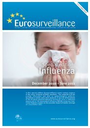
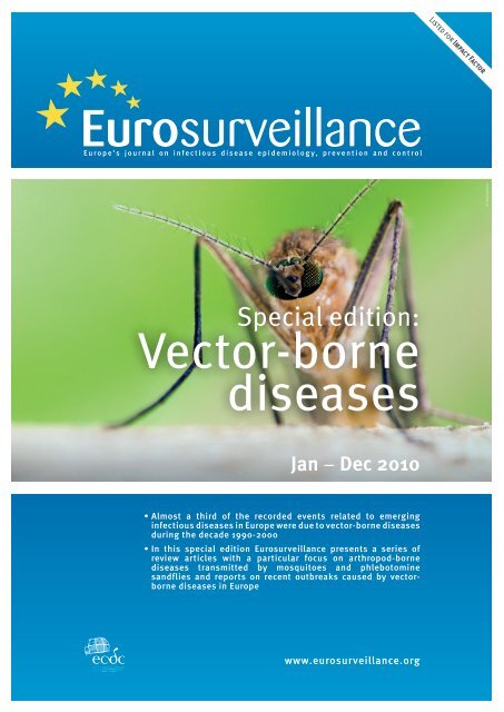
Special edition:<br />
<strong>Vector</strong>-<strong>borne</strong><br />
<strong>diseases</strong><br />
Jan − Dec 2010<br />
• Almost a third of the recorded events related to emerging<br />
infectious <strong>diseases</strong> in Europe were due to vector-<strong>borne</strong> <strong>diseases</strong><br />
during the decade 1990-2000<br />
• In this special edition <strong>Eurosurveillance</strong> presents a series of<br />
review articles with a particular focus on arthropod-<strong>borne</strong><br />
<strong>diseases</strong> transmitted by mosquitoes and phlebotomine<br />
sandflies and reports on recent outbreaks caused by vector<strong>borne</strong><br />
<strong>diseases</strong> in Europe<br />
www.eurosurveillance.org<br />
Listed for Impact Factor<br />
© Karsten Schneider - © Science Istockphoto Photo
Editorial team Editorial board<br />
Based at the European Centre for<br />
Disease Prevention and Control (ECDC),<br />
171 83 Stockholm, Sweden<br />
Telephone number<br />
+46 (0)8 58 60 11 38 or +46 (0)8 58 60 11 36<br />
Fax number<br />
+46 (0)8 58 60 12 94<br />
E-mail<br />
<strong>Eurosurveillance</strong>@ecdc.europa.eu<br />
Editor-in-Chief<br />
Karl Ekdahl<br />
Managing Editor<br />
Ines Steffens<br />
Scientific Editors<br />
Kathrin Hagmaier<br />
Williamina Wilson<br />
Assistant Editors<br />
Alina Buzdugan<br />
Ingela Söderlund<br />
Associate Editors<br />
Andrea Ammon, ECDC, Stockholm, Sweden<br />
Tommi Asikainen, ECDC, Stockholm, Sweden<br />
Mike Catchpole, Health Protection Agency,<br />
London, United Kingdom<br />
Denis Coulombier, ECDC, Stockholm, Sweden<br />
Christian Drosten, Universitätsklinikum Bonn, Bonn, Germany<br />
Johan Giesecke, ECDC, Stockholm, Sweden<br />
Herman Goossens, Universiteit Antwerpen, Antwerp, Belgium<br />
David Heymann, World Health Organisation, Geneva,<br />
Switzerland<br />
Irena Klavs, National Institute of Public Health, Ljubljana,<br />
Slovenia<br />
Karl Kristinsson, Landspitali University Hospital, Reykjavik,<br />
Iceland<br />
Daniel Lévy-Bruhl, Institut de Veille Sanitaire, Paris, France<br />
Richard Pebody, Health Protection Agency, London,<br />
United Kingdom<br />
Panayotis T. Tassios, University of Athens, Athens, Greece<br />
Hélène Therre, Institut de Veille Sanitaire, Paris, France<br />
Henriette de Valk, Institut de Veille Sanitaire, Paris, France<br />
Sylvie van der Werf, Institut Pasteur, Paris, France<br />
Design / Layout<br />
Fabrice Donguy / Martin Wincent<br />
Webmaster<br />
Sami Dufva<br />
www.eurosurveillance.org<br />
© <strong>Eurosurveillance</strong>, 2011<br />
Austria: Reinhild Strauss, Vienna<br />
Belgium: Koen De Schrijver, Antwerp<br />
Belgium: Sophie Quoilin, Brussels<br />
Bulgaria: Mira Kojouharova, Sofia<br />
Croatia: Borislav Aleraj, Zagreb<br />
Cyprus: Olga Poyiadji-Kalakouta, Nicosia<br />
Czech Republic: Bohumir Križ, Prague<br />
Denmark: Peter Henrik Andersen, Copenhagen<br />
England and Wales: Neil Hough, London<br />
Estonia: Kuulo Kutsar, Tallinn<br />
Finland: Hanna Nohynek, Helsinki<br />
France: Judith Benrekassa, Paris<br />
Germany: Jamela Seedat, Berlin<br />
Greece: Rengina Vorou, Athens<br />
Hungary: Ágnes Csohán, Budapest<br />
Iceland: Haraldur Briem, Reykjavik<br />
Ireland: Lelia Thornton, Dublin<br />
Italy: Paola De Castro, Rome<br />
Latvia: Jurijs Perevoščikovs, Riga<br />
Lithuania: Milda Zygutiene, Vilnius<br />
Luxembourg: Robert Hemmer, Luxembourg<br />
FYR of Macedonia: Elisaveta Stikova, Skopje<br />
Malta: Tanya Melillo Fenech, Valletta<br />
Netherlands: Paul Bijkerk, Bilthoven<br />
Norway: Hilde Klovstad, Oslo<br />
Poland: Malgorzata Sadkowska-Todys, Warsaw<br />
Portugal: Judite Catarino, Lisbon<br />
Romania: Daniela Pitigoi, Bucharest<br />
Scotland: Norman Macdonald, Glasgow<br />
Slovakia: Lukáš Murajda, Martin<br />
Slovenia: Alenka Kraigher, Ljubljana<br />
Spain: Elena Rodríguez Valín, Madrid<br />
Sweden: Aase Sten, Stockholm<br />
Turkey: Aysegul Gozalan, Istanbul<br />
European Commission: Paolo Guglielmetti, Luxembourg<br />
World Health Organization Regional Office for Europe: Nedret<br />
Emiroglu, Copenhagen
Contents<br />
Special focus: <strong>Vector</strong>-<strong>borne</strong> <strong>diseases</strong><br />
Editorials<br />
A perspective on emerging mosquito and<br />
phlebotomine-<strong>borne</strong> <strong>diseases</strong> in Europe 3<br />
by G Hendrickx, R Lancelot<br />
West Nile virus: the need to strengthen<br />
preparedness in Europe 5<br />
by H Zeller, A Lenglet, W Van Bortel<br />
Surveillance and outbreak reports<br />
West Nile virus circulation in Emilia-Romagna,<br />
Italy: the integrated surveillance system 2009 7<br />
by P Angelini, M Tamba, A C Finarelli, R Bellini, A Albieri,<br />
P Bonilauri, F Cavrini, M Dottori, P Gaibani, E Martini,<br />
A Mattivi, A M Pierro, G Rugna, V Sambri, G Squintani, P Macini<br />
Phylogenetic analysis in a recent controlled<br />
outbreak of Crimean-Congo haemorrhagic fever in<br />
the south of Iran, December 2008 12<br />
by S Chinikar, S Mojtaba Ghiasi, M Moradi, M M Goya,<br />
M Reza Shirzadi, M Zeinali, E Mostafavi, M Pourahmad, A Haeri<br />
Reviews<br />
West Nile virus in Europe: understanding the<br />
present to gauge the future 16<br />
by P Reiter<br />
Yellow fever and Dengue: a threat to Europe? 23<br />
by P Reiter<br />
Rift valley fever - a threat for Europe? 30<br />
by V Chevalier, M Pépin, L Plée, R Lancelot<br />
Leishmaniasis emergence in Europe 41<br />
by P D Ready<br />
Arthropod-<strong>borne</strong> viruses transmitted by<br />
phlebotomine sandflies in Europe: a review 52<br />
by J Depaquit, M Grandadam, F Fouque, PE Andry, C Peyrefitte<br />
Perspectives<br />
Crimean-Congo hemorrhagic fever in Europe:<br />
current situation calls for preparedness 60<br />
by H C Maltezou, L Andonova, R Andraghetti, M Bouloy,<br />
O Ergonul, F Jongejan, N Kalvatchev, S Nichol, M Niedrig,<br />
A Platonov, G Thomson, K Leitmeyer, H Zeller<br />
Rapid communications<br />
Chikungunya infection in a French traveller<br />
returning from the Maldives, October, 2009 64<br />
by M Receveur, K Ezzedine, T Pistone, D Malvy<br />
Chikungunya fever in two German tourists<br />
returning from the Maldives, September, 2009 66<br />
by M Pfeffer, I Hanus, T Löscher, T Homeier, G Dobler<br />
www.eurosurveillance.org<br />
A case of dengue type 3 virus infection imported<br />
from Africa to Italy, October 2009 70<br />
by C Nisii, F Carletti, C Castilletti, L Bordi, S Meschi,<br />
M Selleri, R Chiappini, D Travaglini, M Antonini, S Castorina,<br />
F N Lauria, P Narciso, M Gentile, L Martini, G Di Perri,<br />
S Audagnotto, R Biselli, M Lastilla, A Di Caro,<br />
M Capobianchi, G Ippolito<br />
Recent expansion of dengue virus serotype 3 in<br />
West Africa 73<br />
by L Franco, A Di Caro, F Carletti, O Vapalahti, C Renaudat,<br />
H Zeller, A Tenorio<br />
Dengue type 3 virus infections in European<br />
travellers returning from the Comoros and<br />
Zanzibar, February-April 2010 77<br />
by P Gautret, F. Simon, H Hervius Askling, O Bouchaud,<br />
I Leparc-Goffart, L Ninove, P Parola, for EuroTravNet<br />
Fatal and mild primary dengue virus infections<br />
imported to Norway from Africa and south-east<br />
Asia, 2008-2010 80<br />
by K Vainio, S Noraas, M Holmberg, H Fremstad,<br />
M Wahlstrøm, G Ånestad, S Dudman<br />
First two autochthonous dengue virus infections in<br />
metropolitan France, September 2010 84<br />
by G La Ruche, Y Souarès, A Armengaud, F Peloux-Petiot,<br />
P Delaunay, P Desprès, A Lenglet, F Jourdain, I Leparc-<br />
Goffart, F Charlet, L Ollier, K Mantey, T Mollet, J P Fournier,<br />
R Torrents, K Leitmeyer, P Hilairet, H Zeller, W Van Bortel,<br />
D Dejour-Salamanca, M Grandadam, M Gastellu-Etchegorry<br />
Dengue virus infection in a traveller returning from<br />
Croatia to Germany 89<br />
by J Schmidt-Chanasit, M Haditsch, I Schöneberg,<br />
S Günther, K Stark, C Frank<br />
Relapsing vivax malaria cluster in Eritrean refugees,<br />
Israel, June 2010 91<br />
by E Kopel, E Schwartz, Z Amitai, I Volovik<br />
First autochthonous malaria case due to<br />
plasmodium vivax since eradication, Spain,<br />
October 2010 94<br />
by P Santa-Olalla Peralta, M C Vazquez-Torres, E Latorre-<br />
Fandós, P Mairal-Claver, P Cortina-Solano, A Puy-Azón,<br />
B Adiego Sancho, K Leitmeyer, J Lucientes-Curdi, M J Sierra-Moros<br />
Infection with Mayaro virus in a French traveller<br />
returning from the Amazon region, Brazil,<br />
January, 2010 97<br />
by M C Receveur, M Grandadam, T Pistone, D Malvy<br />
Epidemiological analysis of mosquito-<strong>borne</strong><br />
pogosta disease in Finland, 2009 100<br />
by J Sane, S Guedes, S Kurkela, O Lyytikäinen, O Vapalahti<br />
Ongoing outbreak of West Nile virus infections in<br />
humans in Greece, July – August 2010 103<br />
by A Papa, K Danis, A Baka, A Bakas, G Dougas, T Lytras,G<br />
Theocharopoulos, D Chrysagis, E Vassiliadou, F Kamaria, A<br />
Liona, K Mellou, G Saroglou, T Panagiotopoulos<br />
A head on view of a Culex pipiens mosquito, this species is an important vector in West Nile Virus.<br />
© Istockphoto<br />
1
Retrospective screening of solid organ donors in<br />
Italy, 2009, reveals unpredicted circulation of West<br />
Nile virus 108<br />
by M R Capobianchi, V Sambri, C Castilletti, A M Pierro,<br />
G Rossini, P Gaibani, F Cavrini, M Selleri, S Meschi, D Lapa,<br />
A Di Caro, P Grossi, C De Cillia, S Venettoni, M P Landini,<br />
G Ippolito, A Nanni Costa, on behalf of the Italian Transplant<br />
Network<br />
Case report: West-Nile virus infection in two Dutch<br />
travellers returning from Israel 113<br />
by N Aboutaleb, M FC Beersma, H F Wunderink,<br />
A CTM Vossen, L G Visser<br />
Introduction and control of three invasive<br />
mosquito species in the Netherlands,<br />
July-October 2010 114<br />
by E J Scholte, W Den Hartog, M Dik, B Schoelitsz, M Brooks,<br />
F Schaffner, R Foussadier, M Braks, J Beeuwkes<br />
2 www.eurosurveillance.org
Editorials<br />
A perspective on emerging mosquito and phlebotomine<strong>borne</strong><br />
<strong>diseases</strong> in Europe<br />
G Hendrickx (info@avia-gis.be) 1 , R Lancelot 2<br />
1. Avia-GIS, Agro-Veterinary Information and Analysis, Zoersel, Belgium<br />
2. CIRAD, French Agricultural Research Centre for International Development, UMR CMAEE (Control of emerging and exotic animal<br />
<strong>diseases</strong>), Montpellier, France<br />
Citation style for this article:<br />
Citation style for this article: Hendrickx G, Lancelot R. A perspective on emerging mosquito and phlebotomine-<strong>borne</strong> <strong>diseases</strong> in Europe. Euro Surveill.<br />
2010;15(10):pii=19503. Available online: http://www.eurosurveillance.org/ViewArticle.aspx?ArticleId=19503<br />
Emerging infectious <strong>diseases</strong> are of increasing concern<br />
worldwide and in particular in Europe. In a review,<br />
Jones et al. have shown that between 1940 and 2004,<br />
the majority of emerging infectious <strong>diseases</strong> occurred<br />
in areas with both a high mobility and high density of<br />
population, notably in Western Europe. Furthermore,<br />
nearly a third (29%) of the recorded events related to<br />
emerging infectious <strong>diseases</strong> were due to vector-<strong>borne</strong><br />
<strong>diseases</strong> during the decade 1990-2000[1].<br />
This issue of <strong>Eurosurveillance</strong> presents a series of<br />
review articles with a particular focus on arthropod<strong>borne</strong><br />
<strong>diseases</strong> transmitted by mosquitoes and phlebotomine<br />
sandflies. Each of the papers addresses a<br />
series of issues of common interest such as the relevance<br />
of the disease in the given context, its transmission<br />
and epidemiology, including the current<br />
geographical distribution, and clinical symptoms,<br />
diagnostic methods, treatment strategies and prevention<br />
methods. Furthermore, the papers describe factors<br />
triggering changes in distribution of the vectors<br />
and disease and risk prediction models.<br />
A review on West Nile virus by Reiter includes a fresh<br />
and innovative viewpoint on the epidemiology and<br />
transmission of the disease [2], and the same author<br />
contributed further with a twin-review on two <strong>diseases</strong><br />
which have much in common: yellow fever and<br />
dengue [3]. Most importantly, both have a history of<br />
occurrence in Europe and vectors and pathogens are<br />
spreading through increased movement of persons and<br />
transport of goods. Chevalier et al. present a review on<br />
Rift Valley fever a mosquito-<strong>borne</strong> disease at Europe’s<br />
fringes. The epidemiology of Rift Valley fever is fascinating<br />
because of its complex cycle where the virus<br />
may remain dormant for many years and outbreaks<br />
involve both vector related and direct transmission.<br />
The disease may become a risk in the future in countries<br />
bordering the Mediterranean Sea, mainly through<br />
increased livestock trade.<br />
Two authors have contributed reviews on viruses transmitted<br />
by phlebotomine sandflies. Ready has written<br />
www.eurosurveillance.org<br />
This article has been published on 11 March 2010<br />
about Leishmania, a parasite of particular relevance<br />
to Europe because it is currently established around<br />
the Mediterranean Sea, but known to be spreading<br />
north [4]. Depaquit et al. have contributed a review on<br />
a number of less well known Phlebo-, Vesiculo- and<br />
Orbiviruses such as Sandfly fever Sicilian and Naples<br />
virus, Toscana virus Chandipura virus and others that<br />
are transmitted by sandflies in Europe and more specifically<br />
around the Mediterranean Sea [5]. The paper<br />
summarizes the current knowledge on these viruses<br />
which have a potential to spread throughout the distribution<br />
zone of their phlebotomine vectors in Europe.<br />
Both reviews provide a series of maps displaying<br />
country based information on the distribution of the<br />
disease.<br />
In addition to these papers, the issue features a perspective<br />
paper by Maltezou et al. presenting the<br />
present situation of Crimean-Congo hemorrhagic fever,<br />
a tick-<strong>borne</strong> disease, in Europe and emphasizing relevant<br />
aspects for preparedness concerning the potential<br />
spread of this disease in Europe in the future [6].<br />
As will become clear from reading these reviews, often<br />
crucial knowledge is still missing which is needed to<br />
anticipate, prevent or prepare for the establishment<br />
and spread of vector-<strong>borne</strong> <strong>diseases</strong>. One of these is<br />
reliable information on the continent wide distribution<br />
of potential disease vectors. National presenceabsence<br />
maps, as shown by Ready [4] and Depaquit<br />
[5] are a first step in this direction and need further<br />
refinement.<br />
In 2007/08, the European Centre for Disease Prevention<br />
and Control funded the V-<strong>borne</strong> project with the aim to<br />
identify and document vector-<strong>borne</strong> <strong>diseases</strong> relevant<br />
for public health in Europe, provide an overview of<br />
the existing relevant resources, carry out a qualitative<br />
multi-disciplinary risk assessment within the limits of<br />
the available information and data, and identify priorities<br />
for future prevention and control of vector <strong>borne</strong><br />
<strong>diseases</strong> in Europe. Building on the expertise from<br />
this network, ECDC created a European network for<br />
3
arthropod vector surveillance for human public health,<br />
the VBORNET in 2009 [7]. The network will establish<br />
pan-European state of the art maps of validated vector<br />
distributions that can be used as basis for risk<br />
assessment studies thus contributing to preparedness<br />
for the emerge or re-emerge of vector-<strong>borne</strong> disease in<br />
Europe.<br />
References<br />
1. Jones KE, Pate NG, Levy MA, Storeygard A, Balk D, Gittleman<br />
JL et al. Global trends in emerging infectious <strong>diseases</strong>. Nature.<br />
2008;451:990-3. Available from: http://dx.doi.org/10.1038/<br />
nature06536<br />
2. Reiter P. West Nile virus in Europe: understanding the present<br />
to gauge the future. Euro Surveill. 2010;15(10):pii=19508.<br />
Available from: http://www.eurosurveillance.org/ViewArticle.<br />
aspx?ArticleId=19508<br />
3. Paul Reiter Dengue and Yellow fever <strong>Eurosurveillance</strong>. Euro<br />
Surveill. 2010;15(10):pii=19509. Available online: http://www.<br />
eurosurveillance.org/ViewArticle.aspx?ArticleId=19509<br />
4. Chevalier V, Pépin M, Plée L, Lancelot R. Rift Valley fever - a<br />
threat for Europe?. Euro Surveill. 2010;15(10):pii=19506.<br />
Available from: http://www.eurosurveillance.org/ViewArticle.<br />
aspx?ArticleId=19506<br />
5. Depaquit J, Grandadam M, Fouque F, Andry P,<br />
Peyrefitte C. Arthropod-<strong>borne</strong> viruses transmitted by<br />
Phlebotomine sandflies in Europe: a review . Euro Surveill.<br />
2010;15(10):pii=19507. Available from: http://www.<br />
eurosurveillance.org/ViewArticle.aspx?ArticleId=19507<br />
6. Maltezou HC, Andonova L, Andraghetti R, Bouloy M, Ergonul<br />
O, Jongejan F, Kalvatchev N, Nichol S, Niedrig M, Platonov A,<br />
Thomson G, Leitmeyer K, Zeller H. Crimean-Congo hemorrhagic<br />
fever in Europe: current situation calls for preparedness. Euro<br />
Surveill. 2010;15(10):pii=19504. Available online: http://www.<br />
eurosurveillance.org/ViewArticle.aspx?ArticleId=19504<br />
7. European Centre for Disease prevention and Control (ECDC).<br />
Network of medical entomologists and public health experts<br />
(VBORNET). Available from: http://www.ecdc.europa.eu/en/<br />
activities/diseaseprogrammes/Pages/VBORNET.aspx<br />
4 www.eurosurveillance.org
Editorials<br />
West Nile virus: the need to strengthen preparedness in<br />
Europe<br />
H Zeller (evd@ecdc.europa.eu) 1 , A Lenglet 1 , W Van Bortel 1<br />
1. European Centre for Disease Prevention and Control, Stockholm, Sweden<br />
Citation style for this article:<br />
Zeller H, Lenglet A, Van Bortel W. West Nile virus: the need to strengthen preparedness in Europe. Euro Surveill. 2010;15(34):pii=19647. Available online: http://<br />
www.eurosurveillance.org/ViewArticle.aspx?ArticleId=19647<br />
The ongoing outbreak of West Nile virus (WNV) infections<br />
in humans in Greece described in this issue of<br />
<strong>Eurosurveillance</strong> is a timely reminder that WNV is a reemerging<br />
pathogen in Europe [1]. So far, WNV has been<br />
documented in animals and humans in several countries<br />
across Europe, mainly in central Europe and in<br />
the Mediterranean region. Over the last 15 years, outbreaks<br />
in horses and/or humans were reported from<br />
Romania, Hungary and Portugal, Spain, France, Italy<br />
and Greece [2].<br />
In 2010, a single probable human case was reported<br />
in July in Portugal. Outside the European Union (EU),<br />
WNV circulation has been documented in horses in<br />
Morocco and human cases have occurred in Russia<br />
(Volgograd Oblast) and in Israel. All these regions are<br />
located along the main routes of migratory birds. The<br />
current outbreak in humans in northern Greece, is the<br />
first recognised WN fever outbreak in humans in this<br />
country. However, studies suggest that WNV has probably<br />
been circulating in humans in the region of central<br />
Macedonia in northern Greece for many years [3,4].<br />
West Nile fever is a viral disease transmitted by mosquitoes<br />
and is distributed worldwide. The primary cycle<br />
of WNV involves ornithophilic mosquitoes and birds;<br />
some mosquito species mostly from the Culex genus<br />
can bite infectious birds and subsequently transmit the<br />
virus to humans and/or horses during another blood<br />
meal. Humans and horses are considered as deadend<br />
hosts. The vast majority of human cases remain<br />
asymptomatic after infection and severe neuroinvasive<br />
illness is reported in less than 1% of the patients. The<br />
main risk factor for severe clinical presentation is to<br />
be an elderly person. In this age group, reported case<br />
fatality rates may reach 10% [5]. In addition the high<br />
number of non-symptomatic cases may increase the<br />
risk of WNV transmission through blood donation or<br />
organ transplants [6].<br />
Each WNV outbreak is unique in that there is a complex<br />
interaction of different factors in space and time that<br />
contribute to the transmission of the virus to humans.<br />
These factors range from the introduction of infected<br />
migratory birds into native local bird populations, to<br />
www.eurosurveillance.org<br />
Article published on 26 August 2010<br />
climatic factors that increase the abundance of competent<br />
mosquito vectors and bridge vectors, to changes<br />
in human behaviour that favour exposure to infected<br />
mosquitoes. It is this complexity that makes each WNV<br />
outbreak particular and that make development and<br />
implementation of preparedness plans for the prevention<br />
of cases in humans so difficult.<br />
The recently reported probable and confirmed cases<br />
of WNV infection in Portugal and Greece, respectively<br />
reconfirm that this virus is actively circulating in several<br />
countries in the EU and that transmission to humans<br />
can be expected on a regular basis during the mosquito<br />
season. Also, reports of sporadic cases from several<br />
regions in Hungary during previous years indicate<br />
that WNV activity is widely distributed throughout this<br />
country and not limited to a single focus [7]. A recent<br />
study in Italy linked to infected organ donors [8] draws<br />
the same conclusion, that the virus is being transmitted<br />
in areas previously thought to not be at risk or<br />
affected. Furthermore, the case report in this issue of a<br />
Dutch traveller returning from Israel with WN infection<br />
highlights the need for awareness among physicians<br />
and laboratory staff to consider WNV infections as a<br />
differential diagnosis in cases where patients return<br />
from areas where they may have been exposed to the<br />
virus [9].<br />
The events described above strengthen the need for<br />
integrated multidisciplinary surveillance systems and<br />
response plans. This includes raising the awareness of<br />
clinicians and veterinarians of the clinical presentation<br />
of WNV disease in humans and horses (particularly<br />
during the mosquito season from June to October), primarily<br />
in areas considered as at major risk surrounding<br />
irrigated areas and river deltas. Furthermore, strengthening<br />
the understanding of suitable habitats for birds<br />
that would increase the bird-mosquito-human interface<br />
would be of value. In terms of entomology, a thorough<br />
understanding of competent vector species, their<br />
breeding ecology, their abundance and geographic<br />
range is of significant importance in establishing limits<br />
around WNV affected areas and in the identification of<br />
potential new at-risk areas.<br />
5
In addition, there is a need to have a better and more<br />
precise picture of WNV risk areas in Europe and neighbouring<br />
countries in order to implement appropriate<br />
control measures, especially guidelines for blood donation<br />
and organ transplants. Also, studies in Europe are<br />
required to better understand the cycle of transmission<br />
and the maintenance of WNV in the environment<br />
over the years to provide appropriate indicators for risk<br />
assessment.<br />
References<br />
1. Papa A, Danis K, Baka A, Bakas A, Dougas G, Lytras T,<br />
et al. Ongoing outbreak of West Nile virus infections in<br />
humans in Greece, July – August 2010. Euro Surveill.<br />
2010;15(34):pii=19644. Available from: http://www.<br />
eurosurveillance.org/ViewArticle.aspx?ArticleId=19644<br />
2. Calistri P, Giovannini A, Hubalek Z, Ionescu A, Monaco F,<br />
Savini G, et al. Epidemiology of West Nile in Europe and in the<br />
Mediterranean basin. Open Virol J. 2010 Apr 22;4:29-37.<br />
3. Antoniadis A, Alexiou-Daniel S, Malissiovas N, Doutsos J,<br />
Polyzoni T, LeDuc JW, et al. Seroepidemiological survey for<br />
antibodies to arboviruses in Greece. Arch Virol. 1990 [suppl 1]:<br />
277-285.<br />
4. Papa A, Perperidou P, Tzouli A, Castilletti C. West Nile virus<br />
neutralizing antibodies in humans in Greece. <strong>Vector</strong> Borne and<br />
Zoonotic Dis. Epub 2010 Aug 25<br />
5. O’Leary DR, Marfin AA, Montgomery SP, Kipp AM, Lehman<br />
JA, Biggerstaff BJ et al. The epidemic of West Nile virus<br />
in the United States, 2002. <strong>Vector</strong> Borne Zoonotic Dis.<br />
2004;4(1):61-70.<br />
6. Centers for Disease Control and Prevention (CDC). Update:<br />
Investigations of West Nile virus infections in recipients of<br />
organ transplantation and blood transfusion. MMWR Morb<br />
Mortal Wkly Rep. 2002 Sep 20;51(37):833-6.<br />
7. Krisztalovics K, Ferenczi E, Molnár Z, Csohán Á, Bán E, Zöldi<br />
V, Kaszás K. West Nile virus infections in Hungary, August–<br />
September 2008. Euro Surveill. 2008;13(45):pii=19030.<br />
Available from: http://www.eurosurveillance.org/ViewArticle.<br />
aspx?ArticleId=19030<br />
8. Capobianchi M, Sambri V, Castilletti C, Pierro AM, Rossini G,<br />
Gaibani P et al, on behalf of the Italian Transplant Network.<br />
Retrospective screening of solid organ donors in Italy, 2009,<br />
reveals unpredicted circulation of West Nile virus. Euro<br />
Surveill. 2010;15(34):pii=19648. Available from: http://www.<br />
eurosurveillance.org/ViewArticle.aspx?ArticleId=19648<br />
9. Aboutaleb N, Beersma MF, Wunderink HF, Vossen AC, Visser LG.<br />
Case report: West-Nile virus infection in two Dutch travellers<br />
returning from Israel. Euro Surveill. 2010;15(34):pii=19649.<br />
Available from: http://www.eurosurveillance.org/ViewArticle.<br />
aspx?ArticleId=19649<br />
6 www.eurosurveillance.org
Surveillance and outbreak reports<br />
West Nile virus circulation in Emilia-Romagna, Italy:<br />
the integrated surveillance system 2009<br />
P Angelini (pangelini@regione.emilia-romagna.it) 1 , M Tamba 2 , A C Finarelli 1 , R Bellini 3 , A Albieri 3 , P Bonilauri 2 , F Cavrini 4 ,<br />
M Dottori 2 , P Gaibani 4 , E Martini 5 , A Mattivi 1 , A M Pierro 4 , G Rugna 2 , V Sambri 4 , G Squintani 5 , P Macini 1<br />
1. Public Health Service, Emilia-Romagna Region, Bologna, Italy<br />
2. Istituto Zooprofilattico Sperimentale della Lombardia e dell’Emilia-Romagna, Brescia, Italy<br />
3. Centro Agricoltura Ambiente “G Nicoli”, Crevalcore, Italy<br />
4. Regional Reference Centre for Microbiological Emergencies (CRREM), Microbiology Unit, Azienda Ospedaliero-Universitaria di<br />
Bologna, Policlinico S. Orsola–Malpighi, Bologna, Italy<br />
5. Veterinary and Food Hygiene Service, Emilia-Romagna Region, Bologna, Italy<br />
Citation style for this article:<br />
Citation style for this article: Angelini P, Tamba M, Finarelli AC, Bellini R, Albieri A, Bonilauri P, Cavrini F, Dottori M, Gaibani P, Martini E, Mattivi A, Pierro<br />
AM, Rugna G, Sambri V, Squintani G, Macini P. West Nile virus circulation in Emilia-Romagna, Italy: the integrated surveillance system 2009. Euro Surveill.<br />
2010;15(16):pii=19547. Available online: http://www.eurosurveillance.org/ViewArticle.aspx?ArticleId=19547<br />
Following a large West Nile virus (WNV) epidemic in<br />
north-eastern Italy in 2008, human and animal surveillance<br />
activities were implemented in Emilia Romagna.<br />
Human surveillance was performed by serology or<br />
genome detection on blood and cerebrospinal fluid for<br />
all suspected cases suffering from acute meningoencephalitis<br />
in the regional territory. Animal surveillance<br />
consisted of passive and active surveillance of horses<br />
and active surveillance of wild birds and mosquitoes.<br />
Between 15 June and 31 October 2009, nine of 78 possible<br />
cases of West Nile neuroinvasive disease were<br />
confirmed (three fatal). From May to October, 26 cases<br />
of neurological West Nile disease were confirmed<br />
among 46 horses. The overall incidence of seroconversion<br />
among horses in 2009 was 13%. In 2009, 44 of<br />
1,218 wild birds yielded positive PCR results for WNV<br />
infection. The planned veterinary and entomological<br />
surveillance actions detected WNV activity from the<br />
end of July 2009, about 2-3 weeks before the onset of<br />
the first human neurological case. Passive surveillance<br />
of horses seems to be an early and suitable tool for<br />
the detection of WNV activity, but it will be less sensitive<br />
in the future, because an intensive programme of<br />
horse vaccination started in June 2009.<br />
Regional integrated surveillance system<br />
In Italy a national veterinary plan for the surveillance<br />
of West Nile virus (WNV) circulation was set up in 2001<br />
under the coordination of the National Reference Centre<br />
for Exotic Diseases of animals (CEntro Studi Malattie<br />
Esotiche; CESME).<br />
During the late summer of 2008 a large epidemic of<br />
WNV infection occurred in north-eastern Italy over an<br />
area exceeding 7,000 km 2 , in three regions, including<br />
Emilia-Romagna. Twenty-three horse cases and three<br />
human cases of the neuroinvasive form of West Nile<br />
disease (WND) were confirmed by laboratory tests<br />
[1,2]. After the first evidence of WNV circulation in<br />
horses was found, additional surveillance on horses,<br />
birds and mosquitoes was activated.<br />
www.eurosurveillance.org<br />
This article has been published on 22 April 2010<br />
In Emilia-Romagna the WNV surveillance plan 2009<br />
adopted locally the surveillance measures indicated by<br />
the national plan. In particular, among the surveillance<br />
activities, the choice was to monitor wild non-migratory<br />
birds, such as corvids (the crow family), considered<br />
the most sensitive indicators among birds, which<br />
can be captured easily. As regards equine surveillance,<br />
the regional plan emphasised the education of veterinary<br />
practitioners, focusing on the inclusion of WNV<br />
in differential diagnosis and the achievement of rapid<br />
reporting. A major feature of this plan was to establish<br />
an extremely sensitive system of passive surveillance.<br />
In addition to passive surveillance, active monitoring<br />
of horses was implemented in the area involved in the<br />
2008 outbreak, including Ferrara and the neighbouring<br />
provinces [3].<br />
Evidence of WNV circulation in 2008 was found in<br />
animals [1,4], humans [5], and mosquitoes. This<br />
highlighted the need to implement an integrated surveillance<br />
system which would describe the phenomenon<br />
comprehensively. Such a system facilitates the<br />
collection of data to evaluate spatial distribution and<br />
time trends of viral circulation and shares information.<br />
For this reason the 2009 Regional Surveillance Plan<br />
implemented activities beyond those of the National<br />
Plan, revised the surveillance system of human cases,<br />
activated intensive entomological monitoring, and<br />
enlarged the surveillance area to involve all the provinces<br />
along the Po River.<br />
Human surveillance<br />
The aim of the human surveillance system was the<br />
early detection of infection in humans and the estimation<br />
of its diffusion through the systematic analysis of<br />
newly emerging clinical cases, in order to manage specific<br />
interventions.<br />
7
The surveillance was performed throughout the<br />
regional territory, from 15 June to 31 October 2009,<br />
corresponding to the period of vector activity in Emilia-<br />
Romagna and adjusted locally according to weather<br />
conditions and vector activity reports. In 2009, the<br />
2008 case definition [6] was extended to include cases<br />
of all ages and not only those over 15 years of age.<br />
The human surveillance activity was performed<br />
by serology or genome detection on blood and<br />
Figure 1<br />
Map of municipalities with confirmed West Nile<br />
virus circulation and localisation of human West Nile<br />
neuroinvasive disease cases by probable infection site,<br />
Emilia-Romagna, Italy, 2009<br />
WNV: West Nile virus<br />
cerebrospinal fluid for all suspected cases suffering<br />
from acute meningoencephalitis in the regional territory.<br />
Active surveillance of people who live or work<br />
in areas of documented viral circulation was also performed.<br />
In addition blood and cerebrospinal fluid samples<br />
from subjects living or staying for at least one<br />
night in the regional area were sent to the Regional<br />
Reference Centre for Microbiological Emergencies<br />
(Centro di Riferimento Regionale per le Emergenze<br />
Microbiologiche; CRREM). In selected cases, positive<br />
specimens were confirmed by the National Health<br />
Institute (Istituto Superiore di Sanità; ISS) and by<br />
the National Institute for Infectious Diseases (Istituto<br />
Nazionale Malattie Infettive; INMI).<br />
From October 2008 to April 2009 a serosurvey performed<br />
on 9,177 healthy blood donors living in the<br />
province of Ferrara detected a total of 62 IgG positive<br />
subjects, corresponding to a seroprevalence of 0.68%.<br />
Animal surveillance<br />
The regional veterinary WNV surveillance system was<br />
activated from May to October, performing passive and<br />
active surveillance on horses and on non-migratory<br />
wild birds.<br />
Horse passive surveillance<br />
In Italy all suspected signs of WND in horses must be<br />
notified to the official veterinary services. Suspected<br />
cases were confirmed if resulted positive by reverse<br />
transcription – polymerase chain reaction (RT-PCR) performed<br />
on central nervous system [7] or to a WNV virus<br />
Figure 2<br />
Distribution of West Nile virus confirmed cases (mosquito pools, birds, horses, and human West Nile neuroinvasive<br />
disease) by date, Emilia-Romagna, Italy, July-October 2009<br />
Number of cases<br />
18<br />
16<br />
14<br />
12<br />
10<br />
8<br />
6<br />
4<br />
2<br />
0<br />
15/7-31/7 1/8-15/8 16/8-31/8 1/9-15/9 16/9-30/9 1/10-15/10 16/10-31/10<br />
Date 2009<br />
WNND: West Nile neuroinvasive disease<br />
Note: The figure does not include a magpie (found in early May) or a jay (found in early November).<br />
Mosquito pools<br />
8 www.eurosurveillance.org<br />
Birds<br />
Horses<br />
Human WNND
neutralisation (VN) test (cut-off titre 1:10) in microtitre<br />
plates and to IgM enzyme linked immunosorbent assay<br />
(ELISA) [8,9].<br />
Horse active surveillance<br />
In the provinces of Ferrara, Bologna, Modena, Ravenna,<br />
and Reggio Emilia, every 1,600 km 2 , 28 seronegative<br />
unvaccinated equine sentinels, sufficient to detect an<br />
incidence above 10% (CI 95%), were selected in the<br />
spring of 2009. They were serologically tested twice<br />
after the selection, at the beginning of August and<br />
the beginning of September. Samples collected were<br />
screened by a home-made competitive ELISA [10].<br />
Positive samples were confirmed by VN and IgM ELISA<br />
at the CESME in Teramo. A seroconversion was confirmed<br />
if VN titre was at least 1:10 and there was evidence<br />
of IgM antibodies.<br />
Wild bird surveillance<br />
Monitoring was carried out in all the provinces along<br />
the Po River, in the plain area of Emilia-Romagna.<br />
Every 1,600 km 2 , a monthly sample of about 40 wild<br />
birds caught or shot within specific wildlife population<br />
control programmes was collected. Samples of organs<br />
(brain, heart, and kidney) of each bird were pooled and<br />
examined by RT-PCR [7].<br />
Entomological surveillance<br />
The surveillance system was based on the weekly to<br />
monthly (frequency depends on local resources) collection<br />
of mosquitoes in fixed stations and in the sites<br />
where birds, humans, or horses signalled WNV activity.<br />
Mosquito collections for WNV screening were conducted<br />
in six provinces: Ferrara, Ravenna, Bologna,<br />
Modena, Reggio Emilia, and Parma, using 92 CO 2<br />
baited traps positioned in fixed stations. Moreover,<br />
mosquito collections were performed promptly using<br />
CO 2 and gravid traps in sites where positive horses and<br />
human cases had been detected.<br />
The surveillance system was activated in the period 15<br />
April to 10 October. Collected mosquitoes were pooled<br />
www.eurosurveillance.org<br />
(maximum 200) by species, date, and site of collection<br />
and examined by RT-PCR [7]. In addition, overwintered<br />
mosquito females were collected during the period 3<br />
March to 8 April by manual aspirator in rural buildings<br />
in the area where WNV was active in 2008.<br />
Virological analysis<br />
Human samples<br />
The detection of WNV genome in human plasma, cerebrospinal<br />
fluid, and serum samples obtained from<br />
patient suffering from clinical symptoms of meningoecephalitis<br />
was performed by an RT-PCR assay based on<br />
specific TaqMan probes [7].<br />
Animal samples<br />
In horses RNA was extracted starting from 200 µl of<br />
serum or whole blood with EDTA as anticoagulant. In<br />
birds RNA was extracted from 200 µl of phosphate<br />
buffer saline homogenate (about 20% tissue g / buffer<br />
ml) of pooled brain, heart and kidney of each analysed<br />
bird. In mosquitoes RNA was extracted from 200 µl of<br />
a maximum of 200 pooled mosquitoes manual grinded<br />
by using copper stained beads, in 500-800 µl of PBS.<br />
cDNA was submitted to RT-PCR according to the method<br />
of Tang and colleagues [7]. Positive samples were confirmed<br />
by sequencing of partial nucleocapsin and premembrane<br />
protein M amplified according to [11]. Finally<br />
from each RT-PCR positive sample WNV was isolated<br />
on Vero and RK13 cell lines.<br />
Results<br />
Results of the integrated surveillance system are<br />
mapped in figure 1, with the sequence of events summarised<br />
in figure 2 (July-October), and discussed<br />
below.<br />
Human cases<br />
As of 31 October 2009, nine out of 78 possible cases<br />
of West Nile neuroinvasive disease (WNND) notified<br />
in Emilia-Romagna have been confirmed (8/9 males;<br />
median age 72 years, range: 62-78). Three patients<br />
Table 1<br />
Species distribution of wild birds tested for West Nile virus (n=1,218), Emilia-Romagna, Italy, May-October 2009<br />
Species Birds tested WNV RT-PCR positive % WNV positive<br />
European magpie (Pica pica) 607 27 4.4<br />
Carrion crow (Corvus corone cornix) 350 5 1.4<br />
European starling (Sturnus vulgaris) 98 5 5<br />
Eurasian jay (Garrulus glandarius) 96 2 2<br />
Common blackbird (Turdus merula) 30 0 -<br />
Strigiformes 11 2 18<br />
Charadriiformes 8 3 38<br />
Other Passeriformes 7 0 -<br />
Other bird orders 5 0 -<br />
Piciformes 4 0 -<br />
Columbiformes 2 0 -<br />
Total 1,218 44 3.6<br />
RT-PCR: reverse transcription-polymerase chain reaction; WNV: West Nile virus<br />
9
Table 2<br />
Species of mosquitoes tested for West Nile virus (n=190,516), Emilia-Romagna, Italy, May-October 2009<br />
Province Bologna Forlì Ferrara Modena Piacenza Parma Ravenna Reggio Emilia Total<br />
Mos-<br />
Species<br />
Pool<br />
quito<br />
Pool Mos-<br />
Pool<br />
+ quito<br />
Pool Mos-<br />
Pool<br />
+ quito<br />
Pool Mos-<br />
Pool<br />
+ quito<br />
Pool Mos-<br />
Pool<br />
+ quito<br />
Pool Mos-<br />
Pool<br />
+ quito<br />
Pool Mos-<br />
Pool<br />
+ quito<br />
Pool Mos- Pool Mos-<br />
Pool Pool<br />
+ quito + quito<br />
Pool<br />
+<br />
Ae. albop-<br />
169 13 11 4 392 31 227 23 58 11 228 6 142 20 1,227 108 0<br />
ictus<br />
Ae. caspius 1,713 51 14 5 9,953 114 16,915 121 2 1 12 2 606 11 68 9 29,283 314 0<br />
Ae. detritus 1 1 4 1 5 2 0<br />
Ae. dorsalis 13 1 13 1 0<br />
Ae. geniculatus 6 2 2 1 8 3 0<br />
Ae.vexans 2 1 11 2 84 3 4,090 41 1 1 363 3 46 9 4,597 60 0<br />
An. maculip-<br />
4 1 59 3 14 5 1 1 4 4 82 14 0<br />
ennis<br />
An. plumbeus 2 2 2 2 0<br />
Cx. modes-<br />
7 3 114 10 117 12 8 1 246 26 0<br />
tus<br />
Cx. pipiens 84,225 645 9 158 17 52,973 396 5 6,664 78 7 331 13 6,926 50 1 911 10 2,865 50 5 155,053 1,259 27<br />
Total 86,120 714 9 194 28 0 63,575 557 5 28,048 285 7 393 27 0 7,530 62 1 1,517 21 0 3,139 95 5 190,516 1,789 27<br />
died, two living in Ferrara Province and one in Modena.<br />
In addition, not reported in figures, the local health<br />
units of Parma and Modena notified two other confirmed<br />
cases, both 72 year-old women resident in<br />
Mantua province (Lombardy region), treated in hospital<br />
in Emilia-Romagna. Another case of infection was that<br />
of a 78 year-old female liver donor. Before her death,<br />
she had spent two weeks visiting relatives in the WNV<br />
affected area (Reggio Emilia).<br />
Horse cases<br />
Passive surveillance<br />
From May to October, 26 cases of neurological WND<br />
were confirmed among 46 horses in which it was suspected.<br />
Four of the eight provinces involved in the<br />
regional surveillance system had WND cases in horses.<br />
The first symptoms in horses were detected in the second<br />
half of July in the provinces of Ferrara and Reggio<br />
Emilia, but the most cases were notified between mid-<br />
August and mid-September (figure 2).<br />
Active surveillance<br />
Seroconversions in sentinel horses were detected in<br />
three provinces (Ferrara, Modena, and Reggio Emilia).<br />
Early seroconversions were registered among the controls<br />
examined at the beginning of August. Serological<br />
controls around the stables with WND cases also confirmed<br />
recent WNV infections in the province of Parma.<br />
The overall incidence of seroconversion among horses<br />
in 2009 was 13% (95% CI: 10% to 16%), but in Ferrara it<br />
was 28% (95% CI: 19% to 39%).<br />
Wild birds<br />
Six of the eight provinces that took part in the regional<br />
surveillance system reported positive birds. In 2009,<br />
44 wild birds out of 1,218 (tested by PCR) yielded<br />
positive results for WNV infection. With the exception<br />
of a magpie caught in May, positive wild birds were<br />
detected from the end of July (figure 2). Most of the<br />
infected wild birds were corvids (Pica pica, Corvus corone<br />
cornix, Garrulus glandarius), collected within population<br />
control programmes in August and September,<br />
but WNV was detected also in other species, mainly<br />
found dead in wildlife recovery centres (table 1).<br />
Table 1. Species distribution of wild birds tested for<br />
West Nile V (n=1,218), Emilia-Romagna, Italy, May-<br />
October 2009<br />
Mosquitoes<br />
About 190,000 mosquitoes were collected, pooled and<br />
tested using PCR (1,789 pools of ≤200 individuals/<br />
pool). Culex pipiens were the most abundant species<br />
(81.4%) followed by Aedes caspius (15.4%) and Aedes<br />
vexans (2.4%). Other collected species are shown in<br />
table 2.<br />
Twenty-seven pools, all consisting of Culex pipiens,<br />
yielded positive results for WNV. Early positive pools<br />
were collected in the province of Reggio Emilia at<br />
the end of July. Minimum infection rates (MIR: (no. of<br />
10 www.eurosurveillance.org
positive pools/no. of mosquitoes tested) x 1,000) [12]<br />
were calculated, with higher MIR values recorded in<br />
August in the provinces of Reggio Emilia (3.08) and<br />
Modena (1.44).<br />
Referring to the collection of overwintering mosquitoes,<br />
three mosquito species were collected: Culex pipiens<br />
(516 females, 52%), Anopheles maculipennis s.l.<br />
(475 females, 48%), Culiseta annulata (four females,<br />
Surveillance and outbreak reports<br />
Phylogenetic analysis in a recent controlled outbreak<br />
of Crimean-Congo haemorrhagic fever in the south of<br />
Iran, December 2008<br />
S Chinikar (chinikar@pasteur.ac.ir) 1 , S Mojtaba Ghiasi 1 , M Moradi 1 , M M Goya 2 , M Reza Shirzadi 2 , M Zeinali 2 , E Mostafavi 1 ,<br />
M Pourahmad 3 , A Haeri 3,4<br />
1. National Reference Laboratory for Arboviruses and Viral Haemorrhagic Fevers, Pasteur Institute of Iran, Tehran, Iran<br />
2. Centre for Disease Control (CDC), Ministry of Health of Iran, Tehran, Iran<br />
3. Jahrom Faculty of Medicine<br />
4. Shahid Beheshti University of Medical Sciences, Tehran, Iran<br />
Citation style for this article:<br />
Chinikar S, Mojtaba Ghiasi S, Moradi M, Goya MM, Reza Shirzadi M, Zeinali M, Mostafavi E, Pourahmad M, Haeri A. Phylogenetic analysis in a recent controlled<br />
outbreak of Crimean-Congo haemorrhagic fever in the south of Iran, December 2008. Euro Surveill. 2010;15(47):pii=19720. Available online: http://www.<br />
eurosurveillance.org/ViewArticle.aspx?ArticleId=19720<br />
Crimean-Congo haemorrhagic fever (CCHF) is a viral<br />
zoonotic disease with a high mortality rate in humans.<br />
The CCHF virus is transmitted to humans through the<br />
bite of Ixodid ticks or contact with blood or tissues<br />
of CCHF patients or infected livestock. In December<br />
2008, a re-emerging outbreak of CCHF occurred in the<br />
southern part of Iran. Five people were hospitalised<br />
with sudden fever and haemorrhaging, and CCHF was<br />
confirmed by RT-PCR and serological assays. One of<br />
the cases had a fulminant course and died. Livestock<br />
was identified as the source of infection; all animals in<br />
the incriminated herd were serologically analysed and<br />
more than half of them were positive for CCHFV. We<br />
demonstrated that two routes of transmission played<br />
a role in this outbreak: contact with tissue and blood<br />
of infected livestock, and nosocomial transmission.<br />
Phylogenetic analyses helped to identify the origin of<br />
this transmission. This outbreak should be considered<br />
as a warning for the national CCHF surveillance system<br />
to avoid further outbreaks through robust prevention<br />
and control programmes.<br />
Introduction<br />
Crimean-Congo haemorrhagic fever (CCHF) is a viral<br />
zoonotic haemorrhagic fever with up to 13-50% mortality<br />
rate in humans. Infected animals are unsymptomatic.<br />
The disease is caused by Crimean-Congo<br />
haemorrhagic fever virus (CCHFV) that belongs to the<br />
family Bunyaviridae, genus Nairovirus. The of negative<br />
single-stranded RNA genome consists of three segments,<br />
large (L), medium (M) and small (S), coding for<br />
the viral polymerase (L), the envelope glycoproteins (M)<br />
and the viral nucleoprotein (S) [1-5]. The typical course<br />
of CCHF progresses through four distinct phases: incubation,<br />
pre-haemorrhagic phase, haemorrhagic phase<br />
and convalescence [6-8]. After a incubation period of<br />
one to three days, the patient has sudden onset of<br />
fever, myalgia, nausea and severe headache. Within<br />
three to six days of the onset of illness, a petechial<br />
rash and haemorrhagic symptoms such as epistaxis,<br />
Article published on 25 November 2010<br />
haematemesis, and melaena may occur. The most<br />
severely ill patients develop multiorgan failure characterised<br />
by shock, haemorrhaging and coma [9-11].<br />
The virus is transmitted to humans through the bite of<br />
Ixodid ticks or by contact with blood or tissues from<br />
infected livestock [12-14]. In addition to zoonotic transmission,<br />
CCHFV can be spread from person to person<br />
and is one of the rare haemorrhagic fever viruses able<br />
to cause nosocomial outbreaks in hospitals [15-20].<br />
In the period from 1 January 2000 to 12 September<br />
2010, 738 confirmed cases of CCHF and 108 associated<br />
fatalities were notified in Iran [15,21]. The province<br />
reporting most infections was Sistan-va-Baluchistan,<br />
Isfahan and Fars (Figure 1).<br />
Figure 1<br />
Geographical distribution of Crimean-Congo<br />
haemorrhagic fever in Iran, 1 January 2000-12 September<br />
2010 (n=738)<br />
12 www.eurosurveillance.org
Table<br />
History and laboratory results of probable cases of Crimean-Congo haemorrhagic fever, Fars province, Iran, November–December 2008 (n=5)<br />
Clinical and paraclinical signs Ig M Ig G<br />
Date of sampling<br />
RT-PCR<br />
www.eurosurveillance.org<br />
Second sample<br />
First sample<br />
Second sample<br />
First sample<br />
Proteinuria<br />
Thrombocytopenia<br />
Leukocytopenia<br />
Haemorrhage<br />
Petechia<br />
Fever<br />
Death<br />
Second sample<br />
First sample<br />
Date of fever<br />
Contact with suspected patient<br />
Contact with livestock<br />
Age (years)/sex<br />
Profession<br />
Patient<br />
Aa Housewife 46/F Yes No 18 Dec 2008 21 Dec 2008 Not taken Yes Yes No Yes Yes Yes No report Negative Not taken Negative Not taken Positive<br />
B Butcher 21/M Yes No 18 Dec 2008 20 Dec 2008 25 Dec 2008 No Yes No Yes Yes Yes Yes Positive Positive Negative Negative Negative<br />
C Butcher 24/M Yes Yes 19 Dec 2008 20 Dec 2008 25 Dec 2008 No Yes No Yes No Yes Yes Negative Positive Negative Positive Positive<br />
D Self-employed 28/M Yes No 21 Dec 2008 22 Dec 2008 Not taken No Yes No No Yes Yes Yes Positive Not taken Negative Not taken Negative<br />
E Nurse 26/F No Yes 27 Dec 2008 30 Dec 2008 Not taken No Yes Yes Yes No Yes No Positive Not taken Negative Not taken Positive<br />
F: female; M: male.<br />
a Index case.<br />
Outbreak description<br />
Here, we report a CCHF outbreak in Fars province,<br />
Iran, caused by contact of humans with blood or tissues<br />
of infected livestock, with additional nosocomial<br />
transmission. In total, five patients (A-E) were admitted<br />
to the regional hospital with similar presentations<br />
of a haemorrhagic condition in the period from 18<br />
December to 21 December 2008. This period coincides<br />
with the Muslim ceremony Eid-al-Adha (the ceremony<br />
of sacrificing livestock) which is celebrated in Islamic<br />
countries on 9 December.<br />
Patients A and D (who are part of the same family)<br />
bought a calf from a butchery run by two brothers,<br />
patients B and C, and hired them to sacrifice the animal.<br />
On the morning of 18 December 2008, Patient A,<br />
the index case of this outbreak, was admitted to hospital<br />
and died after a fulminant course of CCHF. In the<br />
evening of the same day, Patients B and C were admitted<br />
to the same hospital with fever and chill, severe<br />
headache, dizziness, photophobia. Patient D developed<br />
similar clinical signs on 21 December and was<br />
hospitalised. Nine days after the index case, the nurse<br />
caring of these four patients was also hospitalised<br />
with haemorrhagic symptoms (Patient E).<br />
Materials and methods<br />
Case definition<br />
The case definition for probable cases included patients<br />
admitted between 18 December and 27 December 2008<br />
in the regional hospital and presenting with a clinical<br />
picture compatible with CCHF, or contact with tissues<br />
or blood from a possibly infected animal, or a healthcare<br />
worker with a history of contact with a CCHF case.<br />
Probable cases with positive IgM serology and/or positive<br />
RT-PCR were considered as CCHF confirmed cases.<br />
Laboratory analysis<br />
Human and animal sera were analysed by ELISA for<br />
anti-CCHFV IgM and IgG as described [15,22]. Viral RNA<br />
was extracted from patient’s serum using QIAamp RNA<br />
Mini kit (QIAgen GmbH, Hilden, Germany) and analysed<br />
by gel-based and real-time RT-PCR with a one-step<br />
RT-PCR kit (QIAgen GmbH, Hilden, Germany). A 536 bp<br />
fragment of the S segment of the CCHFV genome was<br />
amplified [4,8,12] and sequenced.<br />
Phylogenetic analysis was performed with the neighbour-joining<br />
method based on Kimura two-parameter<br />
distances by using Mega 4 software. Bootstrap confidence<br />
limits were based on 500 replicates. Evolutionary<br />
divergence, distance matrix and subsequently sets of<br />
phylogenetic trees were calculated by the software<br />
[23].<br />
Results and discussion<br />
As summarised in the Table, five probable CCHF cases<br />
in this outbreak were confirmed by serological and<br />
molecular methods. It is worth mentioning that no<br />
immunological response was detected in the fatal<br />
case that had a fulminant course, Patient A, and CCHF<br />
13
in this case was only confirmed by a strongly positive<br />
RT-PCR. There is evidence of other fatal cases lacking<br />
an immune response to CCHFV [6,24]. Patients C and E<br />
were positive both for viral RNA and antibodies against<br />
CCHFV, while Patients B and D were negative in the PCR<br />
and only confirmed by serological assay. Notably, ribavirin<br />
was administered to the patients in hospital.<br />
At the same time, serum samples were collected from<br />
50 animals in the herd from which the calf had been<br />
bought. In 30 of these samples antibodies to CCHFV<br />
were detected. Although CCHF is an asymptomatic disease<br />
in livestock that does not kill the animals, seroepidemiological<br />
surveys of animal populations in endemic<br />
areas and high risk regions could be useful in that they<br />
may complement the national surveillance system and<br />
serve as an early warning of CCHF in the area.<br />
In this outbreak, it was demonstrated that the main<br />
transmission route of CCHF was through handling<br />
blood or tissues of infected livestock (for patients<br />
A, B, C and D), while patient E had had no contact to<br />
livestock and was infected nosocomially. It is unclear<br />
why Patient A had such a fulminant course of disease<br />
and died. Patients B, C and D were infected through the<br />
same route, by direct contact with tissue and blood of<br />
the same animal, but had a milder course of disease<br />
and recovered. It is important in infectious disease outbreaks<br />
to investigate what factors determine the severity<br />
of the disease in different individuals [6]. There are<br />
published reports on the influence of cytokine levels<br />
on the immune response to CCHFV in different patients<br />
[24,25]. It has been shown that patients infected with<br />
a higher dose of virus develop more severe disease<br />
symptoms and outcomes [26-28]. Although we did not<br />
use quantitative RT-PCR, the band density of the PCR<br />
product obtained from patient A was much higher than<br />
that of the other patients. On the other hand, it seems<br />
likely that patients B and C presented a mild form of the<br />
disease because they may already have had antibodies<br />
against CCHFV due to their professional exposure.<br />
Moreover, no anti-CCHFV IgG antibody response was<br />
detected for the patient B, whereas a normal serological<br />
and molecular pattern was seen in patient C, which<br />
Figure 2<br />
Phylogenetic comparison of Crimean-Congo haemorrhagic fever virus isolates from Iran with isolates form patients in the<br />
recent outbreak in Fars province, Iran, December 2008<br />
A<br />
B<br />
Divergence<br />
1 2 3 4<br />
Percent identity<br />
5 6 7 8 9<br />
1 99.9 99.9 99.9 99.9 99.8 97.6 97.6 97.7 1 AY366373(partial)<br />
2 0.2 100.0 99.9 99.9 99.9 97.6 97.6 97.7 2 AY366379(partial)<br />
3 0.2 0.0 99.9 99.9 99.9 97.6 97.6 97.7 3 AY366378(partial)<br />
4 0.2 0.4 0.4 99.8 99.8 97.5 97.6 97.6 4 AY366375(partial)<br />
5 0.4 0.2 0.2 0.6 99.9 97.6 97.6 97.7 5 AY366374(partial)<br />
6 0.6 0.4 0.4 0.8 0.2 97.5 97.5 97.6 6 AY366376(partial)<br />
7 1.0 0.8 0.8 1.2 1.0 1.2 99.9 99.8 7 Patient A<br />
8 1.0 0.8 0.8 1.3 1.0 1.3 0.2 99.9 8 Patient C<br />
9 0.8 0.6 0.6 1.0 0.8 1.0 0.4 0.2 9 Patient E<br />
1 2 3 4 5 6 7 8 9<br />
A. The phylogeny tree of nucleotide sequences spanning described regions of the S-segment of CCHF virus genome which is detected in the<br />
outbreak. B. Nucleotide identity and divergence of CCHF virus genomes isolated from patients of the outbreak.<br />
14 www.eurosurveillance.org
might be interpreted to indicate that patient B was<br />
infected with a very low viral dose. Recent investigations<br />
have concluded a relationship linking the severity<br />
and outcome of CCHFV infections with the strength<br />
of the host immune response and the initial viral load<br />
[24,25,27].<br />
Phylogenetic analysis of alignments of three partial<br />
genomic sequences (536 bp) of the CCHFV S segment<br />
indicates that the viruses isolated from patients A, C<br />
and E can be differentiated into two distinct branches,<br />
with a slightly lower identity between patients A and<br />
E. As illustrated in Figure 2, the sequences obtained<br />
in this outbreak are not clustered with other CCHFV<br />
sequences isolated in Iran (about 97.5% identity). It<br />
is possible that a new strain occurred in the outbreak<br />
region, and further phylogenetic analyses are required<br />
to identify the precise origin of this genetic variant.<br />
However, comparison of the isolates from our patients<br />
with isolates from other areas may give some indications<br />
as to the origin of this outbreak [12].<br />
One of the factors that contributed to the control of<br />
this outbreak was the well-coordinated and efficient<br />
surveillance system for CCHF and other viral haemorrhagic<br />
fevers that is in place in Iran. The system is not<br />
only responsible for continuous monitoring of these<br />
<strong>diseases</strong> but also deals with outbreaks. Rapid and precise<br />
laboratory diagnosis of CCHF allowed controlling<br />
this outbreak. Nevertheless, a higher level of training<br />
and precautionary measures for healthcare workers<br />
(such as use of isolation chambers in hospital wards,<br />
mask and other medical shields during contact to CCHF<br />
patients) and other high risk professions could help<br />
to decrease the outbreak rate in the endemic areas.<br />
In conclusion, with Iran being an endemic country for<br />
CCHF in the Middle East and neighbouring Turkey an<br />
endemic country in Europe, efficient surveillance and<br />
control programmes on CCHF in Iran could prove beneficial<br />
also for the European region.<br />
Acknowledgements<br />
We would like to thank the other members of the National<br />
Reference Laboratory for Arboviruses and Viral Haemorrhagic<br />
Fevers at Pasteur Institute of Iran for their technical support.<br />
This study was performed funded by the budget of<br />
the National Reference Laboratory for Arboviruses and Viral<br />
Haemorrhagic Fevers at Pasteur Institute of Iran.<br />
References<br />
1. Donets MA, Chumakov MP, Korolev MB, Rubin SG.<br />
Physicochemical characteristics, morphology and<br />
morphogenesis of virions of the causative agent of Crimean<br />
hemorrhagic fever. Intervirology. 1977;8(5):294-308.<br />
2. Marriott AC, Nuttall PA. Comparison of the S RNA segments<br />
and nucleoprotein sequences of Crimean-Congo hemorrhagic<br />
fever, Hazara and Dugbe viruses. Virology. 1992;189(2):795-9.<br />
3. Martin ML, Lindsey-Regnery H, Sasso DR, McCormick JB,<br />
Palmer E. Distinction between Bunyaviridae genera by surface<br />
structure and comparison with Hantaan Virus using negative<br />
stain electron microscopy. Arch Virol. 1985;86(1-2):17-28.<br />
www.eurosurveillance.org<br />
4. Papa A, Bozovi B, Pavlidou V, Papadimitriou E, Pelemis M,<br />
Antoniadis A. Genetic detection and Isolation of Crimean-<br />
Congo hemorrhagic fever virus Kosovo, Yugoslavia. Emerg<br />
Infect Dis. 2002;8(8):852-4.<br />
5. Sanchez AJ, Vincent MJ, Nichol ST. Characterization of the<br />
glycoproteins of Crimean-Congo hemorrhagic fever virus. J<br />
Virol. 2002;76(14):7263-75.<br />
6. Ergonul O, Celikbas A, Dokuzoguz B, Eren S, Baykam N, Esener<br />
H. Characteristics of patients with Crimean-Congo hemorrhagic<br />
fever in a recent outbreak in Turkey and impact of oral ribavirin<br />
therapy. Clin Infect Dis. 2004;39(2):284-7.<br />
7. Ergonul O, Whitehouse CA. Crimean-Congo Hemorrhagic<br />
Fever. A Global Perspective. The Netherlands:Springer. 2007:<br />
250-470.<br />
8. Whitehouse CA. Crimean-Congo hemorrhagic fever. Antiviral<br />
Res. 2004;64(3):145-60.<br />
9. Paragas J, Whitehouse CA, Endy TP, Bray M. A simple assay<br />
for determining antiviral activity against Crimean-Congo<br />
hemorrhagic fever virus. Antiviral Res. 2004;62(1):21-5.<br />
10. Swanepoel R, Gill DE, Shepherd AJ, Leman PA, Mynhardt<br />
JH, Harvey S. The clinical pathology of Crimean-Congo<br />
hemorrhagic fever. Rev Infect Dis. 1989;11 Suppl 4:S794-800.<br />
11. Swanepoel R. Nairovirus infections. In: Porterfield JS,<br />
editor. Exotic viral infections. London: Chapman and Hall;<br />
1995;285-93.<br />
12. Chinikar S, Persson SM, Johansson M, Bladh L, Goya M,<br />
Houshmand B, et al. Genetic analysis of Crimean-Congo<br />
hemorrhagic fever virus in Iran. J Med Virol. 2004;73(3):<br />
404-11.<br />
13. Logan TM, Linthicum KJ, Bailey CL, Watts DM, Moulton JR.<br />
Experimental transmission of Crimean-Congo hemorrhagic<br />
fever virus by Hyalomma truncatum Koch. Am J Trop Med Hyg.<br />
1989;40(2):207-12.<br />
14. Shepherd AJ, Swanepoel R, Shepherd SP, Leman PA, Mathee<br />
O. Viremic transmission of Crimean-Congo hemorrhagic fever<br />
virus to ticks. Epidemiol Infect. 1991;106(2):373-82.<br />
15. Chinikar S, Ghiasi SM, Hewson R, Moradi M, Haeri A. Crimean-<br />
Congo hemorrhagic fever in Iran and neighboring countries. J<br />
Clin Virol. 2010;47(2):110-14.<br />
16. Fisher-Hoch SP, Khan JA, Rehman S, Mirza S, Khurshid M,<br />
McCormick JB. Crimean-Congo hemorrhagic fever treated with<br />
oral ribavirin. Lancet. 1995; 346(8973):472-5.<br />
17. Gonzalez JP, Camicas JL, Cornet JP, Faye O, Wilson ML. Sexual<br />
and transovarian transmission of Crimean-Congo hemorrhagic<br />
fever virus in Hyalomma truncatum ticks. Res Virol.<br />
1992;143(1):23-8.<br />
18. Hoogstraal H. The epidemiology of tick-born Crimean-Congo<br />
hemorrhagic fever in Asia, Europe, and Africa. J Med Entomol.<br />
1979;15(4):307-417.<br />
19. Van Eeden PJ, Joubert JR, Van de Wal BW, King JB, de Kock<br />
A, Groenewald JH. A nosocomial outbreak of Crimean-Congo<br />
haemorrhagic fever at Tygerberg Hospital. Part 1. Clinical<br />
Features. S Afr Med J. 1985;68(10):711-7.<br />
20. Vorou R, Pierroutsakos IN, Maltezou HC. Crimean-Congo<br />
hemorrhagic fever. Curr Opin Infect Dis. 2007;20(5):495-500.<br />
21. Chinikar S, Goya MM, Shirzadi MR, Ghiasi SM, Mirahmadi R,<br />
Haeri A, et al. Surveillance and Laboratory Detection System of<br />
Crimean–Congo Hemorrhagic Fever in Iran. Transbound Emerg<br />
Dis. 2008;55(5-6):200-4.<br />
22. Garcia S, Chinikar S, Coudrier D, Billecocq A, Hooshmand B,<br />
Crance JM, et al. Evaluation of a Crimean-Congo hemorrhagic<br />
fever virus recombinant antigen expressed by Semliki Forest<br />
Suicide virus for IgM and IgG antibody detection in human and<br />
animal sera collected in Iran. J Clin Virol. 2006;35(2):154-9.<br />
23. Tamura K, Dudley J, Nei M, Kumar S. MEGA4: Molecular<br />
Evolutionary Genetics Analysis (MEGA) software version 4.0.<br />
Mol Biol Evol. 2007;24(8):1596-9.<br />
24. Papa A, Bino S, Velo E, Harxhi A, Kota M, Antoniadis A.<br />
Cytokine levels in Crimean-Congo hemorrhagic fever. J Clin<br />
Virol. 2006;36(4):272-6.<br />
25. Weber F, Mirazimi A. Interferon and cytokine responses<br />
to Crimean-Congo hemorrhagic fever virus; an emerging<br />
and neglected viral zoonosis. Cytokine Growth Factor Rev.<br />
2008;19(5-6):395-404.<br />
26. Duh D, Saksida A, Petrovec M, Ahmeti S, Dedushaj I, Panning<br />
M, et al. Viral load as predictor of Crimean-Congo hemorrhagic<br />
fever outcome. Emerg Infect Dis. 2007;13(11):1769-72.<br />
27. Papa A, Drosten C, Bino S, Papadimitriou E, Panning M, Velo E,<br />
et al. Viral load and Crimean-Congo hemorrhagic fever. Emerg<br />
Infect Dis. 2007;13(5): 805-6.<br />
28. Wölfel R, Paweska JT, Petersen N, Grobbelaar AA, Leman PA,<br />
Hewson R, et al. Virus detection and monitoring of viral load in<br />
Crimean-Congo hemorrhagic fever virus patients. Emerg Infect<br />
Dis. 2007;13(7):1097-100.<br />
15
Review articles<br />
West Nile virus in Europe: understanding the present to<br />
gauge the future<br />
P Reiter (paul.reiter@pasteur.fr) 1<br />
1. Insects and Infectious Disease Unit, Institut Pasteur, Paris, France<br />
Citation style for this article:<br />
Citation style for this article: Reiter P. West Nile virus in Europe: understanding the present to gauge the future. Euro Surveill. 2010;15(10):pii=19508. Available<br />
online: http://www.eurosurveillance.org/ViewArticle.aspx?ArticleId=19508<br />
The appearance of West Nile virus in New York in 1999<br />
and the unprecedented panzootic that followed, have<br />
stimulated a major research effort in the western hemisphere<br />
and a new interest in the presence of this virus<br />
in the Old World. This review considers current understanding<br />
of the natural history of this pathogen, with<br />
particular regard to transmission in Europe.<br />
Background<br />
West Nile virus (WNV) is by far the most widely distributed<br />
arbovirus. It belongs to the Japanese encephalitis<br />
antigenic complex of the family Flaviviridae, transmitted<br />
in an avian cycle by ornithophilic mosquitoes,<br />
chiefly of the genus Culex [1]. Mammals can also be<br />
infected, but are considered dead end hosts because<br />
viraemia is generally too low to infect mosquitoes [2].<br />
Mosquitoes acquire infection by feeding on a viraemic<br />
host. Virus passes through the gut wall into the<br />
haemolymph, the ‘blood’ of the insect, after which replication<br />
occurs in most of the internal tissues. When<br />
the salivary glands are infected, the virus can pass to a<br />
new host via saliva injected into the skin by the insect<br />
when it takes a blood meal. The period from the infective<br />
blood meal to infectivity, the extrinsic incubation<br />
period, lasts 10-14 days depending on temperature.<br />
Ornithophilic vectors that also bite and infect mammals,<br />
including humans, are termed bridge vectors.<br />
Human infections attributable to WNV have been<br />
reported in many countries in the Old World for more<br />
than 50 years [3-5]. In recent years these have included<br />
Algeria 1994 (eight deaths) [6], Romania 1996-2000<br />
(21 deaths) [7], Tunisia 1997 (eight deaths) [8], Russia<br />
1999 (40 deaths) [9], Israel 2000 (42 deaths) [10], and<br />
Sudan 2004 (four deaths) [11]. By far the largest outbreaks<br />
occurred in Bucharest in 1996 (393 hospitalised<br />
cases, 17 deaths) and Volgograd in 1999 (826 hospitalised<br />
cases, 40 deaths). Both occurred in urban areas<br />
and were associated with cellars flooded with sewagepolluted<br />
water in poorly maintained apartment blocks,<br />
a highly productive breeding site for an effective vector,<br />
Culex pipiens [7,9,12]. Outbreaks on this scale have<br />
also occurred in Israel [13]. All three sites are on major<br />
migratory routes of birds that overwinter in Africa.<br />
This article has been published on 11 March 2010<br />
In its original range, WNV is enzootic throughout Africa,<br />
parts of Europe, Asia and Australia, but it received little<br />
attention until 1999, when a topotype circulating<br />
in Tunisia and Israel appeared in the Bronx, New York<br />
[14,15], probably imported in a live bird. The epizootic<br />
that followed was spectacular and unprecedented:<br />
within five years, the virus appeared ubiquitous, sometimes<br />
common, in nearly all counties of every state east<br />
of the Rocky Mountains, as well as parts of western<br />
Nevada and southern California. Sizeable outbreaks<br />
were also observed in six Canadian provinces. It is now<br />
widely established from Canada to Venezuela. To date<br />
(1999-2009), 29,606 clinical cases and 1,423 deaths<br />
have been reported in humans, and more than 27,000<br />
cases in horses, with a case fatality rate of about 33%<br />
[16]. Two thirds of the horse population in the United<br />
States are now vaccinated, but no vaccine is available<br />
for humans.<br />
The virus<br />
Two lineages of WNV are widely recognised that are<br />
about 30% divergent [14]. Lineage I includes WNV<br />
strains from Africa, the Middle East, Europe, India,<br />
Australia (formerly Kunjin virus) and the Americas.<br />
The close relationship between isolates from Kenya,<br />
Romania and Senegal are evidence of the geographic<br />
mobility of the virus in migratory birds [17]. The virus<br />
isolated in the Bronx, New York in 1999 was closely<br />
related to Lineage I strains circulating in Israel and<br />
Tunisia a year earlier [18] and most probably imported<br />
in a wild bird. Until recently, all isolates of Lineage II<br />
were from Sub-Saharan Africa and Madagascar, but in<br />
2004, it was isolated from a goshawk in Hungary, and<br />
from several birds of prey in 2005 [19].<br />
At least five new lineages have been proposed for<br />
strains isolated in central Europe, Russia and India [20-<br />
23]. This is not surprising, given that the original range<br />
of the virus spans Europe, Africa, Asia and Australia,<br />
the increasing accessibility of sequencing technology,<br />
and the enormous interest in the virus since its<br />
appearance in North America. Lastly, a new genotype<br />
was identified in the US in 2003 and may now be the<br />
dominant strain in North America [24,25].<br />
16 www.eurosurveillance.org
Pathology<br />
Only a small portion of human infections are symptomatic,<br />
with the headache, tiredness, body aches and<br />
swollen lymph glands typical of many febrile <strong>diseases</strong>.<br />
Occasionally there is an abdominal rash. About one<br />
in 150 patients develop one or multiple indicators of<br />
neuro-invasive disease; neck stiffness, stupor, disorientation,<br />
coma, tremors, convulsions, muscle weakness,<br />
and paralysis. This can occur in people of any age,<br />
but those over 50 years are at highest risk [26]. In the<br />
past five years, 4.8% of laboratory-confirmed clinical<br />
infections reported in the US were fatal. Symptomatic<br />
infections in horses are also rare and generally mild,<br />
but can cause neurologic disease including fatal<br />
encephalomyelitis [27].<br />
In the Old World, mortality in birds associated with<br />
WNV infection is rare [28], although significant numbers<br />
of storks and domestic geese died during epizootics<br />
in Israel [29]. In striking contrast, the virus is highly<br />
pathogenic for New World birds; the appearance of<br />
large numbers of dead or dying birds is often an indicator<br />
of local transmission [30]. In the early days following<br />
appearance of the virus in the US, it appeared that<br />
members of the crow family (Corvidae) were particularly<br />
susceptible, but virus has been detected in dead<br />
and dying birds of more than 250 species – with viraemia<br />
as high as 109 pfu/ml – as well as various species<br />
of mammals and even in alligators [1]. In rural Europe,<br />
in the absence of large-scale bird mortality, neurologic<br />
symptoms in horses are often the sole indication of<br />
local presence of the virus.<br />
<strong>Vector</strong>s<br />
Mosquitoes of the genus Culex are generally considered<br />
the principal vectors of WNV, both in the Old World and<br />
in the Americas. Studies with bird-baited traps in the<br />
wetlands of Mediterranean Europe indicate four such<br />
species are dominant. For example, in a region of the<br />
Danube delta that has enormous populations of resident<br />
and migrant birds, 82% of mosquito captures in<br />
2008 (>10,000 mosquitoes, 17 species) were of three<br />
species: Cx. pipiens (44%), Cx. torrentium (27%) and<br />
Cx. modestus (11%). Coquillettidia richardii (14%) and<br />
Anopheles maculipennis (3%) made up all but 1% of the<br />
remaining species (F-L Prioteasa, personal communication).<br />
In contrast, Cx. modestus and C. richardii were<br />
the dominant species captured on humans in the same<br />
area (35% and 34%, respectively), while Cx. pipiens<br />
was one of five species that contributed less than 2%<br />
of the catch. On the other hand, 93% of all mosquitoes<br />
captured by bird-baited traps in a village were Cx. pipiens,<br />
5% were Cx. torrentium, and neither Cx. modestus<br />
nor C. richardii were present. Similarly, in many urban<br />
areas, Cx. pipiens is the dominant species, and bloodmeal<br />
analysis confirms that it is highly ornithophilic<br />
[31]. These data illustrate the complex relationship<br />
between abundance, species composition, host preference<br />
and vector competence. It has been suggested<br />
that a decline in bird populations in the autumn migration<br />
season augments the incidence of mammal-biting,<br />
www.eurosurveillance.org<br />
but this is not <strong>borne</strong> out by field studies in Chicago,<br />
Illinois, an area of intense transmission [32].<br />
In a study in the Danube delta study WNV was indicated<br />
by RAMP kit (Response BiomedicaL Corporation,<br />
Canada; a commercial kit based on WNV-specific antibodies<br />
with high specificity and sensitivity, [33,34])<br />
in 14 pools of mosquitoes: 11 of Cx. pipiens, two of<br />
Cx. torrentium and one of An. maculipennis (F-L<br />
Prioteasa, personal communication). In a laboratory<br />
study of mosquitoes from the Rhone delta, France,<br />
infection and transmission rates were 89.2% and<br />
54.5%, respectively, for Cx. modestus, and 38.5% and<br />
15.8%, respectively, for Cx. pipiens [35,36]. Coupled<br />
with this high potential as a vector, Cx. modestus is<br />
abundant in reed-beds that are very probably an important<br />
ecotope for WNV transmission.<br />
In New York following the appearance of the virus in<br />
the US, WNV RNA was detected in three pools of overwintering<br />
Cx. pipiens, and virus was isolated from<br />
one of these [37]. In the Czech Republic, virus was<br />
detected in overwintering Cx. pipiens by PCR, but not<br />
confirmed by isolation (Z Hubalek and Iwo Rolf, personal<br />
communication), and in the Danube delta region,<br />
four pools of Cx. pipiens and one of An. maculipennis<br />
tested positive by RAMP (F-L Prioteasa, personal communication).<br />
These results, although not confirmed by<br />
virus isolation, are particularly interesting because a<br />
field study of Culex species in Massachusetts, US, confirmed<br />
that females do not feed on blood before overwintering<br />
(P. Reiter, unpublished data). This implies<br />
that these insects acquire their infection by vertical<br />
transmission between generations via the egg stage.<br />
Moreover, in the spring of the year of the study, a few<br />
days after mosquitoes had exited their overwintering<br />
sites, a number of WNV-positive crows were collected<br />
in a neighbouring states, circumstantial evidence that<br />
infected overwintering females had transmitted virus<br />
to these birds in their first (post-winter) blood meal.<br />
Lastly, WNV has been isolated from male Cx. pipiens in<br />
Connecticut, US, further evidence of vertical transmission<br />
[38], and from larvae of Cx. univittatus s.l. in the<br />
Rift Valley, Kenya [39].<br />
Transmission between vertebrates<br />
In a landmark study, 25 bird species representing<br />
17 families and 10 orders were exposed to WNV by<br />
infectious mosquito bite. Only four of 87 individuals<br />
did not develop a detectable viraemia [40]. The most<br />
competent species, judged by magnitude and duration<br />
of viraemia, were passerines (perching birds, 11<br />
species, including members of the crow family) and<br />
charadriiformes (a wader and a gull). In surviving<br />
birds, the infection persisted in certain organs in 16<br />
of 41 infected birds until euthanised on day 14 after<br />
infection. In addition, five of 15 species (representing<br />
11 families) became infected when virus was placed in<br />
the back of the oral cavity (either in suspension or as<br />
a single infected mosquito) and crows were infected<br />
17
when fed a dead infected sparrow. Furthermore, virus<br />
was observed in the faeces of 17 of 24 species and in<br />
the oral cavity of 12 of 14 species for up to 10 days after<br />
infection. Moreover, contact transmission between<br />
cage mates was observed in four species. In summary,<br />
birds can be infected by a variety of routes other than<br />
mosquito bites, and different species may have different<br />
potential for maintaining the transmission cycle.<br />
In the light of this complexity, the spectacular conquest<br />
of the New World by WNV demands attention.<br />
Mosquito-<strong>borne</strong> transmission involves both the extrinsic<br />
and intrinsic incubation periods; even at high ambient<br />
temperatures this takes a minimum of 10-14 days,<br />
so it is hard to imagine that the virus could have traversed<br />
an entire continent in a period of four or five<br />
summers by this mechanism alone. Importation by<br />
infected migrant birds returning from their overwintering<br />
grounds could explain the distribution. Indeed, a<br />
new region of transmission, separate from the northern<br />
states, did appear in Florida and adjoining states two<br />
years after the initial New York infestation, presumably<br />
introduced by infected migrant birds, but thereafter the<br />
virus progressed rapidly westward along a broad front<br />
stretching from Canada to the Gulf of Mexico [41]. By<br />
2003, by far the majority of counties east of the Rocky<br />
Mountains had reported confirmed WNV-positive dying<br />
birds.<br />
An alternative explanation for dispersal rests on oral<br />
infection: crows are scavengers and feed on carrion,<br />
including dead crows. They are social birds, roost in<br />
large crowded colonies, have a wide daily dispersal<br />
range of up to 20 km in all directions, and their feeding<br />
grounds overlap with crows from other roosts. They<br />
also exhibit “kin-based cooperative breeding” in which<br />
grown offspring remain with their parents to rear new<br />
young [42]. It is conceivable that: (i) crows that die away<br />
from their roost relay virus by oral infection to birds<br />
from neighbouring roosts; (ii) faecal-oral transmission<br />
is significant in crowded roosts, (iii) crows feeding on<br />
carcasses of other infected species/animals introduce<br />
the infection to others in their roosts, and (iv) viraemic<br />
adult and juvenile birds infect nestlings per os. In<br />
this way, bird-to-bird transmission, particularly among<br />
social birds, could be a major, even the principal driver<br />
of amplification and dispersal, with mosquito-<strong>borne</strong><br />
transmission active at the local level. Modelling studies<br />
give some support for this hypothesis [43].<br />
There is also good evidence that oral and faecal-oral<br />
infection may be important in transmission dynamics<br />
in other species. In the New World, mortality in many<br />
species of raptors is out of all proportion to their abundance<br />
in nature [44-46]. Fatal infections in Imperial<br />
Eagles in Spain and goshawks in Hungary [47], high<br />
seroprevalence in kestrels in Egypt [48], and high mortality<br />
in flocks of domestic geese in Israel [49] and<br />
Hungary [47] point to the same mechanism.<br />
Oral infection is not limited to consumption of dead<br />
or dying birds. For example, adult hamsters are readily<br />
infected by ingestion of infected material as well as<br />
by mosquito bite [50]. In these animals, virus is rapidly<br />
cleared from the blood, but can survive in the central<br />
nervous system for at least 86 days [51]. Moreover, as<br />
a chronic renal infection, virus is excreted in the urine<br />
for at least eight months [52]. Thus, even if viraemia<br />
in mammals is insufficient to infect mosquitoes, it may<br />
still contribute to infection of scavengers and raptors.<br />
Circumstantial evidence for this may be the high mortality<br />
of owls, which largely feed on nocturnal rodents<br />
and other small mammals. For example, an epizootic<br />
of 64 dead or dying Great Horned Owls received by a<br />
wildlife rehabitation centre in Ohio in the space of six<br />
weeks was attributed to WNV [53], and there are similar<br />
reports from other sites in the US. Lastly, large dieoff<br />
in an alligator farm in Georgia has been attributed<br />
to the alligators’ diet of horse meat [54].<br />
In the 1950s, up to 100% of hooded crows (Corvus corone<br />
sardonius) and more than 80% of the human population<br />
sampled in a group of villages at the southern<br />
end of the Nile delta, 50 km north of Cairo, Egypt, were<br />
seropositive for WNV, and more than 80% of the human<br />
population were also seropositive [55]. Laboratory<br />
studies confirmed that the birds were highly susceptible<br />
to WNV infection with consistently high titres of<br />
viraemia. The African species is not markedly social in<br />
habits, but it may be that, as in the New World, carrion<br />
feeding contributes to the high infection rate, and<br />
it is tempting to speculate that the virus is particularly<br />
adapted to corvids and raptors. Moreover, these birds<br />
feed by tearing shreds of meat from carrion or prey and<br />
packing them into a large storage bolus in the crop,<br />
after which fragments of the bolus are moved, piece<br />
by piece, to the stomach. Virus will be destroyed by<br />
the low pH of the stomach, but presumably until then,<br />
infection can occur by contact with the walls of the<br />
crop.<br />
The contrast in pathogenicity between the Old and the<br />
New World is indicative of a long association between<br />
the virus and its avian hosts in its original range.<br />
Indeed, bird species with low mortality in the Americas<br />
are those that, like the virus, are exotics imported from<br />
the Old World. In this context, there is a clear parallel<br />
with another Old World flavivirus, yellow fever virus,<br />
which was transported to the Americas from Africa in<br />
the slave trade. In its original range, infections in wild<br />
primates, the enzootic hosts, are asymptomatic, but<br />
in the Americas, the virus is lethal to monkeys; local<br />
inhabitants recognise an epizootic when the rain forest<br />
goes ‘silent’ because of mass mortality among<br />
Howler monkeys. In both cases, the introduction of<br />
an exotic zoonotic virus that is not pathogenic in its<br />
original range (presumably because it has a long history<br />
of contact with its hosts) has had a catastrophic<br />
impact on the local fauna in its new habitat. This is an<br />
important point: it is probably inappropriate to suggest<br />
that WNV will emerge as a serious pathogen in the Old<br />
18 www.eurosurveillance.org
World on the basis of what has happened in the past<br />
decade in the Americas.<br />
Bird migration<br />
In a serosurvey along the entire Nile valley, from southern<br />
Sudan to the Nile delta, seropositive humans were<br />
present at 39 of 40 locations [48]. The river is one of<br />
the world’s major routes for migrating birds, and the<br />
continuation of this flyway into Europe, via the Levant,<br />
the Bosporus and into eastern Europe, is a pathway<br />
with a consistent history of equine and human cases of<br />
WNV. Indeed, more than 130 records of suspected and<br />
confirmed WNV infection, dating back to the 1950s,<br />
have recently been collected from archives of health<br />
reports in Romania (G. Nicolescu, personal communication).<br />
It is interesting, however, that although the<br />
seasonal pattern of West Nile fever cases in Egypt and<br />
Romania is roughly synchronous, the seroprevalence<br />
data suggest a much higher and more consistent rate<br />
of transmission south of the Mediterranean. Moreover,<br />
the Nile valley study, there was little indication of significant<br />
mortality in humans; WNV appeared to be a<br />
childhood infection and the majority of people in older<br />
cohorts, who are more vulnerable to central nervous<br />
system complications, were already immune.<br />
In a study of 25 species of birds captured in the<br />
Guadialquivir delta, southern Spain, trans-Saharan<br />
migrant species had higher seroprevalence and<br />
higher antibody titres than resident and short-distance<br />
migrants, evidence that the migrants are primarily<br />
exposed to WNV in areas with higher circulation<br />
of virus, rather than in Europe. Indeed, a study in<br />
Senegal, where several of these species overwinter,<br />
revealed seroprevalence in horses as high as 90% [56],<br />
recalling the high seroprevalence observed along the<br />
river Nile [48]. An interesting point regards infections<br />
in horses: morbidity and mortality has not been documented<br />
in Egypt or Senegal, perhaps an indication of<br />
innate immunocompetence in areas of high circulation.<br />
The same may to apply to Romania, which has a population<br />
of about a million horses but little evidence of<br />
symptomatic infections.<br />
Transport of virus<br />
As already stated, commonality between viral<br />
sequences in different geographic areas is clear evidence<br />
of transportation in birds. This raises the question:<br />
how is it possible that a bird, in which viraemia<br />
lasts at most seven our eight days, can carry virus over<br />
distances of thousands of kilometres in a flight that<br />
lasts many weeks? The simplest explanation is that<br />
migrants en route have refuelling stops where they rest<br />
and feed before continuing their journey; at these sites,<br />
virus could be transmitted between migrants, and to<br />
local resident species, so that stopovers become foci<br />
of infection. This is plausible at certain sites, for example<br />
at desert oases, but transmission in, for example,<br />
the Nile valley occurs in mid-summer, after the passage<br />
of spring migrants [48]. An alternative explanation<br />
is that ectoparasites, such as hippoboscids and<br />
www.eurosurveillance.org<br />
ticks, may constitute the real reservoir, carrying the<br />
virus on their avian hosts, and somehow transferring<br />
it to new birds at the migration destination. Lastly, it<br />
has been suggested that migration is stressful, and<br />
that this stress may cause a recrudescence of virus in<br />
birds with chronic infection. There is no physiological<br />
evidence for such stress, and indeed corticosterone<br />
levels rise after migration is complete [57]. Moreover, it<br />
is unlikely that viraemia in immunocompromised birds<br />
would attain levels sufficient to infect mosquitoes. A<br />
more likely possibility is that latent virus enters the<br />
transmission cycles when migrants are consumed by<br />
scavengers or raptors, or when feeding their young.<br />
<strong>Vector</strong> control<br />
In the US, ultra-low-volume fogging with adulticidal<br />
aerosols of insecticides delivered from road vehicles is<br />
widely used to combat WNV vectors and nuisance mosquitoes<br />
in residential areas. Unfortunately, the efficacy<br />
of this technique is affected by spacing between buildings,<br />
distance between roads, amount and type of vegetation,<br />
wind, convection and many other factors, and<br />
realistic field evaluations have given markedly variable<br />
results [58,59]. Moreover, aerosols do not affect the<br />
aquatic stages of the insects, and mortality of adults is<br />
restricted to those that are in flight and exposed in the<br />
short time, a matter of minutes, that the aerosol is air<strong>borne</strong><br />
in lethal concentrations. For this and many other<br />
reasons, the epidemiological impact of fogging is hard<br />
to assess [60], and may be minor at best.<br />
In Europe, most transmission is associated with wetland<br />
areas of high biodiversity where, apart from difficulty<br />
of access, the use of insecticides is undesirable.<br />
In urban areas, a logical approach is the elimination<br />
of larval habitat. Graded drainage systems and other<br />
measures of basic sanitation are key to eliminating<br />
the problem at source, but this is not always straightforward.<br />
For example, water in catch-basins (settling<br />
tanks below street-drains) can be a major source of<br />
Cx. pipiens during dry weather, but they are difficult to<br />
treat effectively because they are flushed by rainfall.<br />
The problem in poorly constructed apartment buildings,<br />
such as occurred in Bucharest, will require major<br />
reconstruction.<br />
Weather and WNV recrudescence<br />
A great deal of attention has been paid to the potential<br />
impact of climate change on the prevalence and incidence<br />
of mosquito-<strong>borne</strong> disease [61]. Given that WNV<br />
is rarely evident in the Old World, however, it is hard<br />
to assess the role of climatic factors in its transmission.<br />
In this context it is therefore pertinent to review<br />
knowledge about Saint Louis encephalitis virus (SLEV),<br />
a closely related counterpart in the New World that has<br />
been the subject of research in the Americas since the<br />
1930s, for the similarities to WNV are striking:<br />
• Both are flaviviruses in the Japanese encephalitis<br />
complex.<br />
19
• Both are transmitted between birds by ornithophilic<br />
mosquitoes, mainly of the genus Culex.<br />
• Transmission of SLEV is rarely evident because<br />
infections in birds are asymptomatic, as is the<br />
case with WNV in the Old World.<br />
• As in Europe, urban epidemics of Saint Louis<br />
encephalitis have occurred in areas of poor sanitation,<br />
where sewage-polluted ditches and other<br />
collections of organically rich water lead to large<br />
numbers of Culex mosquitoes.<br />
• Infection of humans and horses can cause encephalitis,<br />
sometimes fatal.<br />
• Mammals appear to be dead-end hosts; viraemia is<br />
insufficient to infect mosquitoes.<br />
In common with WNV and indeed many other arboviruses,<br />
SLEV can remain undetected over long periods<br />
of time. It is only at erratic intervals, sometimes separated<br />
by several decades, that a sudden recrudescence<br />
is observed, occasionally developing into a significant<br />
epizootic. For example, the last major outbreak of<br />
Saint Louis encephalitis in North America was in 1976,<br />
yet despite a massive increase in surveillance of mosquitoes<br />
and birds for WNV (with simultaneous testing<br />
for SLEV), only a few small outbreaks have been documented<br />
in the past 25 years.<br />
Attempts have been made to associate Saint Louis<br />
encephalitis outbreaks with specific weather conditions.<br />
In regions of the US where Cx. pipiens and a second<br />
species, Cx. restuans, are the principal vectors, a<br />
pattern of mild winters, cold wet springs and hot dry<br />
summers has been associated with epizootics and<br />
human cases [62]. In the period since the first major<br />
epidemics to be recognised (more than 1,000 cases in<br />
St. Louis, Missouri, in neighbourhoods with primitive<br />
sanitation and extensive sewage-polluted ditches in<br />
1932 and 1933) the pattern holds true for some, but by<br />
no means all outbreaks, nor for years with such conditions<br />
but no outbreaks. Hot dry summers are liable<br />
to result in large accumulations of organically polluted<br />
stagnant water, favoured breeding habitat for<br />
Cx. pipiens, but although a number of summers have<br />
fitted this description since the appearance of WNV,<br />
and despite an enormous increase in vigilance for<br />
WNV, evidence of SLEV transmission has been unusually<br />
low. Similarly in Europe, the summers of 1996 and<br />
1999 were unusually hot and dry and coincided with<br />
outbreaks in Bucharest and Volgograd, but even hotter<br />
and drier years have occurred since then without<br />
any accompanying transmission. In short, the causes<br />
for recrudescence of both viruses remain enigmatic,<br />
and it may well be impossible to associate periods of<br />
transmission with specific patterns of weather. Indeed,<br />
given that the cradle of transmission is almost certainly<br />
south of the Sahara, we may need to look to the African<br />
continent for clues; transmission in Europe may represent<br />
the tip of the iceberg which has its main mass in<br />
the tropics.<br />
Future of WNV in Europe<br />
As already stated, the spectacular panzootic of WNV<br />
in the Americas has drawn attention to this virus,<br />
and it has been suggested that it is also an emerging<br />
pathogen in the Old World. It is important to put<br />
this into perspective: even if we include the urban outbreaks<br />
in Romania and Russia, less than 200 deaths<br />
in humans have been recorded over the past decade,<br />
and the number of equine cases is in the same order of<br />
magnitude. While it is true that an increasing number<br />
of small outbreaks, mainly among horses, have been<br />
reported, at least part of this increase was probably<br />
due to increased awareness of the virus, and major<br />
improvements in surveillance and diagnostic facilities.<br />
One point is clear: the importation and establishment<br />
of vector-<strong>borne</strong> pathogens that have a relatively low<br />
profile in their current habitat is a serious danger to<br />
Europe and throughout the world. It is a direct result of<br />
the revolution of transport technologies and increasing<br />
global trade that has taken place in the past three decades.<br />
Modern examples include the global circulation<br />
of dengue virus serotypes [63], the intercontinental<br />
dissemination of Aedes albopictus and other mosquitoes<br />
in used tires [64,65], the epidemic of chikungunya<br />
virus in Italy [66], and the importation of bluetongue<br />
virus and trypanosomiasis into Europe [67,68]. Thus, if<br />
for example SLEV were to be introduced into the Old<br />
World, there is every reason to believe that it would<br />
spark a panzootic analogous to that of WNV in the<br />
western hemisphere. In short, globalisation is potentially<br />
a far greater challenge to public health in Europe<br />
than any future changes in climate [69].<br />
Acknowledgements<br />
I am grateful to Guy Hendrickx and Sarah Randolph for<br />
helpful suggestions. This review is based on a literature<br />
study conducted as part of the European Centre for Disease<br />
Prevention and Control funded V-<strong>borne</strong> project “Assessment<br />
of the magnitude and impact of vector-<strong>borne</strong> <strong>diseases</strong> in<br />
Europe”, tender n° OJ/2007/04/13-PROC/2007/003. It was<br />
compiled, edited and reviewed according to the required<br />
Euro-Surveillance scientific review format using EDEN funding,<br />
EU grant GOCE-2003-010284 EDEN. The paper is catalogued<br />
by the EDEN Steering Committee as EDEN0196 (http://<br />
www.eden-fp6project.net/). The contents of this publication<br />
are the responsibility of the authors and do not necessarily<br />
reflect the views nor of the European Centre for Disease<br />
Prevention and Control, nor of the European Commission.<br />
References<br />
1. Hayes E, Komar N, Nasci R, Montgomery S, O’Leary D,<br />
Campbell G. Epidemiology and transmission dynamics of West<br />
Nile virus disease. Emerg Infect Dis. 2005;11(8):1167-73.<br />
2. Dauphin G, Zientara S, Zeller H, Murgue B. West Nile:<br />
worldwide current situation in animals and humans. Comp<br />
Immunol Microbiol Infect Dis. 2004;27(5):343-55.<br />
3. Bernkopf H, Levine S, Nerson R. Isolation of West Nile virus in<br />
Israel. J Infect Dis. 1953;93(3):207-18.<br />
4. Hubalek Z, Halouzka J, Juricova Z. West Nile fever in Czechland.<br />
Emerg Infect Dis. 1999;5(4):594-5.<br />
5. Murgue B, Murri S, Triki H, Deubel V, Zeller HG. West Nile<br />
in the Mediterranean basin: 1950-2000. Ann N Y Acad Sci.<br />
2001;951:117-26.<br />
20 www.eurosurveillance.org
6. Le Guenno B, Bougermouh A, Azzam T, Bouakaz R. West Nile: a<br />
deadly virus? The Lancet. 1996;348(9037):1315.<br />
7. Tsai TF, Popovici F, Cernescu C, Campbell GL, Nedelcu NI. West<br />
Nile encephalitis epidemic in southeastern Romania. Lancet.<br />
1998;352(9130):767-71.<br />
8. Feki I, Marrakchi C, Ben Hmida M, Belahsen F, Ben Jemaa<br />
M, Maaloul I, et al. Epidemic West Nile virus encephalitis in<br />
Tunisia. Neuroepidemiology. 2005;24(1-2):1-7.<br />
9. Platonov AE, Shipulin GA, Shipulina OY, Tyutyunnik EN,<br />
Frolochkina TI, Lanciotti RS, et al. Outbreak of West Nile virus<br />
infection, Volgograd Region, Russia, 1999. Emerg Infect Dis.<br />
2001;7(1):128-32.<br />
10. Green MS, Weinberger M, Ben-Ezer J, Bin H, Mendelson E,<br />
Gandacu D, et al. Long-term Death Rates, West Nile virus<br />
epidemic, Israel, 2000. Emerg Infect Dis. 2005;11(11):1754-7.<br />
11. Depoortere E, Kavle J, Keus K, Zeller H, Murri S, Legros D.<br />
Outbreak of West Nile virus causing severe neurological<br />
involvement in children, Nuba Mountains, Sudan, 2002. Trop<br />
Med Int Health. 2004;9(6):730-6.<br />
12. Savage H, Ceianu C, Nicolescu G, Karabatsos N, Lanciotti R.<br />
Entomologic and avian investigations of an epidemic of West<br />
Nile fever in Romania in 1996, with serologic and molecular<br />
characterization of a virus isolate from mosquitoes. Am J Trop<br />
Med Hyg. 1999;61(4):600-11. Erratum. Am J Trop Med Hyg.<br />
2000;62(1):162.<br />
13. Weinberger M, Pitlik SD, Gandacu D, Lang R, Nassar F,<br />
Ben David D, et al. West Nile fever outbreak, Israel, 2000:<br />
epidemiologic aspects. Emerg Infect Dis. 2001;7(4):686-91.<br />
14. Lanciotti RS, Roehrig JT, Deubel V, Smith J, Parker M, Steele K,<br />
et al. Origin of the West Nile virus responsible for an outbreak<br />
of encephalitis in the northeastern United States. Science.<br />
1999;286(5448):2333-7.<br />
15. Hayes CG. West Nile virus: Uganda, 1937, to New York City,<br />
1999. Ann N Y Acad Sci. 2001;951:25-37.<br />
16. Centers for Disease Control and Prevention; December 2009,<br />
available from http://www.cdc.gov/ncidod/dvbid/westnile/<br />
surv&controlCaseCount09_detailed.htm.<br />
17. Charrel RN, Brault AC, Gallian P, Lemasson JJ, Murgue B, Murri<br />
S, et al. Evolutionary relationship between Old World West Nile<br />
virus strains. Evidence for viral gene flow between Africa, the<br />
Middle East, and Europe. Virology. 2003;315(2):381-8.<br />
18. Lanciotti R, Ebel G, Deubel V, Kerst A, Murri S. Complete<br />
genome sequences and phylogenetic analysis ofWest Nile<br />
virus strains isolated from the United States, Europe, and the<br />
Middle East. Virology. 2002;298(1):96-105.<br />
19. Erdelyi K, Ursu K, Ferenczi E, Szeredi L, Ratz F, Skare J, et al.<br />
Clinical and pathologic features of lineage 2 West Nile virus<br />
infections in birds of prey in Hungary. <strong>Vector</strong> Borne Zoonotic<br />
Dis. 2007 Summer;7(2):181-8.<br />
20. Bakonyi T, Hubalek Z, Rudolf I, Nowotny N. Novel flavivirus or<br />
new lineage of West Nile virus, central Europe. Emerg Infect<br />
Dis. 2005;11(2):225-31.<br />
21. Lvov D, Butenko A, Gromashevsky V, Kovtunov A, Prilipov A,<br />
Kinney R, et al. West Nile virus and other zoonotic viruses in<br />
Russia: examples of emerging reemerging situations. Arch<br />
Virol Suppl. 2004;(18):85-96.<br />
22. Bondre VP, Jadi RS, Mishra AC, Yergolkar PN, Arankalle VA.<br />
West Nile virus isolates from India: evidence for a distinct<br />
genetic lineage. J Gen Virol. 2007;88(Pt 3):875-84.<br />
23. Kramer LD, Styer LM, Ebel GD. A global perspective on<br />
the epidemiology of West Nile virus. Annu Rev Entomol.<br />
2008;53:61-81.<br />
24. Davis C, Ebel G, Lanciotti R, Brault A, Guzman H. Phylogenetic<br />
analysis of North American West Nile virus isolates, 2001-<br />
2004: evidence for the emergence of a dominant genotype.<br />
Virology. 2005;342(2):252-65.<br />
25. Snapinn KW, Holmes EC, Young DS, Bernard KA, Kramer LD,<br />
Ebel GD. Declining growth rate of West Nile virus in North<br />
America. J Virol. 2007;81(5):2531-4.<br />
26. Hayes EB, Sejvar JJ, Zaki SR, Lanciotti RS, Bode AV, Campbell<br />
GL. Virology, pathology, and clinical manifestations of West<br />
Nile virus disease. Emerg Infect Dis. 2005;11(8):1174-9.<br />
27. Cantile C, Di Guardo G, Eleni C, Arispici M. Clinical and<br />
neuropathological features of West Nile virus equine<br />
encephalomyelitis in Italy. Equine Vet J. 2000;32(1):31-5.<br />
28. Hubalek Z, Halouzka J. West Nile fever--a reemerging<br />
mosquito-<strong>borne</strong> viral disease in Europe. Emerg Infect Dis.<br />
1999;5(5):643-50.<br />
29. Malkinson M, Banet C, Weisman Y, Pokamunski S, King<br />
R, Drouet MT, et al. Introduction of West Nile virus in the<br />
Middle East by migrating white storks. Emerg Infect Dis.<br />
2002;8(4):392-7.<br />
www.eurosurveillance.org<br />
30. Steele KE, Linn MJ, Schoepp RJ, Komar N, Geisbert TW,<br />
Manduca RM, et al. Pathology of fatal West Nile virus<br />
infections in native and exotic birds during the 1999 outbreak<br />
in New York City, New York. Vet Pathol. 2000;37(3):208-24.<br />
31. Hamer GL, Kitron UD, Brawn JD, Loss SR, Ruiz MO, Goldberg TL,<br />
et al. Culex pipiens (Diptera: Culicidae): a bridge vector of West<br />
Nile virus to humans. J Med Entomol. 2008;45(1):125-8.<br />
32. Loss SR, Hamer GL, Walker ED, Ruiz MO, Goldberg TL,<br />
Kitron UD, et al. Avian host community structure and<br />
prevalence of West Nile virus in Chicago, Illinois. Oecologia.<br />
2009;159(2):415-24.<br />
33. Burkhalter KL, Lindsay R, Anderson R, Dibernardo A, Fong W,<br />
Nasci RS. Evaluation of commercial assays for detecting West<br />
Nile virus antigen. J Am Mosq Control Assoc. 2006;22(1):64-9.<br />
34. Stone WB, Therrien JE, Benson R, Kramer L, Kauffman EB,<br />
Eldson M, et al. Assays to detect West Nile virus in dead birds.<br />
Emerg Infect Dis. 2005;11(11):1770-3.<br />
35. Balenghien T, Vazeille M, Grandadam M, Schaffner F, Zeller H,<br />
Reiter P, et al. <strong>Vector</strong> competence of some French Culex and<br />
Aedes mosquitoes for West Nile virus. <strong>Vector</strong> Borne Zoonotic<br />
Dis. 2008;8(5):589-95.<br />
36. Balenghien T, Vazeille M, Reiter P, Schaffner F, Zeller H,<br />
Bicout DJ. Evidence of laboratory vector competence of Culex<br />
modestus for West Nile virus. J Am Mosq Control Assoc.<br />
2007;23(2):233-6.<br />
37. Nasci RS, Savage HM, White DJ, Miller JR, Cropp BC, Godsey<br />
MS, et al. West Nile virus in overwintering Culex mosquitoes,<br />
New York City, 2000. Emerg Infect Dis. 2001;7(4):742-4.<br />
38. Anderson JF, Andreadis TG, Main AJ, Ferrandino FJ, Vossbrinck<br />
CR. West Nile virus from female and male mosquitoes (Diptera:<br />
Culicidae) in subterranean, ground, and canopy habitats in<br />
Connecticut. J Med Entomol. 2006;43(5):1010-9.<br />
39. Miller BM, Nasci RS, Godsey MS, Savage HM, Lutwama JJ,<br />
Lanciotti RS, et al. First field evidence for natural vertical<br />
transmission of West Nile virus in Culex univittatus complex<br />
mosquitoes from Rift Valley Province, Kenya. Am J Trop Med<br />
Hyg. 2000;62(2):240-6.<br />
40. Komar N, Langevin S, Hinten S, Nemeth N, Edwards E, Hettler<br />
D, et al. Experimental infection of North American birds with<br />
the New York 1999 Strain of West Nile virus. Emerg Infect Dis.<br />
2003;9(3):311-22.<br />
41. Rappole JH, Hubalek Z. Migratory birds and West Nile virus. J<br />
Appl Microbiol. 2003;94 Suppl:47S-58S.<br />
42. Baglione V, Canestrari D, Marcos JM, Griesser M, Ekman J.<br />
History, environment and social behaviour: experimentally<br />
induced cooperative breeding in the carrion crow. Proc Biol<br />
Sci. 2002;269(1497):1247-51.<br />
43. Hartemink NA, Davis SA, Reiter P, Hubalek Z, Heesterbeek JA.<br />
Importance of bird-to-bird transmission for the establishment<br />
of West Nile virus. <strong>Vector</strong> Borne Zoonotic Dis. 2007<br />
Winter;7(4):575-84.<br />
44. Nemeth NM, Kratz GE, Bates R, Scherpelz JA, Bowen RA, Komar<br />
N. Naturally induced humoral immunity to West Nile virus<br />
infection in raptors. Ecohealth. 2008;5(3):298-304.<br />
45. Nemeth N, Kratz G, Edwards E, Scherpelz J, Bowen R, Komar<br />
N. Surveillance for West Nile virus in clinic-admitted raptors,<br />
Colorado. Emerg Infect Dis. 2007 Feb;13(2):305-7.<br />
46. Joyner PH, Kelly S, Shreve AA, Snead SE, Sleeman JM, Pettit<br />
DA. West Nile virus in raptors from Virginia during 2003:<br />
clinical, diagnostic, and epidemiologic findings. J Wildl Dis.<br />
2006;42(2):335-44.<br />
47. Glavits R, Ferenczi E, Ivanics E, Bakonyi T, Mato T, Zarka P, et<br />
al. Co-occurrence of West Nile Fever and circovirus infection in<br />
a goose flock in Hungary. Avian Pathol. 2005;34(5):408-14.<br />
48. Taylor RM, Work TH, Hurlbut HS, Rizk F. A study of the ecology<br />
of west nile virus in Egypt. American Journal of Tropical<br />
Medicine and Hygiene. 1956;5:579-620.<br />
49. Banet-Noach C, Simanov L, Malkinson M. Direct (non-vector)<br />
transmission of West Nile virus in geese. Avian Pathol.<br />
2003;32(5):489-94.<br />
50. Sbrana E, Tonry JH, Xiao SY, da Rosa AP, Higgs S, Tesh RB. Oral<br />
transmission of West Nile virus in a hamster model. Am J Trop<br />
Med Hyg. 2005;72(3):325-9.<br />
51. Siddharthan V, Wang H, Motter NE, Hall JO, Skinner RD,<br />
Skirpstunas RT, et al. Persistent West Nile virus associated<br />
with a neurological sequela in hamsters identified by motor<br />
unit number estimation. J Virol. 2009;83(9):4251-61.<br />
52. Tesh RB, Siirin M, Guzman H, Travassos da Rosa AP, Wu X,<br />
Duan T, et al. Persistent West Nile virus infection in the golden<br />
hamster: studies on its mechanism and possible implications<br />
for other flavivirus infections. J Infect Dis. 2005;192(2):287-95.<br />
53. Castalia, Ohio; October 2002, available from http://<br />
backtothewild.com/birdofpreyproblem.html<br />
21
54. Miller DL, Mauel MJ, Baldwin C, Burtle G, Ingram D, Hines ME,<br />
et al. West Nile virus in farmed alligators. Emerg Infect Dis.<br />
2003;9(7):794-9.<br />
55. Taylor RM, Work TH, Hurlbut HS, Rizk F. A study of the ecology<br />
of west nile virus in Egypt. American Journal of Tropical<br />
Medicine and Hygiene. 1956;5:579-620.<br />
56. Chevalier V, Lancelot R, Diaite A, Mondet B, Sall B, De<br />
Lamballerie X. Serological assessment of West Nile fever virus<br />
activity in the pastoral system of Ferlo, Senegal. Ann N Y Acad<br />
Sci. 2006;1081:216-25.<br />
57. Reneerkens J, Morrison RI, Ramenofsky M, Piersma T, Wingfield<br />
JC. Baseline and stress-induced levels of corticosterone during<br />
different life cycle substages in a shorebird on the high arctic<br />
breeding grounds. Physiol Biochem Zool. 2002;75(2):200-8.<br />
58. Reiter P, Eliason DA, Francy DB, Moore CG, Campos EG.<br />
Apparent influence of the stage of blood meal digestion on<br />
the efficacy of ground applied ULV aerosols for the control of<br />
urban Culex mosquitoes. I. Field evidence. J Am Mosq Control<br />
Assoc. 1990;6(3):366-70.<br />
59. Reddy MR, Spielman A, Lepore TJ, Henley D, Kiszewski AE,<br />
Reiter P. Efficacy of resmethrin aerosols applied from the road<br />
for suppressing Culex vectors of West Nile virus. <strong>Vector</strong> Borne<br />
Zoonotic Dis. 2006 Summer;6(2):117-27.<br />
60. Newton EA, Reiter P. A model of the transmission of dengue<br />
fever with an evaluation of the impact of ultra-low volume<br />
(ULV) insecticide applications on dengue epidemics. Am J Trop<br />
Med Hyg. 1992;47(6):709-20.<br />
61. Reiter P. A field trial of expanded polystyrene balls for the<br />
control of Culex mosquitoes breeding in pit latrines. J Am Mosq<br />
Control Assoc. 1985;1(4):519-21.<br />
62. Reiter P. Weather, vector biology and arboviral recrudescence.<br />
Monath TP, editor. Boca Raton, Fl: CRC Press; 1988.<br />
63. Gubler DJ. The global pandemic of dengue/dengue<br />
haemorrhagic fever: current status and prospects for the<br />
future. Ann Acad Med Singapore. 1998;27(2):227-34.<br />
64. Hawley WA, Reiter P, Copeland RS, Pumpuni CB, Craig<br />
GB, Jr. Aedes albopictus in North America: probable<br />
introduction in used tires from northern Asia. Science.<br />
1987;29;236(4805):1114-6.<br />
65. Reiter P. Aedes albopictus and the world trade in used tires,<br />
1988-1995: the shape of things to come? J Am Mosq Control<br />
Assoc. 1998;14(1):83-94.<br />
66. Angelini R, Finarelli AC, Angelini P, Po C, Petropulacos K,<br />
Macini P, et al. An outbreak of chikungunya fever in the<br />
province of Ravenna, Italy. Euro Surveill. 2007;12(36).<br />
pii=3260. Available from: http://www.eurosurveillance.org/<br />
ViewArticle.aspx?ArticleId=3260<br />
67. Meroc E, Faes C, Herr C, Staubach C, Verheyden B, Vanbinst<br />
T, et al. Establishing the spread of bluetongue virus at<br />
the end of the 2006 epidemic in Belgium. Vet Microbiol.<br />
2008;131(1-2):133-44.<br />
68. Moretti G. [African trypanosomiasis detected in France.<br />
Difficulties of diagnosis]. Presse Med. 1969;77(41):1404.<br />
[French].<br />
69. Tatem AJ, Hay SI, Rogers DJ. Global traffic and disease vector<br />
dispersal. Proc Natl Acad Sci U S A. 2006;103(16):6242-7.<br />
22 www.eurosurveillance.org
Review articles<br />
Yellow fever and dengue: a threat to Europe?<br />
P Reiter (paul.reiter@pasteur.fr) 1<br />
1. Insects and Infectious Disease Unit, Institut Pasteur, Paris, France<br />
Citation style for this article:<br />
Citation style for this article: Reiter P. Yellow fever and dengue: a threat to Europe?. Euro Surveill. 2010;15(10):pii=19509. Available online: http://www.<br />
eurosurveillance.org/ViewArticle.aspx?ArticleId=19509<br />
The introduction and rapidly expanding range of Aedes<br />
albopictus in Europe is an iconic example of the growing<br />
risk of the globalisation of vectors and vector<strong>borne</strong><br />
<strong>diseases</strong>. The history of yellow fever and dengue<br />
in temperate regions confirms that transmission of<br />
both <strong>diseases</strong> could recur, particularly if Ae. aegypti,<br />
a more effective vector, were to be re-introduced. The<br />
article is a broad overview of the natural history and<br />
epidemiology of both <strong>diseases</strong> in the context of these<br />
risks.<br />
Background<br />
There is logic in dealing with yellow fever and dengue<br />
together, for they have much in common:<br />
• Both are caused by viruses of the family<br />
Flaviviridae, genus Flavivirus.<br />
• Both viruses are strictly primatophilic – they only<br />
infect primates, including man.<br />
• In their original habitat, both are zoonotic infections<br />
transmitted by forest-dwelling mosquitoes.<br />
• Both can cause haemorrhagic illness in humans,<br />
often with fatal consequences.<br />
• Both owe their importance as human pathogens to<br />
two forest mosquitoes that have become closely<br />
associated with the peridomestic environment.<br />
• The viruses and their urban vectors owe their<br />
worldwide distribution to transportation of goods<br />
and people.<br />
• Both <strong>diseases</strong> have a history of transmission in<br />
temperate regions, including Europe.<br />
According to the World Health Organization, there are<br />
currently 200,000 worldwide cases and 30,000 deaths<br />
from yellow fever per year, 90% of them in Africa [1],<br />
and as many as 50 million cases of dengue [2].<br />
Epidemics of yellow fever, sometimes catastrophic,<br />
were once common in North America as far north as<br />
New York and Boston (Table), and in European ports<br />
as far north as Cardiff and Dublin [3]. Large epidemics<br />
of dengue occurred in the same regions from the 18th<br />
century onwards. A massive epidemic, estimated at<br />
one million cases, with at least 1,000 deaths, occurred<br />
in Greece in 1927-28 [4,5]<br />
www.eurosurveillance.org<br />
This article has been published on 11 March 2010<br />
Aedes aegypti, the primary urban vector for both<br />
viruses, was once established as far north in Europe<br />
as Brest and Odessa (Figure 1). It disappeared from<br />
the entire Mediterranean region in the mid-20th century,<br />
for reasons that are not clear. Ae. albopictus, generally<br />
regarded as a less important vector of dengue<br />
[7], is also capable of transmitting yellow fever. It was<br />
introduced to Europe in the 1970s, is well established<br />
in at least twelve countries (Figure 2) [8], and is likely<br />
to spread northwards, perhaps as far as Scandinavia.<br />
The number of persons who visit countries endemic for<br />
dengue and yellow fever is continually rising [11,12]. It<br />
is therefore cogent to consider whether introduction<br />
of these viruses is likely to lead to autochthonous and<br />
even endemic transmission in Europe.<br />
Transmission<br />
Five factors are key to the epidemiology of vector-<strong>borne</strong><br />
<strong>diseases</strong>: the ecology and behaviour of the host, the<br />
ecology and behaviour of the vectors, and the degree<br />
of immunity in the population. A holistic view of this<br />
complexity is key to assessing the likelihood of transmission<br />
in Europe [13].<br />
Origin of the viruses<br />
There is little doubt that the yellow fever virus (YFV)<br />
originated in Africa, and that viruses circulating in the<br />
New World are of African origin. Curiously, yellow fever<br />
has never been recorded in Asia, although Ae. aegypti<br />
is widespread there.<br />
There are four antigenically distinct DENV serotypes<br />
that cause very similar disease in humans. It is widely<br />
accepted that all four are of Asian origin [14], although<br />
DENV-2 is enzootic in Africa [15].<br />
Zoonotic vectors and hosts<br />
In the Old World, the sylvatic vectors of yellow fever<br />
and dengue are canopy-dwelling mosquitoes of the<br />
genus Aedes and three subgenera, Stegomyia, Finlaya,<br />
and Diceromyia, that feed exclusively on monkeys. In<br />
the Americas, the principal zoonotic vectors of yellow<br />
fever are Sabethes and Haemogogus species; both are<br />
also strictly primatophilic [3].<br />
23
Sylvatic transmission to humans<br />
Sylvatic infections are acquired when humans enter<br />
woodland where there is zoonotic transmission. In<br />
recent years, a number of unvaccinated tourists have<br />
died of yellow fever after visiting enzootic areas [16,17].<br />
<strong>Vector</strong>-host specificity<br />
Host specificity is a characteristic of many vectors; it<br />
is conceivable that it improves the chances of locating<br />
hosts. This may be particularly useful in the sylvatic<br />
environment, where bands of monkeys roam between<br />
established sleeping sites.<br />
The specificity of DENV and YFV to primatophilic vectors<br />
may have evolved to exploit this relationship,<br />
and/or to surmount barriers to infection in the insect.<br />
Table<br />
Major epidemics of yellow fever in North America, north of Mexico<br />
Whatever the reason, given the absence of wild primates,<br />
it is unlikely that any vector species native to<br />
Europe is able to transmit these viruses.<br />
Peridomestic transmission<br />
Neither YFV nor DENV would have major importance as<br />
human pathogens in the absence of two mosquito species,<br />
Ae. (Stegomyia) aegypti and Ae. (S.) albopictus,<br />
both of which have become closely associated with the<br />
peridomestic environment. Infected humans returning<br />
from an enzootic area may initiate transmission to<br />
humans in human settlements if either of these species<br />
is present (although to date, no yellow fever infections<br />
have been attributed to Ae. albopictus).<br />
Year Year<br />
1668 New York, Philadelphia and other settlements 1803 Boston, Philadelphia<br />
1690 Charleston 1804 Philadelphia<br />
1691 Boston 1805 Philadelphia<br />
1693 Charleston, Philadelphia, Boston 1807 Charleston<br />
1694 Philadelphia, New York, Boston 1811 New Orleans, Florida, New Jersey<br />
1699 Charleston, Philadelphia 1817 New Orleans, Charleston, Baltimore<br />
1702 New York 1819 New Orleans, Charleston, Baltimore, Philadelphia, New York<br />
1703 Charleston 1820 New Orleans, Philadelphia<br />
1728 Charleston 1821<br />
New Orleans, Mississippi Valley, Alabama, Charleston,<br />
Baltimore, Philadelphia, New York, Boston<br />
1732 Charleston 1822 New Orleans, New York<br />
1734 Charleston, Philadelphia, New York, Albany, Boston 1823 Key West<br />
1737 Virginia 1824 New Orleans, Charleston<br />
1739 Charleston 1825 Mobile, Natchez, Washington<br />
1741 Virginia, Philadelphia, New York 1827 New Orleans, Mobile<br />
1743 Virginia, New York 1828 New Orleans, Memphis<br />
1745 Charleston, New York 1829 Key West, Mobile, Natchez<br />
1747 New Haven 1837 New Orleans, Mobile, Natchez<br />
1748 Charleston 1839 Galveston, Mobile, Charleston<br />
1751 Philadelphia, New York 1841 Key West, New Orleans<br />
1762 Philadelphia 1843 Galveston, Mobile, Mississippi Valley, Charleston<br />
1778 Philadelphia 1847 New Orleans, Mobile, Natchez<br />
1780 Philadelphia 1852 Charleston<br />
1783 Baltimore 1853 New Orleans<br />
1791 Philadelphia, New York 1854 New Orleans, Mobile, Alabama, Charleston<br />
1792 Charleston 1855 Mississippi Valley, Norfolk<br />
1793 Philadelphia 1856 New Orleans, Charleston<br />
1794 Philadelphia 1858 Charleston<br />
1795 Philadelphia 1867 Key West, Galveston, New Orleans, Mobile, Philadelphia<br />
1796 Philadelphia 1870 New York<br />
1797 Philadelphia 1873 New Orleans, Mississippi Valley, Alabama, Memphis<br />
1798 Philadelphia 1876 Charleston<br />
1799 Philadelphia 1877 Port Royal SC<br />
1800 Philadelphia 1878<br />
New Orleans, Memphis, Mississippi Valley to St Louis, Chattanooga,<br />
many other cities<br />
1801 Norfolk, New York, Massachussetts 1879 Memphis<br />
1802 Philadelphia 1905 New Orleans<br />
Reproduced from [6] with permission from Environmental Health Perspectives.<br />
24 www.eurosurveillance.org
Figure 1<br />
Historical distribution of Aedes aegypti<br />
Dark grey areas: maximum range distribution of Ae. aegypti, black<br />
lines: January 10°C isotherm in the northern hemisphere; mid grey<br />
lines: the July 10°C isotherm in the southern hemisphere.<br />
The distribution limit broadly fits the 10°C isotherm in the southern<br />
hemisphere, but far less so in the northern hemisphere.<br />
Source: adapted from a map published by Christophers [9].<br />
Figure 2<br />
Current (2009) distribution of Aedes albopictus in Europe by administrative unit<br />
Overwintering expanding populations<br />
No recent data on mosquito fauna<br />
Source: [10].<br />
www.eurosurveillance.org<br />
Dengue is endemic in many urban and rural populations<br />
throughout the tropics. ‘Virgin soil’ epidemics in<br />
large cities are often explosive. In 1988, for example,<br />
there were an estimated 420,000 cases in four months<br />
in the coastal city of Guayaquil, Ecuador [18]<br />
The large urban outbreaks of yellow fever that were<br />
common until the early 20th century remain a real<br />
and constant danger in enzootic countries that do not<br />
enforce routine vaccination. Moreover, it is reasonable<br />
to assume that areas that are prone to dengue transmission<br />
are equally prone to yellow fever, so areas<br />
without history of the latter, including those in southeast<br />
Asia, may well be at risk.<br />
<strong>Vector</strong>s<br />
The yellow fever mosquito, Aedes aegypti<br />
Ae. aegypti is the quintessential urban vector of yellow<br />
fever and dengue. It is a remarkable species because<br />
Populations only observed indoors (in glass houses)<br />
No information on any mosquito studies<br />
Not detected in past 5 years<br />
Not included in this study<br />
25
the ‘domesticated’ form is rarely found more than 100<br />
m from human habitation and feeds almost exclusively<br />
on human blood. Nevertheless, like its forest ancestor,<br />
it remains day-active with a preference for heavy<br />
shade. It freely enters homes and other buildings and<br />
spends much of its time hidden in dark places, often<br />
among clothing, a stable microclimate with few predators.<br />
Its human host is abundant and lives under the<br />
same roof, an arrangement that minimises the hazards<br />
of questing for a blood meal. It lays eggs in man-made<br />
objects that contain water, from discarded tires and<br />
buckets to the saucers under flowerpots and waterstorage<br />
barrels. In short, humans are the perfect host:<br />
they provide safe shelter, plentiful food and abundant<br />
sites for procreation. Indeed, in most cities of the tropics,<br />
homes are so close together and breeding sites so<br />
abundant that they can be regarded as a single factory<br />
for mosquitoes in an urban jungle. In the past three<br />
decades, attempts to reduce populations of the species<br />
have rarely been successful and never sustained<br />
[19,20].<br />
The Asian Tiger mosquito, Aedes albopictus<br />
Ae. albopictus is often abundant in the peridomestic<br />
environment, particularly in areas with plentiful vegetation.<br />
However, in addition to humans, it feeds freely<br />
on animals and birds, and so can exist far from human<br />
habitation. Since non-primates are not susceptible to<br />
the viruses, such blood meals do not contribute to the<br />
transmission cycle, and for this reason, Ae. albopictus<br />
has generally been regarded as a secondary vector [7].<br />
Nevertheless, dengue epidemics have been recorded in<br />
places where Ae. albopictus is the only vector [21], and<br />
in recent years, the species has proved highly effective<br />
in urban transmission of another African sylvatic virus,<br />
chikungunya virus [22,23].<br />
Globalisation of vectors and viruses<br />
Aedes aegypti<br />
Ae. aegypti and yellow fever arrived in the New World<br />
together, as passengers in the slave trade. Slave<br />
ships generally made the passage from Africa to the<br />
Americas in four to six weeks. The virus was enzootic<br />
in regions where the slave caravans captured local<br />
inhabitants, and urban transmission was rife in the<br />
ports of dispatch. The casks used for shipboard storage<br />
of water must have been prolific breeding sites for<br />
the mosquito, and the slaves were an abundant source<br />
of blood. With the slaves and the mosquito came the<br />
virus, and it was not uncommon for ships to arrive<br />
in port with large numbers of dying persons aboard,<br />
hence the yellow flag of quarantine.<br />
In the United States, the species has been recorded<br />
from 21 states (Alabama, Arkansas, Florida, District of<br />
Colombia, Georgia, Illinois, Indiana, Kansas, Kentucky,<br />
Louisiana, Maryland, Missouri, Mississippi, New York,<br />
North Carolina, Ohio, Oklahoma, South Carolina,<br />
Tennessee, Texas, and Virginia) [24]. In many of these,<br />
winter temperatures below -20°C are not unusual.<br />
Presumably the mosquitoes survive in sheltered sites,<br />
for they are not resistant to freezing. Thus there is no<br />
obvious climatic reason why the species, were it to<br />
be re-introduced, could not survive in most areas in<br />
Europe.<br />
Aedes albopictus<br />
In its original range, Ae. albopictus was present from<br />
Beijing and northern Japan to tropical Asia [25]. In<br />
1983, however, the mosquito was found in Memphis,<br />
Tennessee [26], and, two years later, a survey revealed<br />
that it was widely distributed, often common, in the<br />
southern United States. Investigation revealed a global<br />
trade in used tyres that were frequently infested<br />
with eggs and larvae of the species [27]. Japan was the<br />
principal exporter, and a study of winter diapause at<br />
various latitudes in Asia confirmed that the day-length<br />
that triggered diapause was identical in the southern<br />
United States and in southern Japan [28]. The mosquito<br />
is now widespread in the United States, and is a<br />
major nuisance species as far north as Nebraska and<br />
Illinois, where winter snowfall can be well above 200<br />
cm, average January night-time temperatures are -10 0 C,<br />
and temperatures as low as -33 0 C have been recorded.<br />
It is also established in Mexico and all the countries of<br />
Central and South America except Chile. In Africa it is<br />
well established in Nigeria, Gabon, Equatorial Guinea<br />
and Cameroon [29,30], and in Europe it has been<br />
reported from 16 countries [8]. Recent infestations in<br />
the Netherlands have been traced to imports of ‘lucky<br />
bamboo’ from sub-tropical China [31], but these mosquitoes<br />
do not appear to have survived the winter, perhaps<br />
because they have no winter diapause.<br />
Clinical features<br />
Yellow fever<br />
As with most viral <strong>diseases</strong>, yellow fever can present<br />
with a wide spectrum of symptoms, from mild to fatal.<br />
In clinical cases, there is generally a sudden onset of<br />
fever with severe headache, arthralgias, and myalgia.<br />
The striking yellowing of the eyes and skin, caused by<br />
hepatic dysfunction, may appear on the third day and<br />
indicates a poor prognosis. The fever often follows a<br />
‘saddleback’ curve, with a brief drop in temperature<br />
and symptoms after the third day, followed by a return<br />
with increased severity that can lead to spontaneous<br />
haemorrhage (‘coffee ground’ vomit), delirium, renal<br />
failure, coma and death. Fatality rates of clinical cases<br />
can be as high as 80% [3], on a par with Ebola, Marburg<br />
and other haemorrhagic viral infections.<br />
Dengue<br />
As many as 80% of all dengue infections are asymptomatic.<br />
Among clinical cases, early stages are similar<br />
to those of yellow fever, although with excruciating<br />
arthralgia and myalgia, hence the term ‘break-bone<br />
fever’. Fever and other symptoms rarely last more<br />
than seven days, but convalescence can be prolonged<br />
and debilitating. The later stages of the illness often<br />
include a widespread rash [32].<br />
26 www.eurosurveillance.org
A portion of dengue cases, usually less than 5%, can be<br />
severe and a fraction of these may be fatal [33]. Severe<br />
dengue, commonly referred to as dengue haemorrhagic<br />
fever/dengue shock syndrome (DHF/DSS) to distinguish<br />
it from ‘classic’ dengue, is associated with spontaneous<br />
haemorrhage and an increase of vascular permeability<br />
that can lead to life-threatening hypovolemic<br />
shock. The causes of this condition have been debated<br />
for decades, but remain unresolved [34-36]. A widely<br />
held but hotly contested hypothesis is that after infection<br />
with one serotype, secondary infections by one or<br />
more of the others can precipitate the syndrome by a<br />
process referred to as antibody-dependent enhancement,<br />
but the occurrence of severe dengue in epidemics<br />
of primary infection, such as the Greek epidemic<br />
and a recent epidemic in Cape Verde [37], contradicts<br />
this hypothesis. An associated controversy is the validity<br />
of graded sets of criteria to categorise severity that<br />
are recommended by the World Health Organization,<br />
and these have been revised several times in recent<br />
years [38]. Both issues are of prime importance for the<br />
management and treatment of patients.<br />
It is a common misconception that DHF/DSS first<br />
appeared in the 1950s in south-east Asia. It is certainly<br />
true that the syndrome became a serious public health<br />
problem in that period, but it was not a new phenomenon:<br />
significant mortality associated with haemorrhagic<br />
symptoms had been described in the earliest epidemic<br />
of dengue-like disease on record, in Philadelphia in<br />
1789, as well as in later epidemics in East Africa and in<br />
Australia [14,39]. Moreover, as already mentioned, at<br />
least 1,000 people died in the Greek epidemic in 1927-<br />
28. In the years after the Second World War, however,<br />
rapid expansion of densely populated urban areas,<br />
coupled with enormous infestations of Ae. aegypti, led<br />
to a massive increase in the prevalence and incidence<br />
of the disease in south-east Asia, so a plausible explanation<br />
for the emergence of this ‘new’ syndrome is that<br />
escalating numbers of classic infections simply led to<br />
an increased awareness of the relatively rare manifestations<br />
– the ‘iceberg effect’.<br />
Treatment<br />
There is no specific treatment for yellow fever or dengue<br />
virus infections; supportive therapy is the only<br />
option, although there is active research into antiviral<br />
drugs against these <strong>diseases</strong> [40]. For dengue fevers,<br />
intravenous fluids are used to counter haemoconcentration,<br />
and platelet transfusions in the event of severe<br />
thrombocytopaenia [41]. Strict avoidance of anticoagulants,<br />
including aspirin, is important.<br />
Prevention<br />
Vaccination<br />
Yellow fever<br />
A safe, effective yellow fever vaccine, based on a live<br />
attenuated strain, has been available for more than half<br />
a century, and mass vaccination is a highly effective<br />
approach to prevent urban transmission, but the incidence<br />
of the disease, particularly in Africa, confirms<br />
www.eurosurveillance.org<br />
that coverage is inadequate, and there is a real and<br />
present danger of a major urban epidemic. Moreover,<br />
there is good reason to believe that the 2.5 billion people<br />
who live in regions at risk of dengue infection are<br />
also at risk of yellow fever; if so, then, given the lax<br />
attitude towards vaccination of travellers in most countries,<br />
the danger of a catastrophic epidemic beyond<br />
regions generally associated with transmission is also<br />
real, and this could include parts of Europe infested<br />
with Ae. albopictus. If such an event were to occur, current<br />
stocks of vaccine would probably be inadequate to<br />
respond to worldwide demand.<br />
Dengue<br />
No vaccine against dengue is available, but attenuated<br />
virus vaccines and second-generation recombinant<br />
vaccines are in active development [42]. A large-scale<br />
trial (phase IIb) of a chimeric tetravalent vaccine [43]<br />
has been under way since February 2009 [44]. If successful,<br />
then a vaccine might be licenced within five<br />
years.<br />
<strong>Vector</strong> control<br />
At the beginning of the 20th century, urban yellow fever<br />
was eliminated from many countries by energetic campaigns<br />
to eliminate Ae. aegypti breeding sites. After<br />
the Second World War, focal application of the synthetic<br />
pesticide dichlorodiphenyltrichloroethane (DDT)<br />
to infested containers and their surroundings was an<br />
outstanding success; according to the Pan American<br />
Health Organization, the species was eradicated from<br />
22 countries in the Americas [45]. The reason for the<br />
efficacy of this method has only recently become<br />
apparent: ‘skip-oviposition’ (the deposition of small<br />
numbers of eggs in many different sites) made it highly<br />
probable that they would encounter treated sites [19].<br />
No substitute for DDT is currently available, so many<br />
authorities resort to spraying insecticidal aerosols<br />
(ultra-low-volume) of organophosphates or pyrethroids<br />
from hand-held machines, road vehicles or aircraft.<br />
Unfortunately, the method is expensive and generally<br />
ineffective, at least against Ae. aegypti, because<br />
the species spends much of its time indoors at sites<br />
that are inaccessible to the aerosol [20,46]. Moreover,<br />
even if a large number of mosquitoes were to be eliminated<br />
by this treatment, the impact on adult mosquito<br />
populations would probably be too short for an effective<br />
impact on transmission [47]. Although the World<br />
Health Organization recommends that health authorities<br />
evaluate the technique under local circumstances<br />
[6], their principal strategy is community-based source<br />
reduction, the elimination of breeding sites by the community.<br />
Unfortunately, there is no evidence that this<br />
approach has been successful in any part of the world.<br />
Control of Ae. albopictus is probably even more difficult<br />
than for Ae. aegypti, given its ability to breed away<br />
from human habitation, but insecticidal aerosols may<br />
be more effective for Ae. albopictus because the mosquito<br />
tends to rest outdoors.<br />
27
The future in Europe<br />
Dengue is essentially an urban disease because of<br />
the urban ecology of its vectors and the behaviour of<br />
its hosts. Rapid urbanisation has made it an increasingly<br />
serious public health problem in the tropics [48].<br />
Millions of people travel from the tropics to Europe<br />
and North America each year (for example, 1.2 million<br />
people who live in the UK visit the Indian subcontinent,<br />
with average stays of 29 days) and, after malaria, dengue<br />
infection is the second most frequent reason for<br />
hospitalisation after their return [11,12].<br />
The history of dengue and yellow fever in Europe<br />
is evidence that conditions are already suitable<br />
for transmission. The establishment of<br />
Ae. albopictus has made this possible, and the possibility<br />
will increase as the species expands northwards, or<br />
if Ae. aegypti is re-established. The epidemic of chikungunya<br />
in northern Italy in 2007 [8,49] confirms that<br />
Ae. albopictus is capable of supporting epidemic transmission,<br />
although laboratory studies indicate that the<br />
strain of virus involved was particularly adapted to this<br />
species [50,51]. Nevertheless, it is not unreasonable<br />
to assume that climatic conditions that permit malaria<br />
transmission will also support transmission of yellow<br />
fever and dengue, in which case transmission could<br />
extend into northern Europe [52].<br />
Lastly, it is widely stated that the incidence of vector<strong>borne</strong><br />
<strong>diseases</strong> will increase if global temperatures<br />
increase. While there is no doubt that temperature<br />
and rainfall play a role in their transmission, it is clear<br />
that many other factors are involved [6]. A more urgent<br />
emerging problem is the quantum leap in the mobility<br />
of vectors and pathogens that has taken place in<br />
the past four decades, a direct result of the revolution<br />
of transport technologies and global travel [53]. The<br />
potential impact of this globalisation of vector-<strong>borne</strong><br />
<strong>diseases</strong> is a challenge for the future.<br />
Acknowledgements<br />
This review is based on a literature study conducted as part<br />
of the European Centre for Disease Prevention and Control<br />
funded V-<strong>borne</strong> project “Assessment of the magnitude<br />
and impact of vector-<strong>borne</strong> <strong>diseases</strong> in Europe”, tender n°<br />
OJ/2007/04/13-PROC/2007/003. It was compiled, edited and<br />
reviewed according to the required Euro-Surveillance scientific<br />
review format using EDEN funding, EU grant GOCE-2003-<br />
010284 EDEN. The paper is catalogued by the EDEN Steering<br />
Committee as EDEN0198 (http://www.eden-fp6project.net/).<br />
The contents of this publication are the responsibility of the<br />
authors and don’t necessarily reflect the views nor of the<br />
European Centre for Disease Prevention and Control, nor of<br />
the European Commission.<br />
References<br />
1. World Health Organization (WHO). Yellow Fever. Geneva,<br />
December 2009. Fact sheet No 100. Available from: http://<br />
www.who.int/mediacentre/factsheets/fs100/en/<br />
2. World Health Organization (WHO). Dengue and dengue<br />
haemorrhagic fever. Geneva, March 2009. Fact sheet No 117.<br />
Available from: http://www.who.int/mediacentre/factsheets/<br />
fs117/en/index.html<br />
3. Monath TP. Yellow fever as an endemic/epidemic disease<br />
and priorities for vaccination. Bull Soc Pathol Exot.<br />
2006;99(5):341-7.<br />
4. Papaevangelou G, Halstead SB. Infections with two<br />
dengue viruses in Greece in the 20th century. Did dengue<br />
hemorrhagic fever occur in the 1928 epidemic? J Trop Med Hyg.<br />
1977;80(3):46-51.<br />
5. Rosen L. Dengue in Greece in 1927 and 1928 and the<br />
pathogenesis of dengue hemorrhagic fever: new data and a<br />
different conclusion. Am J Trop Med Hyg. 1986;35(3):642-53.<br />
6. Reiter P. Climate change and mosquito-<strong>borne</strong> disease. Environ<br />
Health Perspect. 2001;109 Suppl 1:141-61.<br />
7. Reiter P, Fontenille D, Paupy C. Aedes albopictus as an<br />
epidemic vector of chikungunya virus: another emerging<br />
problem? Lancet Infect Dis. 2006;6(8):463-4.<br />
8. Scholte E-J, Schaffner F. Waiting for the tiger: establishment<br />
and spread of the Aedes albopictus mosquito in Europe.<br />
Takken WaKB, editor. Wageningen, The Netherlands:<br />
Wageningen Academic Publishers; 2007.<br />
9. Christophers SR. Aëdes aegypti (L.) the yellow fever mosquito,<br />
its life history, bionomics and structure. 1960. Cambridge<br />
University Press Cambridge, MA.<br />
10. Aedes albopictus distribution maps. European Centre for<br />
Disease Prevention and Control (ECDC). Stockholm; March 2010<br />
Available from: http://ecdc.europa.eu/en/activities/pages/<br />
programme_on_emerging_and_vector-<strong>borne</strong>_<strong>diseases</strong>_maps.<br />
aspx<br />
11. Wichmann O, Gascon J, Schunk M, Puente S, Siikamaki H,<br />
Gjorup I, et al. Severe dengue virus infection in travelers:<br />
risk factors and laboratory indicators. J Infect Dis.<br />
2007;195(8):1089-96.<br />
12. Wilder-Smith A, Schwartz E. Dengue in travelers. N Engl J Med.<br />
2005;353(9):924-32.<br />
13. Reiter P. Climate change and mosquito-<strong>borne</strong> disease:<br />
knowing the horse before hitching the cart. Rev Sci Tech.<br />
2008;27(2):383-98.<br />
14. Gubler D. Dengue and dengue hemorrhagic fever: its history<br />
and resurgence as a global public health problem. Gubler<br />
D, Kuno G, editors. Dengue and dengue hemorrhagic fever.<br />
Wallingford, Oxon, UK; New York: CAB International; 1997, p.<br />
478.<br />
15. Diallo M, Ba Y, Sall AA, Diop OM, Ndione JA, Mondo M, et al.<br />
Amplification of the sylvatic cycle of dengue virus type 2,<br />
Senegal, 1999-2000: entomologic findings and epidemiologic<br />
considerations. Emerg Infect Dis. 2003;9(3):362-7.<br />
16. McFarland JM, Baddour LM, Nelson JE, Elkins SK, Craven RB,<br />
Cropp BC, et al. Imported yellow fever in a United States<br />
citizen. Clin Infect Dis. 1997;25(5):1143-7.<br />
17. World Health Organization (WHO). One imported case of<br />
confirmed yellow fever detected in Belgium. Wkly Epidemiol<br />
Rec. 2001;76:357.<br />
18. Centers for Disease Control and Prevention (CDC). International<br />
Notes Dengue Epidemic -- Ecuador, 1988. MMWR Morb Mortal<br />
Wkly Rep. 1989;38(24);419-421. Available from: http://www.<br />
cdc.gov/mmwr/preview/mmwrhtml/00001411.htm<br />
19. Reiter P. Oviposition, dispersal and survival in Aedes aegypti;<br />
implications for the efficacy of control strategies. <strong>Vector</strong> Borne<br />
Zoonotic Dis. 2007: Summer;7(2):261-73.<br />
20. Reiter P, Gubler DJ. Surveillance and control of urban dengue<br />
vectors. Gubler DJ, Kuno G, editors. Dengue and Dengue<br />
Hemorrhagic Fever. New York: CAB International; 1997. p.<br />
425-62.<br />
21. Effler PV, Pang L, Kitsutani P, Vorndam V, Nakata M, Ayers<br />
T, et al. Dengue fever, Hawaii, 2001-2002. Emerg Infect Dis.<br />
2005;11(5):742-9.<br />
22. Pages F, Peyrefitte CN, Mve MT, Jarjaval F, Brisse S, Iteman I,<br />
et al. Aedes albopictus mosquito: the main vector of the 2007<br />
Chikungunya outbreak in Gabon. PLoS ONE. 2009;4(3):e4691.<br />
23. Renault P, Solet JL, Sissoko D, Balleydier E, Larrieu S, Filleul<br />
L, et al. A major epidemic of chikungunya virus infection<br />
on Reunion Island, France, 2005-2006. Am J Trop Med Hyg.<br />
2007;77(4):727-31.<br />
24. Darsie R, Ward R. Identification and geographical distribution<br />
of the mosquitoes of North America, north of Mexico. Salt Lake<br />
City, Utah, USA: American Mosquito Control Association; 1981.<br />
25. Hawley WA. The biology of Aedes albopictus. J Am Mosq<br />
Control Assoc Suppl. 1988;1:1-39.<br />
26. Reiter P, Darsie R. Aedes albopictus in Memphis, Tennessee<br />
(USA): An achievement of modern transportation? Mosquito<br />
News. 1984;44:396-9.<br />
27. Reiter P, Sprenger D. The used tire trade: a mechanism for the<br />
worldwide dispersal of container breeding mosquitoes. J Am<br />
Mosq Control Assoc. 1987;3(3):494-501.<br />
28 www.eurosurveillance.org
28. Hawley WA, Reiter P, Copeland RS, Pumpuni CB, Craig GB, Jr.<br />
Aedes albopictus in North America: probable introduction in<br />
used tires from northern Asia. Science. 1987;236(4805):1114-6.<br />
29. Paupy C, Ollomo B, Kamgang B, Moutailler S, Rousset D,<br />
Demanou M, et al. Comparative Role of Aedes albopictus and<br />
Aedes aegypti in the Emergence of Dengue and Chikungunya in<br />
Central Africa. <strong>Vector</strong> Borne Zoonotic Dis. 2009 Sep 2.<br />
30. Simard F, Nchoutpouen E, Toto JC, Fontenille D. Geographic<br />
distribution and breeding site preference of Aedes albopictus<br />
and Aedes aegypti (Diptera: culicidae) in Cameroon, Central<br />
Africa. J Med Entomol. 2005;42(5):726-31.<br />
31. Scholte E, Jacobs F, Linton Y, Dijkstra E, Fransen J, Takken<br />
W. First record of Aedes (Stegomyia) albopictus in the<br />
Netherlands. European Mosquito Bulletin. 2007;22:5-9.<br />
Available from: http://e-m-b.org/sites/e-m-b.org/files/<br />
European_Mosquito_Bulletin_Publications811/EMB22/<br />
EMB22_02.pdf<br />
32. Farrar J. Clinical features of dengue. Halstead SB, editor.<br />
Dengue. London: Imperial College Press; 2008. p. 171-91.<br />
33. Morens DM. Dengue fever and dengue hemorrhagic fever.<br />
Pediatr Infect Dis J. 2009;28(7):635-6.<br />
34. Halstead SB, Lan NT, Myint TT, Shwe TN, Nisalak A, Kalyanarooj<br />
S, et al. Dengue hemorrhagic fever in infants: research<br />
opportunities ignored. Emerg Infect Dis. 2002;8(12):1474-9.<br />
35. Rosen L. The Emperor’s New Clothes revisited, or reflections on<br />
the pathogenesis of dengue hemorrhagic fever. Am J Trop Med<br />
Hyg. 1977;26(3):337-43.<br />
36. Murgue B. Severe dengue: questioning the paradigm. Microbes<br />
Infect. 2010;12(2):113-8.<br />
37. World Health Organization (WHO). Dengue fever in Cape Verde<br />
- update 1. Geneva, November 2009. Available from: http://<br />
www.who.int/csr/don/2009_11_18/en/index.html<br />
38. Deen JL, Harris E, Wills B, Balmaseda A, Hammond SN, Rocha<br />
C, et al. The WHO dengue classification and case definitions:<br />
time for a reassessment. Lancet. 2006;368(9530):170-3.<br />
39. Carey DE. Chikungunya and dengue: a case of mistaken<br />
identity? J Hist Med Allied Sci. 1971;26(3):243-62.<br />
40. Monath TP. Treatment of yellow fever. Antiviral Res.<br />
2008;78(1):116-24.<br />
41. Wills B. Management of dengue. Halstead SB, editor. Dengue.<br />
London: Imperial College Press; 2008. p. 193-217.<br />
42. Webster DP, Farrar J, Rowland-Jones S. Progress towards a<br />
dengue vaccine. Lancet Infect Dis. 2009;9(11):678-87.<br />
43. Guy B, Nougarede N, Begue S, Sanchez V, Souag N, Carre M,<br />
et al. Cell-mediated immunity induced by chimeric tetravalent<br />
dengue vaccine in naive or flavivirus-primed subjects. Vaccine.<br />
2008;26(45):5712-21.<br />
44. Efficacy and Safety of Dengue Vaccine in Healthy Children.<br />
Washington: National Institutes of Health; February<br />
2010, available from: http://clinicaltrials.gov/ct2/show/<br />
NCT00842530<br />
45. PAHO. Dengue and dengue hemorrhagic fever in the Americas:<br />
guidelines for prevention and control. Washington, DC: Pan<br />
American Health Organization; 1994.<br />
46. Reiter P, Nathan M. Guidelines for assessing the efficacy of<br />
insecticidal space sprays for control of the dengue vector,<br />
Aedes aegypti. Geneva 2001. World Health Organization<br />
(WHO).<br />
47. Newton EA, Reiter P. A model of the transmission of dengue<br />
fever with an evaluation of the impact of ultra-low volume<br />
(ULV) insecticide applications on dengue epidemics. Am J Trop<br />
Med Hyg. 1992;47(6):709-20.<br />
48. Gubler DJ. Epidemic dengue/dengue hemorrhagic fever as a<br />
public health, social and economic problem in the 21st century.<br />
Trends Microbiol. 2002;10(2):100-3.<br />
49. European Centre for Disease prevention and Control (ECDC).<br />
Mission Report: Chikungunya in Italy. Joint ECDC/WHO<br />
visit for a European risk assessment 17-21 September 2007;<br />
Available from: http://ecdc.europa.eu/en/publications/<br />
Publications/0709_MIR_Chikungunya_in_Italy.pdf.<br />
50. Tsetsarkin KA, McGee CE, Volk SM, Vanlandingham DL, Weaver<br />
SC, Higgs S. Epistatic roles of E2 glycoprotein mutations in<br />
adaption of chikungunya virus to Aedes albopictus and Ae.<br />
aegypti mosquitoes. PLoS One. 2009;4(8):e6835.<br />
51. Vazeille M, Moutailler S, Coudrier D, Rousseaux C, Khun H,<br />
Huerre M, et al. Two Chikungunya isolates from the outbreak<br />
of La Reunion (Indian Ocean) exhibit different patterns of<br />
infection in the mosquito, Aedes albopictus. PLoS One.<br />
2007;2(11):e1168.<br />
52. Reiter P. From Shakespeare to Defoe: malaria in England in the<br />
Little Ice Age. Emerg Infect Dis. 2000;6(1):1-11.<br />
53. Reiter P. A mollusc on the leg of a beetle: human activities and<br />
the global dispersal of vectors and vector<strong>borne</strong> pathogens.<br />
Infectious disease movement in a borderless world.<br />
Washington, DC: The National Academies Press; 2010. p. 150.<br />
www.eurosurveillance.org<br />
29
Review articles<br />
Rift Valley fever - a threat for Europe?<br />
V Chevalier (veronique.chevalier@cirad.fr) 1 , M Pépin 2 , L Plée 3 , R Lancelot 4<br />
1. Centre International de Recherche Agronomique pour le Développement (CIRAD, International Centre of Agricultural Research<br />
for Development), Unit for animal and integrated risk management (UR AGIRs), Montpellier, France<br />
2. Agence française pour la sécurité sanitaire des aliments (AFSSA, French Agency for Food Safety), Lyon, France<br />
3. Agence française pour la sécurité sanitaire des aliments (AFSSA, French Agency for Food Safety), Unit for the evaluation of<br />
risks associated with food and animal health, Maisons-Alfort, France<br />
4. Centre International de Recherche Agronomique pour le Développement (CIRAD, International Centre of Agricultural Research<br />
for Development), Unit for the control of exotic and emerging animal <strong>diseases</strong> (UMR CMAEE), Montpellier, France<br />
Citation style for this article:<br />
Citation style for this article: Chevalier V, Pépin M, Plée L, Lancelot R. Rift Valley fever - a threat for Europe?. Euro Surveill. 2010;15(10):pii=19506. Available online:<br />
http://www.eurosurveillance.org/ViewArticle.aspx?ArticleId=19506<br />
Rift Valley fever (RVF) is a severe mosquito-<strong>borne</strong><br />
disease affecting humans and domestic ruminants,<br />
caused by a Phlebovirus (Bunyaviridae). It is widespread<br />
in Africa and has recently spread to Yemen<br />
and Saudi Arabia. RVF epidemics are more and more<br />
frequent in Africa and the Middle East, probably in<br />
relation with climatic changes (episodes of heavy rainfall<br />
in eastern and southern Africa), as well as intensified<br />
livestock trade. The probability of introduction<br />
and large-scale spread of RVF in Europe is very low,<br />
but localised RVF outbreaks may occur in humid areas<br />
with a large population of ruminants. Should this happen,<br />
human cases would probably occur in exposed<br />
individuals: farmers, veterinarians, slaughterhouse<br />
employees etc. Surveillance and diagnostic methods<br />
are available, but control tools are limited: vector control<br />
is difficult to implement, and vaccines are only<br />
available for ruminants, with either a limited efficacy<br />
(inactivated vaccines) or a residual pathogenic effect.<br />
The best strategy to protect Europe and the rest of the<br />
world against RVF is to develop more efficient surveillance<br />
and control tools and to implement coordinated<br />
regional monitoring and control programmes.<br />
Relevance of Rift Valley fever to public<br />
health in the European Union<br />
Rift Valley fever (RVF) is a zoonotic disease of domestic<br />
ruminants and humans caused by an arbovirus belonging<br />
to the Phlebovirus genus (family Bunyaviridae). It<br />
causes high mortality rates in newborn ruminants,<br />
especially sheep and goats, and abortion in pregnant<br />
animals. Human infection by the RVF virus (RVFV) may<br />
result from mosquito bites, exposure to body fluids of<br />
livestock or to carcasses and organs during necropsy,<br />
slaughtering, and butchering [1].<br />
The public health impact of RVF can be severe. In Egypt<br />
in 1976, 200,000 people were infected and 600 fatal<br />
cases officially reported, among others in the River<br />
Nile delta [2]. Over 200 human deaths were reported in<br />
Mauritania in 1987 [3]. In 2007-2008, 738 human cases<br />
were officially reported in Sudan, including 230 deaths<br />
This article has been published on 11 March 2010<br />
[4]. It is likely that the number of cases was underreported<br />
because RVF mostly affects rural populations<br />
living far from public health facilities. The occurrence<br />
of RVF in northern Egypt is evidence that RVF may occur<br />
in Mediterranean countries, thus directly threatening<br />
Europe. In the Indian Ocean, RVF has been introduced<br />
in the French island of Mayotte, with several clinical<br />
cases reported in humans [5]<br />
Transmission, epidemiology<br />
and clinical symptoms<br />
The RVFV transmission cycle involves ruminants and<br />
mosquitoes. Host sensitivity depends on age and animal<br />
species [6] (Table 1). Humans are dead-end hosts.<br />
The epidemiological cycle is made more complex by<br />
direct transmission from infected ruminants to healthy<br />
ruminants or humans, by transovarian transmission<br />
in some mosquito species, and by a large number of<br />
potential vectors with different bio-ecology [6]. The<br />
existence of wild reservoir hosts has not been clearly<br />
demonstrated to date (Figure 1).<br />
Transmission mechanisms<br />
The bite of infected mosquitoes is the main transmission<br />
mechanism of RVF in ruminants during inter-epizootic<br />
periods. More than 30 mosquito species were found to<br />
be infected by RVFV [6,7] (Table 2), belonging to seven<br />
genera of which Aedes and Culex are considered as the<br />
most important from the point of view of vector competence<br />
(other genera are Anopheles, Coquillettidia,<br />
Eretmapodite, Mansonia and Ochlerotatus).<br />
In mosquitoes, transovarian RVFV transmission has<br />
been observed in Aedes mcintoshi. It appears to be a<br />
likely phenomenon in several other species, including<br />
the widespread Ae. vexans species complex. In some<br />
of these Aedes species, infected, diapaused eggs may<br />
survive in dried mud during inter-epizootic and/or dry/<br />
cold periods [8] and hatch infected imagos.<br />
Ruminant-to-human transmission is the main infection<br />
route for humans, although they can also be infected<br />
30 www.eurosurveillance.org
y mosquito bites [9]. Body fluids such as the blood<br />
(during slaughtering and butchering), foetal membranes<br />
and amniotic fluid of viraemic ruminants are<br />
highly infective for humans. Fresh and raw meat may<br />
be a source of infection for humans, but the virus is<br />
destroyed rapidly during meat maturation. Empirical<br />
Table 1<br />
Species susceptibility and sensibility to the Rift Valley fever virus<br />
www.eurosurveillance.org<br />
field observations indicate that ruminants can also<br />
become infected by contact with material containing<br />
virus (e.g. fetus and fetal membranes after abortion),<br />
however, this route of transmission has not yet been<br />
confirmed [10].<br />
Mortality >70% Mortality 10-70%<br />
Severe disease with low<br />
fatality rate (
Table 2<br />
Arthropods naturally infected by Rift Valley fever virus<br />
Genus Species Country (year)<br />
cumminsii<br />
Kenya (1981-1984)<br />
Burkina Faso (1983)<br />
dalzieli Senegal (1974, 1983)<br />
dentatus Zimbabwe (1969)<br />
Aedes (Aedimorphus)<br />
durbanensis Kenya (1937)<br />
ochraceus Senegal (1993)<br />
tarsalis Uganda (1944)<br />
vexans arabiensis<br />
Senegal (1993)<br />
Saudi Arabia (2000)<br />
circumluteolus<br />
Uganda (1955)<br />
South Africa (1955, 1981)<br />
Aedes (Neomelaniconion)<br />
mcintoshi<br />
Zimbabwe (1969)<br />
South Africa (1974-1975)<br />
Kenya (1981-1984)<br />
palpalis Central African Republic (1969)<br />
caballus South Africa (1953)<br />
Ochlerotatus (Ochlerotatus)<br />
caspius Suspected, Egypt (1993)<br />
juppi South Africa (1974-1975)<br />
Aedes (Stegomya)<br />
africanus<br />
demeilloni<br />
Uganda (1956)<br />
Uganda (1944)<br />
Aedes (Diceromya) furcifer group Burkina Faso (1983)<br />
Anopheles (Anopheles)<br />
coustani<br />
Zimbabwe (1969)<br />
Madagascar (1979)<br />
fuscicolor Madagascar (1979)<br />
chrityi Kenya (1981-1984)<br />
Anopheles (Cellia)<br />
cinereus<br />
pauliani<br />
South Africa (1974-1975)<br />
Madagascar (1979)<br />
pharoensis Kenya (1981-1984)<br />
spp. Madagascar (1979)<br />
antennatus<br />
Nigeria(1967-1970)<br />
Kenya (1981-1984)<br />
neavi South Africa (1981)<br />
pipiens Egypt (1977)<br />
Culex (Culex)<br />
poicilipes<br />
theileri<br />
Senegal (1998, 2003)<br />
South Africa (1970)<br />
Zimbabwe (1969)<br />
tritaeniorhynchus Saudi Arabia (2000)<br />
vansomereni Kenya (1981-1984)<br />
zombaensis<br />
South Africa (1981)<br />
Kenya (1981-1984, 1989)<br />
Culex (Eumelanomya) rubinotus Kenya (1981-1984)<br />
chrysogaster Uganda (1944)<br />
Eretmapodites<br />
quinquevittatus<br />
South Africa (1971)<br />
Kenya (1981-1984)<br />
Coquillettidia<br />
fuscopennata<br />
grandidieri<br />
Uganda (1959)<br />
Madagascar (1979)<br />
Uganda (1959, 1968)<br />
africana<br />
Central African Republic (1969)<br />
Mansonia (Mansoniodes)<br />
Kenya (1989)<br />
uniformis<br />
Uganda (1959)<br />
Madagascar (1979)<br />
Other diptera Culicoides spp. Nigeria (1967)<br />
Adapted from [1].<br />
32 www.eurosurveillance.org
Direct human-to-human transmission has not been<br />
reported, and RVF is not considered to be a nosocomial<br />
disease. Transplacental RVFV transmission may occur<br />
in vertebrates, including humans. It results in abortion<br />
and high newborn mortality rates [11].<br />
Rodents may be infected during epizootic periods [12-<br />
15] but their epidemiological role in virus transmission<br />
and maintenance is not clear. Bat species also have<br />
been suspected [16]. Finally, wild ruminants may play<br />
a role in the epidemiology of RVF in areas where their<br />
population density is high [17].<br />
Clinical features<br />
Animals<br />
Clinical manifestations vary depending on age and<br />
animal species. In sheep, a fever of up to 41-42°C is<br />
observed after a short incubation period. Newborn<br />
lambs (and sometimes kids) usually die within 36 to<br />
40 hours after the onset of symptoms, with mortality<br />
rates sometimes reaching 95%. Older animals (from<br />
two weeks to three months-old) either die or develop<br />
only a mild infection. In pregnant ewes, abortions are<br />
frequent, ranging from 5% to 100%. Twenty per cent of<br />
the aborting ewes die. Vomiting may be the only clinical<br />
sign presented by adult sheep and lambs older than<br />
three months. However, these animals may experience<br />
fever with depression, haemorrhagic diarrhoea, bloodstained<br />
muco-purulent nasal discharge, and icterus.<br />
Case-fatality rates vary between 20% and 30%. Adult<br />
goats develop a mild form of the disease, but abortions<br />
are frequent (80%). Mortality rates are generally<br />
low [10]. Calves often develop acute illness, with fever,<br />
fetid diarrhoea, and dyspnoea. Mortality rates may<br />
vary from 10% to 70%. Abortion is often the only clinical<br />
sign and mortality rates are low (10-15%).<br />
Humans<br />
In most cases, human infections remain unapparent, or<br />
with mild, influenza-like symptoms. However, infected<br />
people may experience an undifferentiated, severe,<br />
influenza-like syndrome and hepatitis with vomiting<br />
and diarrhoea. Complications may occur. Severe<br />
forms are manifested in three different clinical syndromes.<br />
The most frequent one is a maculo-retinitis,<br />
with blurred vision and a loss of visual acuity due to<br />
retinal haemorrhage and macular oedema. Encephalitis<br />
may also occur, accompanied by confusion and coma.<br />
This form is rarely fatal but permanent sequelae are<br />
encountered. The third and most severe form is a<br />
haemorrhagic fever, with hepatitis, thrombocytopenia,<br />
icterus, and multiple haemorrhages. This form is often<br />
fatal [10,18,19]. Human case-fatality rates have been<br />
lower than 1% in the past, however, an increase has<br />
been reported since 1970 [19]. In the RVF epidemic in<br />
Saudi Arabia in the year 2000, the fatality rate reached<br />
14% [20].<br />
www.eurosurveillance.org<br />
Diagnostic methods<br />
RVFV presents a high biohazard for livestock farmers,<br />
veterinarians, butchers, slaughterhouse employees,<br />
and laboratory staff handling infected biological samples.<br />
International public health agencies have set a<br />
bio-safety level (BSL) of BSL3 for facilities in Europe<br />
handling the virus and of BSL4 for facilities in the<br />
United States (US).<br />
Appropriate diagnostic samples are peripheral blood<br />
collected on EDTA, plasma or serum of infected animals<br />
or patients, and the liver, brain, spleen or lymph nodes<br />
of dead animals. When samples can be conveyed rapidly<br />
to a diagnostic laboratory (
100% (n = 502), 95% confidence interval: 99.3 to 100%<br />
[29].<br />
Treatments<br />
There is no specific treatment for either humans or<br />
animals.<br />
Prevention<br />
Vaccines<br />
A human vaccine (inactivated with beta-propiolactone)<br />
has been produced in the US and was used to protect<br />
laboratory staff and military troops. However, its production<br />
has been stopped [30].<br />
Given that domestic ruminants are involved in the epidemiological<br />
cycle and that humans mostly become<br />
infected after contact with viraemic animals, the vaccination<br />
of ruminants is the method of choice to prevent<br />
human disease. Both live and inactivated vaccines are<br />
available for livestock.<br />
The Smithburn vaccine is a live attenuated vaccine. It<br />
is inexpensive to prepare and immunogenic for sheep,<br />
goats, and cattle. It protects these species against<br />
abortion caused by a wild RVFV, and post-vaccinal<br />
immunity is life long. However, it has a residual pathogenic<br />
effect and may induce foetal abnormalities and/or<br />
abortion in ruminants. It is also pathogenic for humans<br />
(febrile syndrome). Despite these drawbacks, it is recommended<br />
by the Food and Agriculture Organization<br />
of the United Nations (FAO) [31] and remains the most<br />
widely used vaccine against RVF in Africa.<br />
The inactivated RVF vaccine provides a lower level<br />
of protection and its production is more expensive.<br />
Moreover, it requires at least two inoculations and<br />
frequent booster shots to induce the desired level of<br />
protection, rendering it inappropriate in countries<br />
where large portions of ruminant herds are nomadic.<br />
However, it was used by the Israeli veterinary services<br />
to prevent RVF introduction to Israel after the 1977-<br />
1978 epidemic in Egypt [32], as well as by the Egyptian<br />
veterinary services to prevent re-introduction of RVF<br />
from Sudan after an epidemic hit that country in 2007.<br />
Other candidate vaccines are being evaluated such as<br />
the so-called “clone 13” which is an attenuated strain of<br />
RVFV that was isolated from a moderately ill patient in<br />
the Central African Republic [33]. This vaccine induces<br />
neutralising antibodies against RVFV. New-generation<br />
vaccines are also under study: recombinant vaccines<br />
using a poxvirus or an Alphavirus-based vector [34,35]<br />
and DNA vaccines [34*,36*]. However, these vaccines<br />
are still in the preliminary stages of development.<br />
Smithburn and inactivated vaccines are produced and<br />
commercially available in Egypt, South Africa, and<br />
Kenya. There is no Community pharmaceutical legislation<br />
prohibiting companies from producing RVF<br />
vaccines on EU territory and there is no obligation to<br />
notify such production to the European Commission.<br />
Moreover, quoting Council Directive 2001/82/EC (EC<br />
2OO1b), “in the event of serious epizootic <strong>diseases</strong>,<br />
Member States may provisionally allow the use of<br />
immunological veterinary medicinal products without a<br />
marketing authorisation, in the absence of a suitable<br />
medicinal product and after informing the Commission<br />
of the detailed conditions of use (article 8)” [37*].<br />
Insecticide treatments<br />
Larvicide treatments may provide a control alternative<br />
where mosquito breeding sites are well identified and<br />
cover limited surface areas. Both Methoprene, a hormonal<br />
larval growth inhibitor, and Bacillus thuringiensis<br />
israeliensis (BTI) preparations, a microbial larvicide,<br />
are commercially available and can be used successfully<br />
to treat temporary ponds and watering places<br />
where mosquitoes proliferate. Adulticide treatments<br />
(e.g. using pyrethroids) are expensive and difficult to<br />
implement. Moreover, because this usually involves<br />
treating large areas, the environmental and ecological<br />
consequences may be important.<br />
Other measures<br />
Preventive measures should also include restrictions<br />
on animal movements, the avoidance or control of<br />
the slaughter and butchering of ruminants, the use of<br />
insect repellents and bed nets during outbreaks, information<br />
campaigns, and increased and targeted surveillance<br />
of animals, humans and vectors.<br />
Current geographical distribution<br />
RVF is either enzootic, or is reported in most sub-Saharan<br />
African countries, Egypt and Madagascar (Figure<br />
2).<br />
During the first large epidemic, reported in Egypt in<br />
1977-1978, over 600 people died of RVF [39]. The epidemic<br />
reached the Mediterranean shore (Nile delta) but<br />
did not spread to neighbouring countries. In September<br />
2000, RVF was detected for the first time outside of<br />
the African continent in Saudi Arabia and Yemen, and<br />
led to human deaths and major livestock losses [40].<br />
By the end of 2006, the disease had re-emerged in<br />
Kenya [41], followed by Tanzania and Somalia [42].<br />
Another large epidemic hit the Sudan in 2007 in the<br />
Nile Valley around Khartoum [4]. In May 2007, RVF<br />
was diagnosed on the French island of Mayotte in a<br />
young boy who had been evacuated from Anjouan, one<br />
of the other islands of the Comoros archipelago. The<br />
RVFV was probably introduced there by the trade of<br />
live ruminants imported from Kenya or Tanzania during<br />
the 2006-2007 epidemics. Studies conducted after this<br />
first human case was reported have shown that 10% of<br />
cattle had antibodies against RVFV (ELISA, IgG and/or<br />
IgM) - without any clinical suspicions reported by the<br />
public and private veterinary services. A retrospective<br />
study was then conducted in 2008, using blood samples<br />
collected from clinically suspected human cases<br />
of dengue or chikungunya illness who had tested negative<br />
for these two <strong>diseases</strong>, between 1 September<br />
2007 and 31 May 2008. Ten human RVF cases were<br />
34 www.eurosurveillance.org
found (including IgM- and/or RT-PCR-positive samples),<br />
seven of them (70%) occurring from January to<br />
April, during the hot, rainy season [5]. This study has<br />
Figure 2<br />
Geographical distribution of Rift Valley fever<br />
Latitude (degrees)<br />
40<br />
20<br />
0<br />
-20<br />
Source: United States Centers of Disease Control and Prevention<br />
[38*].<br />
www.eurosurveillance.org<br />
0 20 40 60<br />
Longitude (degrees)<br />
RVF free (or unreported)<br />
Outbreaks and epidemics<br />
Sporadic cases and serological evidences<br />
Table 3<br />
Main outbreaks of Rift Valley fever and factors causing them<br />
demonstrated that RVF had been circulating in Mayotte<br />
at least since early 2007, probably introduced there by<br />
the illegal importation of live infected ruminants from<br />
other Comoros islands.<br />
In 2008, a RVF epidemic occurred in Madagascar with<br />
over 500 human cases [43]. Several outbreaks were<br />
reported in South Africa in late 2007 and 2008 without<br />
any reported human cases [44].<br />
Factors of change<br />
Factors that could cause a change in the epidemiology<br />
of RVF are summarised in Table 3. Irrigated areas,<br />
including rice fields, constitute favourable breeding<br />
sites for many mosquito species. Dambos are temporary<br />
surface water bodies found in semi-arid eastern<br />
Africa. With heavy rainfall and consecutive flooding,<br />
considerable mosquito proliferation may occur (mostly<br />
Aedes and Culex spp.). Wadi are temporary rivers<br />
encountered in arid areas (e.g. Yemen or Saudi Arabia):<br />
when they stop flowing, surface water remains available<br />
in ponds and mosquitoes may proliferate.<br />
Livestock trade and the Mediterranean region<br />
Livestock trade and transport may affect the geographical<br />
distribution of RVF and contribute to a large scale<br />
– sometimes continental - spread of the disease and to<br />
the introduction of the virus into disease-free areas via<br />
livestock movements. RVF cases were reported in irrigated<br />
areas of the Sudan during the 1970s. Antibodies<br />
were detected in camels that crossed the border from<br />
Sudan to Egypt, suggesting that infected camels may<br />
have introduced RVFV into Egypt [39].<br />
Year Country Ecosystem <strong>Vector</strong> Hosts Triggering factor<br />
1975 South Africa ? ? ? ?<br />
1976 Sudan Irrigated area ? Small ruminants Irrigation (?)<br />
1977 Egypt Irrigated area Culex pipiens<br />
Small ruminants, camels,<br />
humans<br />
Irrigation, cattle trade<br />
1987 Mauritania, Senegal Irrigated area Culex pipiens<br />
Small ruminants, cattle,<br />
camels, humans<br />
?<br />
1993 Egypt Irrigated area ? Small ruminants, humans Irrigation<br />
1997 Egypt Irrigated area ? Small ruminants, humans Irrigation<br />
1997-1998 Kenya Dambos<br />
Aedes spp.<br />
Culex zombaensis<br />
Small ruminants Rainfall<br />
2000 Yemen, Saudi Arabia Wadi<br />
Aedes vexans<br />
Culex tritaeniorhynchus<br />
Small ruminants, cattle,<br />
camels, humans<br />
Rainfall and virus introduction<br />
2006-2007<br />
Kenya, Tanzania,<br />
Somalia<br />
Dambos ?<br />
Small ruminants, cattle,<br />
humans<br />
Rainfall<br />
2007 Sudan Irrigated area ?<br />
Small ruminants, cattle,<br />
humans<br />
?<br />
2007-2008 Mayotte Island ?<br />
Small ruminants, cattle,<br />
humans<br />
Virus introduction<br />
2008 Madagascar<br />
Rice field in<br />
highlands<br />
Culex?<br />
Anopheles?<br />
Small ruminants, cattle,<br />
humans<br />
?<br />
35
During the outbreak in Saudi Arabia in 2000, six viral<br />
strains of RVFV were isolated from Aedes mosquitoes.<br />
These strains were genetically close to the strain isolated<br />
in Kenya (1997-1998), suggesting that the virus<br />
was probably introduced into Saudi Arabia from the<br />
Horn of Africa by ruminants [45]. It remains unknown<br />
whether the virus has survived in Saudi Arabia since<br />
2000. In any event, the risk of re-introduction from the<br />
Horn of Africa is high. During the period of religious<br />
festivals in Mecca, 10 to 15 million small ruminants are<br />
imported from there to Saudi Arabia.<br />
A similar pattern in sheep trade is observed between<br />
sub-Saharan Africa and northern Africa. In the coming<br />
years, the Muslim feasts of Eid-ul-Fitr and Eid al-Adha<br />
will occur between September and November, i.e. when<br />
the activity of mosquito populations is high (end of the<br />
rainy season in Sahelian Africa) [46].<br />
Therefore, the introduction of RVF-infected animals on<br />
the eastern and southern shores of the Mediterranean<br />
Sea is a likely event. Once introduced there, RVFV may<br />
find ruminant hosts, as well as competent mosquito<br />
species [47]. However, because livestock trade from<br />
northern Africa and the Middle East to Europe is forbidden,<br />
the introduction of RVF-infected animals to Europe<br />
looks unlikely [48].<br />
Climate<br />
Climate warming is likely to have an impact on the<br />
geographical distribution of RVF. Higher temperatures<br />
increase mosquito feeding frequency and egg production<br />
and decrease the duration of their development<br />
cycle, as well as the extrinsic incubation period of RVFV<br />
in mosquitoes. Therefore, higher temperatures associated<br />
with increased rainfall may result in higher vector<br />
densities and vector competence and, subsequently, a<br />
higher RVFV transmission rate. In addition, transovarian<br />
transmission processes could be altered.<br />
If the virus were introduced to northern Africa or southern<br />
Europe, mosquitoes such as Ae. vexans could play<br />
a role as vectors in many Mediterranean countries.<br />
Several Ochlerotatus species, which breed in wetlands,<br />
might also be able to transmit the virus. Culex pipiens,<br />
a ubiquitous species, is locally abundant (in wetlands,<br />
rice fields, irrigated crops, sewers etc) and may act<br />
as an amplifier in the biological cycle. Increased temperatures<br />
could also have an impact on the vector<br />
competence and capacity of other endemic European<br />
mosquito species [49], although this is difficult to<br />
quantify (it has already been proved in controlled conditions<br />
with other arboviruses). Indeed, if introduced,<br />
several potential vector species that have so far not<br />
been investigated may become involved in the transmission<br />
of the RVFV.<br />
In East Africa, RVFV causes major epidemics at irregular<br />
intervals of 5-15 years. Climate models for this<br />
region predict an increase in the mean annual rainfall<br />
as well as an increase in the frequency and intensity<br />
of extreme rainfall events [50]. These changes may<br />
induce more severe and more frequent outbreaks in<br />
East Africa, which would thus represent a high risk<br />
area for neighbouring regions with livestock trade relationships<br />
such as the Indian Ocean islands.<br />
<strong>Vector</strong>s<br />
The flight capacities of Aedes and Culex mosquitoes are<br />
somewhat limited, ranging from a few hundred meters<br />
to more than 10 km [51,52]. However, these distances<br />
are long enough to allow a local spread of RVF.<br />
Wind transportation of infected mosquitoes has been<br />
reported for other arboviruses [53,54]. Presently, no<br />
information is available for RVFV vectors. Passive<br />
transportation of infected mosquitoes in boats or<br />
planes travelling from Africa has been reported for<br />
Anopheles mosquitoes infected by Plasmodium parasites<br />
[55]. However, for RVFV to be introduced this way,<br />
such infected mosquitoes would need to find susceptible<br />
hosts to initiate a local cycle. This event looks<br />
unlikely.<br />
Predictive models<br />
Risk mapping<br />
East Africa (Kenya)<br />
In Kenya, a correlation has been demonstrated between<br />
heavy rainfall events and the occurrence of RVF outbreaks.<br />
Maps of remotely sensed rainfall as well as<br />
vegetation index maps have been used together with<br />
ground data to monitor and predict vector population<br />
dynamics and RVFV activity and have established a<br />
correlation between these two parameters. The main<br />
advantage of remote sensing for the prediction of RVF<br />
occurrence in East Africa is the relatively low cost. It<br />
is readily available on a country and regional basis<br />
and its use may allow preventive measures to be taken<br />
such as the vaccination of susceptible livestock and<br />
the control of mosquito larvae [56,57].<br />
Predictive models have been improved over the past<br />
decade through the addition of Pacific and Indian<br />
Ocean surface temperature anomalies and rainfall and<br />
normalised difference vegetation index (NDVI) data. An<br />
accuracy of 95-100% was estimated for the prediction of<br />
Kenyan epizootics of RVF, with a lead time of two to five<br />
months [57]. The FAO has used the technology to warn<br />
countries facing an increased risk of RVF. However, the<br />
geographic scope of these models is limited because<br />
ecological and epidemiological processes are different<br />
in other areas of Africa [58]. The outlook for the use<br />
of these models is even worse for the Mediterranean<br />
basin and Europe where climate determinants differ<br />
significantly from those of East Africa and the potential<br />
ecological and epidemiological processes are unknown<br />
as the disease has never been reported in these areas.<br />
West Africa (Senegal)<br />
RVF is endemic in the Ferlo area (northern Senegal)<br />
[59]. This area is characterised by a temporary pond<br />
ecosystem. These ponds are filled at the beginning<br />
36 www.eurosurveillance.org
of the rainy season (July) and dry up from October to<br />
January, according to their size and the intensity of<br />
rainfall, and are favourable environment to the development<br />
of Aedes mosquito populations.<br />
However, the East African model can not be applied<br />
in West Africa: abundant rainfall is not often associated<br />
with RVF outbreaks. The epidemiological process<br />
leading to RVF epidemics looks much more complex,<br />
involving the joint dynamics of hosts movements (tran-<br />
www.eurosurveillance.org<br />
shumance), host immunity, and a vector population<br />
with brief activity during the rainy season.<br />
In this region, the risk of transmission was shown to<br />
be heterogeneous and linked to pond type [59]. A very<br />
high spatial resolution remote sensing image was used<br />
to characterise the temporary ponds and their environment<br />
and derive indices linked to mosquito biology<br />
[60]. However, this work is not advanced enough to be<br />
used in surveillance programmes.<br />
Table 4<br />
Competent mosquito vectors of Rift Valley fever virus with known distribution in the European Union and candidate<br />
countries<br />
Country Aedes vexans vexans Ochlerotatus caspius Culex theileri Culex pipiens Culex perexiguus<br />
Austria X X ? X ?<br />
Belgium X X ? X ?<br />
Bulgaria X X X X X<br />
Croatia 1 X X ? X ?<br />
Cyprus ? X ? X ?<br />
Czech Republic X X ? X ?<br />
Denmark X X ? X ?<br />
Estonia X X ? X ?<br />
Finland X X ? X ?<br />
France (mainland) X X X X ?<br />
France (Corsica) X X X X ?<br />
Germany X X ? X ?<br />
Greece X X X X X<br />
Hungary X X X X ?<br />
Ireland ? X ? X ?<br />
Italy (mainland) X X X X X<br />
Italy (Sardinia) X X X X ?<br />
Italy (Sicily) X X X X X<br />
Latvia X X ? X ?<br />
Lithuania X X ? X ?<br />
Luxembourg ? ? ? ? ?<br />
Former Yugoslav Republic of Macedonia 1 X X X X X<br />
Malta ? X ? X ?<br />
The Netherlands X ? ? X ?<br />
Poland X X ? X ?<br />
Portugal X X X X X<br />
Romania X X X X ?<br />
Slovakia X X X X ?<br />
Slovenia X X ? X ?<br />
Spain (mainland) X X X X X<br />
Spain (Balearic Islands) X ? ? X ?<br />
Sweden X X ? X ?<br />
Turkey 1 X X X X X<br />
United Kingdom X X ? X ?<br />
X: vector present; ?: unknown to the authors, or not found yet.<br />
1EU candidate country.<br />
Adapted from [1].<br />
37
Risk analysis for Europe<br />
A detailed, qualitative risk analysis was performed in<br />
2005 by The European Food Safety Authority (EFSA)<br />
[1]. The main conclusions of this study are summarised<br />
below.<br />
Ruminant importations<br />
The importation of infected ruminants is the greatest<br />
hazard for RVF introduction to the European Union (EU).<br />
Clinical signs may not be observed rapidly in livestock<br />
living in remote, humid areas such as the Camargue<br />
region in France or the Danube delta in Romania. Such<br />
a scenario would allow RVFV to amplify and endemic<br />
foci to develop, if suitable ecological and entomological<br />
conditions were met [1].<br />
Official RVF-free status is required for a country to<br />
export livestock and livestock meat to the EU. Such<br />
a status depends on a country’s ability – relying on<br />
observable evidences - to implement an efficient disease<br />
surveillance system and willingness to report possible<br />
RVF outbreaks. These constraints are the same as<br />
for foot-and-mouth disease and other epizootic <strong>diseases</strong>.<br />
They were instituted in 1972 (directive 72/462/<br />
CEE [61], later modified to be more stringent). The practical<br />
consequence is that any introduction of live ruminants<br />
and their products from Africa and the Middle<br />
East to the European Union is forbidden. However,<br />
illegal and unknown ruminant importations probably<br />
occur between the Middle East and central Europe, and<br />
between northern Africa and southern Europe. This is<br />
also a major component of the risk of introduction of<br />
many other important animal and zoonotic <strong>diseases</strong>,<br />
like peste des petits ruminants, foot–and-mouth disease,<br />
bluetongue disease, Crimean-Congo haemorrhagic<br />
fever, etc. For instance, a risk analysis has<br />
recently been conducted to assess the risk of introduction<br />
of peste des petits ruminants virus (a Morbillivirus)<br />
from Maghreb to France. The conclusion was that the<br />
risk was extremely low, ranging from 0 to 2 on a scale<br />
from 0 (impossible event) to 9 (certain event) [48].<br />
<strong>Vector</strong>s<br />
Several potential RVFV vectors are present in the EU<br />
(Tables 4 and 5). Differences in climate, seasonal variations<br />
of vector and host density, and genetic drift may<br />
result in differences in vector competence (the biological<br />
suitability of the vector to transmit the pathogen)<br />
and vectorial capacity (external factors such as<br />
number and lifespan of the vector, feeding preferences<br />
of the host) compared with the situation in Africa.<br />
Nevertheless, there is almost no doubt that several<br />
of the mosquito species in the EU, e.g. Cx. pipiens,<br />
would be competent vectors for RVF [62]. Moreover,<br />
the introduction and spread of new vector species<br />
represents a further risk. For example, Ae. albopictus<br />
can transmit RVFV [62-64], and many epidemiological<br />
concerns arise from this species’ current distribution<br />
in Europe: Albania, Bosnia and Herzegovina, Croatia,<br />
Italy (including Sicilia and Sardinia), south eastern continental<br />
France and Corsica, limited areas of Germany<br />
(north of the Alps), Greece, Monaco, Montenegro, the<br />
Netherlands (green houses), San Marino, Slovenia,<br />
eastern Spain, southern Switzerland, and the Vatican<br />
city [65].<br />
Virus survival<br />
Blood, organs, fresh meat, fetal fluids and tissues as<br />
well as hides all represent a serious hazard to at-risk<br />
occupational groups (farmers, veterinarians, slaughterhouse<br />
employees, butchers, etc). The virus persists<br />
in the liver, spleen and kidneys, but rapidly disappears<br />
from meat as the pH decreases with meat maturation.<br />
The importance of blood, bone and offal meal products<br />
as a vehicle for RVFV has not been evaluated [4]. Milk<br />
is not considered to constitute a risk. However, due to a<br />
lack of data, transmission by ingestion of milk can not<br />
be definitively ruled out.<br />
Accidental RVF infections have been recorded in laboratory<br />
staff handling blood and tissues from infected<br />
animals.<br />
Conclusion<br />
Several national and Commission-supported analyses<br />
have been conducted to assess the risk of the introduction<br />
and spread of RVF within the EU. The conclusions<br />
have been that the overall risk was low. However,<br />
the recent reappearance of RVF in East Africa, including<br />
Sudan, the Nile Valley, and the Indian Ocean, has<br />
shown that the RVFV is very active and sensitive to<br />
climate and other environmental as well as socio-economic<br />
changes. These changes, together with growing<br />
human populations and an associated increased<br />
demand for meat, will promote greater controlled and<br />
uncontrolled movements of livestock. Consequently,<br />
the Mediterranean basin, central Europe, and the<br />
Middle East will probably be increasingly exposed to<br />
the risk of introduction of RVF. It is important to promote<br />
risk analyses that rely on accurate estimations<br />
of livestock movements between endemic and RVFfree<br />
areas. Moreover, high-risk ecosystems should be<br />
catalogued and the data updated on a regular basis to<br />
account for environmental changes. This latter activity<br />
has been initiated under the EU-funded Emerging<br />
Diseases in a changing European eNvironment (EDEN)<br />
project and should be continued once the project ends<br />
in 2010. Research programmes are needed to better<br />
characterise the bionomics of RVFV vectors in Europe<br />
and to develop RVFV introduction, installation, and<br />
spread models to improve disease surveillance and<br />
provide more efficient decision-making tools.<br />
Furthermore, more efficient vector and disease control<br />
methods are needed to enable the implementation of<br />
efficient contingency plans:<br />
- For vector control, a systematic assessment of existing<br />
methods and tools should be undertaken (laboratory<br />
and field experiments) and research programmes<br />
developing new technologies should be supported,<br />
including options for the development of genetically<br />
modified mosquitoes designed either to reduce<br />
38 www.eurosurveillance.org
population sizes or to replace existing populations<br />
with vectors unable to transmit the disease.<br />
- For disease control in European ruminants, the existing<br />
vaccines should be tested, preferably in collaboration<br />
with pharmaceutical companies. Because the<br />
cheapest and most efficacious existing vaccine (the<br />
Smithburn RVFV strain) has residual pathogenic effects<br />
in ruminants and humans, research on new-generation<br />
vaccines (e.g. recombinant, or reverse-genetic vaccines)<br />
should also be supported, both for human and<br />
animal populations.<br />
- Because a large-scale RVF epidemic appears unlikely<br />
in Europe (where a low proportion of people have direct<br />
contact with ruminants and their body fluids), human<br />
vaccination should target the population subgroups at<br />
high risk of exposure (farmers, veterinarians, slaughterhouse<br />
employees, butchers etc), once human vaccines<br />
have been developed.<br />
- Finally, the most relevant long term strategy is to<br />
control RVF where it is endemic. A substantial effort is<br />
needed to better understand the bio-ecology of RVFV<br />
vectors and viruses and epidemiological processes in<br />
Africa, to develop predictive and quantitative risk models<br />
and maps, and to implement risk-based surveillance<br />
and control methods.<br />
Acknowledgements<br />
This review was conducted in the framework of the European<br />
Centre for Disease Prevention and Control funded V-<strong>borne</strong><br />
project “Assessment of the magnitude and impact of vector-<strong>borne</strong><br />
<strong>diseases</strong> in Europe”, tender no. OJ/2007/04/13-<br />
PROC/2007/003. It was compiled, edited and reviewed using<br />
EDEN and Arbozoonet funding, (EU grant GOCE-2003-010284<br />
EDEN; EU grant agreement 211757). The paper is catalogued<br />
by the EDEN steering committee as EDEN0197 (http://www.<br />
eden-fp6project.net/). The contents of this publication are<br />
the responsibility of the authors and do not necessarily reflect<br />
the views of either the European Centre for Disease<br />
Prevention and Control or the European Commission.<br />
* Erratum: This reference was corrected on 18 March.<br />
References<br />
1. European Food Safety Authority (EFSA). Opinion of the<br />
Scientific Panel on Animal Health and Welfare (AHAW)<br />
on a request from the Commission related to “The Risk<br />
of a Rift Valley Fever Incursion and its Persistence within<br />
the Community”. EFSA-Q-2004-050. The EFSA Journal.<br />
2005;3(10):1-128. Available from: http://www.efsa.europa.eu/<br />
fr/efsajournal/doc/238.pdf<br />
2. Meegan JM, Hoogstraal H, Moussa MI. An epizootic of Rift<br />
Valley fever in Egypt in 1977. Vet Rec. 1979;105(6):124-125.<br />
3. Jouan A, Le Guenno B, Digoutte JP, Philippe B, Riou O, Adam<br />
F. An RVF epidemic in southern Mauritania. Ann Inst Pasteur<br />
Virol. 1988;139(3):307-308.<br />
4. World Health Organization (WHO). Rift Valley fever in<br />
Sudan--Update 4. Available from: http://www.who.int/csr/<br />
don/2007_11_05/<br />
5. Sissoko D, Giry C, Gabrie P, Tarantola A, Pettinelli F, Collet L<br />
et al. Rift Valley fever, Mayotte, 2007-2008. Emerg Infect Dis.<br />
2009;15(4):568-70.<br />
6. Lefèvre PC, Blancou J, Chermette R. Principales maladies<br />
infectieuses et parasitaires du bétail. Europe et régions<br />
chaudes. Vol. 1. Lavoisier, editor. Généralités. Maladies virales.<br />
Londres, Paris, New York; 2003. [French].<br />
7. Mc Intosh BM, Jupp PG. Epidemiological aspects of Rift<br />
Valley fever in South Africa with reference to vectors. Contrib<br />
Epidemiol Biostat. 1981;3:92-9.<br />
www.eurosurveillance.org<br />
8. Linthicum KJ, Davis FG, Kairo A, Bailey, CL. Rift Valley fever<br />
virus (family Bunyaviridae, genus Phlebovirus). Isolations from<br />
Diptera collected during an inter-epizootic period in Kenya. J<br />
Hyg (Lond). 1985;95(1):197-209.<br />
9. Davies FG, Martin V. Recognizing Rift Valley fever. Vet Ital.<br />
2006; 42(1):31-53.<br />
10. Gerdes GH. Rift Valley fever. Rev Sci Tech. 2004;23(2):613-23.<br />
11. Arishi HM, Aqeel AY, Al Hazmi MM. Vertical transmission<br />
of fatal Rift Valley fever in a newborn. Ann Trop Pediatr.<br />
2006;26(3):251-3.<br />
12. Gora D, Yaya T, Jocelyn T, Didier F, Maoulouth D, Amadou S et<br />
al. The potential role of rodents in the enzootic cycle of Rift<br />
Valley fever virus in Senegal. Microbes infect. 2000;2(4):343-6.<br />
13. Weinbren MP, Masson PJ. Rift Valley fever in a wild field rat<br />
(Arvicanthis abyssinicus): a possible natural host. S Afr Med J.<br />
1957;31(18):427-30.<br />
14. Youssef BZ, Donia HA. The potential role of rattus rattus in<br />
enzootic cycle of Rift Valley Fever in Egypt 2-application of<br />
reverse transcriptase polymerase chain reaction (RT-PCR) in<br />
blood samples of Rattus rattus. J Egypt Public Health Assoc.<br />
2002; 77(1-2):133-41.<br />
15. Pretorius A, Oelofsen MJ, Smith MS, van der Ryst E. Rift Valley<br />
fever virus: a seroepidemiologic study of small terrestrial<br />
vertebrates in South Africa. Am J Trop Med Hyg. 1997;<br />
57(6):693-8.<br />
16. Oelofsen M.J., Van der Ryst E. Could bats act as reservoir<br />
hosts for Rift Valley fever virus? Onderstepoort J Vet Res.<br />
1999;66(1):51-4.<br />
17. Evans A, Gakuya F, Paweska JT, Rostal M, Akoolo L, Van Vuren<br />
PJ et al. Prevalence of antibodies against Rift Valley fever virus<br />
in Kenyan wildlife. Epidemiol Infect. 2008;136(9):1261-9.<br />
18. Daubney R, Hudson JR, Garnham PC. Enzootic hepatitis or Rift<br />
Valley fever: an undescribed disease of sheep, cattle and man<br />
from east Africa. J. Pathol Bacteriol. 1931;89:545-579.<br />
19. Gubler DJ. The global emergence/resurgence of arboviral<br />
<strong>diseases</strong> as public health problems. Arch Med Res.<br />
2002;33(4):330-42.<br />
20. Balkhy HH, Memish ZA. Rift Valley Fever: an uninvited<br />
zoonosis in the Arabian peninsula. Int J Antimicrob Agents.<br />
2003;21(2):153-7.<br />
21. Sall A.A.et al. Single-tube and nested reverse transcriptasepolymerase<br />
chain reaction for detection of Rift Valley fever<br />
virus in human and animal sera. J Virol Methods. 2001;<br />
91(1):85-92.<br />
22. Tesh RB, Peters CJ, Meegan JM. Studies on the antigenic<br />
relationship among phleboviruses. Am J Trop Med Hyg.<br />
1982;31(1):149-55.<br />
23. Xu F, Liu D, Nunes MR, DA Rosa AP, Tesh RB, Xiao SY.<br />
Antigenic and genetic relationships among Rift Valley fever<br />
virus and other selected members of the genus Phlebovirus<br />
(Bunyaviridae). Am J Trop Med Hyg. 2007;76(6):1194-200.<br />
24. Paweska JT, Barnard BJ, Williams R. The use of sucroseacetone-extracted<br />
Rift Valley fever virus antigen derived from<br />
cell culture in an indirect enzyme-linked immunosorbent assay<br />
and haemagglutination-inhibition test. Onderstepoort J Vet<br />
Res. 1995;62(4):227-33.<br />
25. Paweska JT, Smith SJ, Wright IM, Williams R, Cohen AS, Van<br />
Dijk AA et al. Indirect enzyme-linked immunosorbent assay<br />
for the detection of antibody against Rift Valley fever virus in<br />
domestic and wild ruminant sera. Onderstepoort J Vet Res.<br />
2003;70(1):49-64.<br />
26. Jansen van Vuren P, Potgieter AC, Paweska JT, van Dijk AA.<br />
Preparation and evaluation of a recombinant Rift Valley fever<br />
virus N protein for the detection of IgG and IgM antibodies<br />
in humans and animals by indirect ELISA. J Virol Methods.<br />
2007;140(1-2):106-14.<br />
27. Fafetine JM, Tijhaar E, Paweska JT, Neves LC, Hendriks J,<br />
Swanepoel R et al. Cloning and expression of Rift Valley<br />
fever virus nucleocapsid (N) protein and evaluation of a<br />
N-protein based indirect ELISA for the detection of specific<br />
IgG and IgM antibodies in domestic ruminants. Vet Microbiol.<br />
2007;121(1-2):29-38.<br />
28. Paweska JT, van Vuren PJ, Kemp A, Buss P, Bengis RG, Gakuya<br />
F et al. Recombinant nucleocapsid-based ELISA for detection<br />
of IgG antibody to Rift Valley fever virus in African buffalo. Vet<br />
Microbiol. 2009;127(1-2):21-8.<br />
29. Cêtre-Sossah C, Billecocq A, Lancelot R, Defernez C,<br />
Favre J, Bouloy M, et al. Evaluation of a commercial ELISA<br />
for the detection of antibodies to Rift Valley fever virus<br />
in sera of domestic ruminants in France. Prev Vet Med.<br />
2009;90(1-2):146-9.<br />
30. Geisbert TW, Jahrling PB. Exotic emerging viral<br />
<strong>diseases</strong>: progress and challenges. Nat Med. 2004;10(12<br />
Suppl):S110-121.<br />
39
31. Geering W, Davis FG, Martin V, Préparations des plans<br />
d’intervention contre la fièvre de la Vallée du Rift. Manuel FAO<br />
de Santé Animale. 2003;15. [French].<br />
32. Shimshony A, Klopfer-Orgad U, Bali S. The influence of<br />
information flow on the veterinary policy of Rift Valley fever<br />
prevention in Israel, 1978-1979. Contributions to Epidemiology<br />
and Biostatistics. 1981; 3:159-171.<br />
33. Muller R, Saluzzo JF, Lopez N, Dreier T, Turell M, Smith J et al.<br />
Characterization of clone 13, a naturally attenuated avirulent<br />
isolate of Rift Valley fever virus, which is altered in the small<br />
segment. Am J Trop Med Hyg. 1995;53(4):405-11.<br />
34. Wallace DB, Ellis CE, Espach A, Smith SJ, Greyling RR, Viljoen<br />
GJ. Protective immune responses induced by different<br />
recombinant vaccine regimes to Rift Valley fever. Vaccine.<br />
2006;24(49-50:7181-9.<br />
35. Gorchakov R, Volkova E, Yun N, Petrakova O, Linde NS,<br />
Paessler S et al. Comparative analysis of the alphavirusbased<br />
vectors expressing Rift Valley fever virus glycoproteins.<br />
Virology. 2007;366(1):212-25.<br />
36. Wang QH, Wang XJ, Hu S, Ge JY, Bu ZG. Study on DNA immune<br />
of envelope protein gene of Rift Valley Fever Virus. Wei Sheng<br />
Wu Xue Bao. 2007;47(4):677-81.<br />
37. Directive 2004/28/EC of the European Parliament and the<br />
Council of the 31 March 2004 amending Directive 2001/82/<br />
EC on the Community code relating to veterinary medical<br />
products. OJ L 136. 2004 April 30. p. 58 -84. Available from:<br />
http://www.echamp.eu/fileadmin/user_upload/Regulation/<br />
Directive_2001-82-EC__-__Consolidated_Version_.pdf<br />
38. Centers for Disease Control and Prevention (CDC). [Internet].<br />
Rift Valley Fever Distribution Map. Available from: http://www.<br />
cdc.gov/ncidod/dvrd/spb/mnpages/dispages/rvfmap.htm<br />
39. Abd el-Rahim IH, el-Hakim U, Hussein M. An epizootic of Rift<br />
Valley fever in Egypt in 1997. Rev Sci Tech. 1999;18(3):741-8.<br />
40. Ahmad K. More deaths from Rift Valley fever in Saudi Arabia<br />
and Yemen. Lancet. 2000;356(9239):1422.<br />
41. Centers for Disease Control and Prevention (CDC). Rift Valley<br />
fever outbreak---Kenya, November 2006-January 2007. MMWR<br />
Morb Mortal Wkly Rep. 2007;56(4):73-6.<br />
42. World Health Organization (WHO). Outbreaks of Rift<br />
Valley fever in Kenya, Somalia, and United Republic of<br />
Tanzania, December 2006-April 2007. Wkly Epidemiol Rec.<br />
2007;82:169-80.<br />
43. Rift Valley fever - Madagascar (02). In: ProMED-mail<br />
[online]. Boston US: International Society for Infectious<br />
Diseases; 16 July 2008. Archive number 20080716.2157.<br />
Available from: http://www.promedmail.org/pls/otn/f?p<br />
=2400:1001:4293294425104239::::F2400_P1001_BACK_<br />
PAGE,F2400_P1001_ARCHIVE_NUMBER,F2400_P1001_USE_<br />
ARCHIVE:1001,20080716.2157,Y<br />
44. Rift Valley fever, buffalo-South Africa: (Mpulalanga). In:<br />
ProMED-mail [online]. Boston US: International Society for<br />
Infectious Diseases; 20 September 2009. Archive number<br />
20090920.3297. Available from: http://www.promedmail.<br />
org/pls/otn/f?p=2400:1001:50943::::F2400_P1001_BACK_<br />
PAGE,F2400_P1001_ARCHIVE_NUMBER,F2400_P1001_USE_<br />
ARCHIVE:1001,20090920.3297,Y<br />
45. Shoemaker T, Boulianne C, Vincent MJ, Pezzanite L, Al-Qahtani<br />
MM, Al-Mazrou Y et al. Genetic analysis of viruses associated<br />
with emergence of Rift Valley fever in Saudi Arabia and Yemen,<br />
2000-01. Emerg Infect Dis. 2002;8(12):1415-20.<br />
46. Chevalier V, de la Rocque S, Baldet T, Vial L, Roger F.<br />
Epidemiological processes involved in the emergence of vector<strong>borne</strong><br />
<strong>diseases</strong>: West Nile fever, Rift Valley fever, Japanese<br />
encephalitis and Crimean-Congo haemorrhagic fever. Rev Sci<br />
Tech. 2005;23(2):535-55.<br />
47. Moutailler S, Krida G, Schaffner F, Vazeille M, Failloux AB.<br />
Potential vectors of Rift Valley fever virus in the Mediterranean<br />
region. <strong>Vector</strong> Borne Zoonotic Dis. 2008;8(6):749-53.<br />
48. Miller M, Etter E, Dufour B, Libeau G, Lancelot, R. Analyse<br />
qualitative du risque d’introduction de la peste des petits<br />
ruminants en France. Epidémiol Santé Anim. 2009;56:217-26.<br />
[French].<br />
49. Turell MJ. Effect of environmental temperature on the vector<br />
competence of Aedes fowleri for Rift Valley fever virus. Res<br />
Virol. 1989;140(2):147-54.<br />
50. Intergovernmental Panel on Climate Change (IPCC).<br />
Climate change 2007: the physical basis. Contribution of<br />
working group I to the fourth assessment report of the<br />
intergovernmental panel on climate change. Solomon, S.,<br />
D. Qin, M. Manning, Z. Chen, M. Marquis, K.B. Averyt et al,<br />
editors. United Kingdom and New York, Cambridge University<br />
Press;2007. Available from: http://www.ipcc.ch/publications_<br />
and_data/publications_ipcc_fourth_assessment_report_wg1_<br />
report_the_physical_science_basis.htm<br />
51. Ba Y, Diallo D, Dia I, Diallo M. Feeding pattern of Rift Valley<br />
fever virus vectors in Senegal. Implications in the disease<br />
epidemiology. Bull Soc Pathol Exot. 2006;99(4):283-9.<br />
52. Bogojević MS, Hengl T, Merdić E. Spatiotemporal monitoring<br />
of floodwater mosquito dispersal in Osijek, Croatia. J Am Mosq<br />
Control Assoc. 2007;23(2):99-108.<br />
53. Kay BH, Farrow RA. Mosquito (Diptera: Culicidae) dispersal:<br />
implications for the epidemiology of Japanese and Murray<br />
Valley encephalitis viruses in Australia. J Med Entomol.<br />
2000:37(6):797-801.<br />
54. Chapman HF, Hughes JM, Ritchie SA, Kay BH. Population<br />
structure and dispersal of the freshwater mosquitoes Culex<br />
annulirostris and Culex palpalis (Diptera: Culicidae) in<br />
Papua New Guinea and northern Australia. J Med Entomol.<br />
2003;40(2):165-9.<br />
55. Guillet P, Germain M, Giacomini T, Chandre F, Akogbeto M, Faye<br />
O et al. Origin and prevention of airport malaria in France. Trop<br />
Med Int Health. 1998;3(9):700-5.<br />
56. Linthicum KJ, Bailey CL, Tucker CJ, Mitchell KD, Logan TM,<br />
Davies FG et al. Application of polar-orbiting, meteorological<br />
satellite data to detect flooding of Rift Valley fever virus<br />
vector mosquito habitats in Kenya. Med Vet Entomol.<br />
1990;4(4):433-8.<br />
57. Linthicum KJ, Anyamba A, Tucker CJ, Kelley PW, Myers MF,<br />
Peters CJ. Climate and satellite indicators to forecast Rift Valley<br />
fever epidemics in Kenya. Science. 1999;285(5426):397-400.<br />
58. Martin V, De Simone L, Lubroth J, Ceccato P, Chevalier V.<br />
Perspectives on using remotely sensed imagery in predictive<br />
veterinary epidemiology and global early warning systems.<br />
Geospat Health. 2007;2(1):3-14.<br />
59. Chevalier V, Lancelot R, Thiongane Y, Sall B, Diaité A, Mondet<br />
B. Rift Valley fever in small ruminants, Senegal, 2003. Emerg<br />
Infect Dis. 2005;11(11):1693-700.<br />
60. Soti V, Chevalier V, Maura J, Tran AL, Etter E, Lelong C et al.<br />
Landscape characterization of Rift Valley fever risk areas using<br />
very high spatial resolution imagery: case study in the Ferlo<br />
area, Senegal. Proceedings of the GISVet Conference 20-24<br />
august 2007. Copenhagen - Denmark.<br />
61. Council Directive 72/462/EEC of 12 December 1972 on health<br />
and veterinary inspection problems upon importation of bovine<br />
animals and swine and fresh meat from third countries. OJ L<br />
302. 1972 Dec 31. p. 28. Available from: http://eur-lex.europa.<br />
eu/LexUriServ/LexUriServ.do?uri=CELEX:31972L0462:EN:HTML<br />
62. Moutailler S, Bouloy M, Failloux AB. Short report: efficient<br />
oral infection of Culex pipiens quinquefasciatus by Rift Valley<br />
fever virus using a cotton stick support. Am J Trop Med Hyg.<br />
2007;76(5):827-9.<br />
63. Turell MJ, Bailey CL, Beaman JR. <strong>Vector</strong> competence of a<br />
Houston, Texas strain of Aedes albopictus for Rift Valley fever<br />
virus. J Am Mosq Control Assoc. 1988;4(1):94-6.<br />
64. Rodhain F. Aedes albopictus: a potential problem in France.<br />
Parassitologia. 1995;37(2-3):115-9.<br />
65. European Centre on Disease Prevention and Control (ECDC).<br />
Areas of possible establishment of Aedes albopictus (the<br />
tiger mosquito) in Europe for 2010 and 2030. In : Impacts of<br />
Europe’s changing climate - 2008 indicator-based assessment,<br />
E.R.N. 4/2008, p. 155. Editor. 2008. The European Environment<br />
Agency (EEA), Copenhagen.<br />
40 www.eurosurveillance.org
Review articles<br />
Leishmaniasis emergence in Europe<br />
P D Ready (P.Ready@nhm.ac.uk) 1<br />
1. Department of Entomology, Natural History Museum, London, United Kingdom<br />
Citation style for this article:<br />
Citation style for this article: Ready PD. Leishmaniasis emergence in Europe. Euro Surveill. 2010;15(10):pii=19505. Available online: http://www.eurosurveillance.<br />
org/ViewArticle.aspx?ArticleId=19505<br />
Leishmaniasis emergence in Europe is reviewed,<br />
based on a search of literature up to and including<br />
2009. Topics covered are the disease, its relevance,<br />
transmission and epidemiology, diagnostic methods,<br />
treatment, prevention, current geographical distribution,<br />
potential factors triggering changes in distribution,<br />
and risk prediction. Potential factors triggering<br />
distribution changes include vectorial competence,<br />
importation or dispersal of vectors and reservoir<br />
hosts, travel, and climatic/environmental change. The<br />
risk of introducing leishmaniasis into the European<br />
Union (EU) and its spread among Member States was<br />
assessed for the short (2-3 years) and long term (15-20<br />
years). There is only a low risk of introducing exotic<br />
Leishmania species because of the absence of proven<br />
vectors and/or reservoir hosts. The main threat comes<br />
from the spread of the two parasites endemic in the EU,<br />
namely Leishmania infantum, which causes zoonotic<br />
visceral and cutaneous leishmaniasis in humans and<br />
the domestic dog (the reservoir host), and L. tropica,<br />
which causes anthroponotic cutaneous leishmaniasis.<br />
The natural vector of L. tropica occurs in southern<br />
Europe, but periodic disease outbreaks in Greece (and<br />
potentially elsewhere) should be easily contained by<br />
surveillance and prompt treatment, unless dogs or<br />
other synanthropic mammals prove to be reservoir<br />
hosts. The northward spread of L. infantum from the<br />
Mediterranean region will depend on whether climate<br />
and land cover permit the vectors to establish<br />
seasonal biting rates that match those of southern<br />
Europe. Increasing dog travel poses a significant risk<br />
of introducing L. infantum into northern Europe, and<br />
the threat posed by non-vectorial dog-to-dog transmission<br />
should be investigated.<br />
Leishmaniasis<br />
Leishmaniasis (or ‘leishmaniosis’) is a complex of<br />
mammalian <strong>diseases</strong> caused by parasitic protozoans<br />
classified as Leishmania species (Kinetoplastida,<br />
Trypanosomatidae) [1,2]. Natural transmission may<br />
be zoonotic or anthroponotic, and it is usually by the<br />
bite of a phlebotomine sandfly species (order Diptera,<br />
family Psychodidae; subfamily Phlebotominae) of<br />
the genera Phlebotomus (Old World) and Lutzomyia<br />
(New World) [2,3]. Primary skin infections (cutaneous<br />
leishmaniasis) sometimes resolve without treatment,<br />
with the host developing acquired immunity<br />
www.eurosurveillance.org<br />
This article has been published on 11 March 2010<br />
through cellular and humoral responses [4], but the<br />
infection can spread to produce secondary lesions in<br />
the skin (including diffuse cutaneous leishmaniasis),<br />
the mucosa (muco-cutaneous leishmaniasis) and the<br />
spleen, liver and bone marrow (visceral leishmaniasis,<br />
which is usually fatal if untreated) [1]. Worldwide, at<br />
least 20 Leishmania species cause cutaneous and/or<br />
visceral human leishmaniasis (HumL) [1,5]. Most foci<br />
occur in the tropics or subtropics, and only zoonotic<br />
L. infantum is transmitted in both the eastern and<br />
western hemispheres [5] (Table 1).<br />
Worldwide and European<br />
relevance of leishmaniasis<br />
The World Health Organization (WHO) reports that<br />
the public health impact of leishmaniasis worldwide<br />
has been grossly underestimated for many years [1].<br />
In 2001 and 2004, Desjeux reported that in the previous<br />
decade endemic regions had spread, prevalence<br />
had increased and the number of unrecorded cases<br />
must have been substantial, because notification was<br />
compulsory in only 32 of the 88 countries where 350<br />
million people were at risk [5,6]. About two million<br />
new cases of HumL (half a million visceral) are considered<br />
to occur every year in the endemic zones of Latin<br />
America, Africa, the Indian subcontinent, the Middle<br />
East and the Mediterranean region [1].<br />
Risks of emergence or re-emergence of leishmaniasis<br />
in Europe are associated with three main scenarios:<br />
1) the introduction of exotic Leishmania species or<br />
strains into Europe via the increasing worldwide travelling<br />
of humans [6] and domestic dogs [7],<br />
2) the natural spread of visceral and cutaneous leishmaniasis<br />
caused by L. infantum and L. tropica from<br />
the Mediterranean region of Europe, where these<br />
species are endemic [1,8,9], to neighbouring temperate<br />
areas where there are vectors without disease [2],<br />
3) the re-emergence of disease in the Mediterranean<br />
region of Europe caused by an increase in the number<br />
of immunosuppressed people.<br />
The high prevalence of asymptomatic human carriers<br />
of L. infantum in southern Europe [10-13] suggests that<br />
this parasite is a latent public health threat. This was<br />
demonstrated by the increase of co-infections with<br />
41
human immunodeficiency virus (HIV) and Leishmania<br />
that has been observed since the 1980s [14], with<br />
leishmaniasis becoming the third most frequent opportunistic<br />
parasitic disease after toxoplasmosis and<br />
cryptosporidiosis [15].<br />
Disease transmission and epidemiology<br />
Visceral leishmaniasis (VL) is usually fatal if untreated,<br />
and so it is distinguished from cutaneous leishmaniasis<br />
(CL) in all sections of the current review. If untreated,<br />
uncomplicated CL is often disfiguring, but not fatal.<br />
In contrast, muco-cutaneous and diffuse cutaneous<br />
disease can lead to fatal secondary infections even<br />
if treated. Patient immunodeficiency is one factor for<br />
this, but in Latin America these <strong>diseases</strong> are associated<br />
with regional strains of the L. braziliensis and<br />
L. mexicana species complexes [1,5].<br />
A female sandfly ingests Leishmania while blood-feeding,<br />
and then transmits the infective stages (usually<br />
accepted to be the metacyclic promastigotes) during a<br />
subsequent blood meal [16]. The infective promastigotes<br />
inoculated by the sandfly are phagocytosed in the<br />
mammalian host by macrophages and related cells, in<br />
which they transform to amastigotes and often provoke<br />
a cutaneous ulcer and lesion at the site of the bite.<br />
There are only two transmission cycles with proven<br />
long-term endemism in Europe [2,17]: zoonotic visceral<br />
and cutaneous HumL caused by L. infantum throughout<br />
the Mediterranean region; and, anthroponotic cutaneous<br />
HumL caused by L. tropica now occurring sporadically<br />
in Greece (Table 1, Figure 1).<br />
Worldwide, most transmission cycles are zoonoses,<br />
involving reservoir hosts such as rodents, marsupials,<br />
edentates, monkeys, domestic dogs and wild canids<br />
[2,5,6,18] (Table 1). However, leishmaniasis can be<br />
anthroponotic, with sandflies transmitting parasites<br />
between human hosts without the involvement of a<br />
reservoir host. Anthroponotic transmission is characteristic<br />
of species of the L. tropica complex and,<br />
except for L. infantum, of the L. donovani complex.<br />
One species (L. donovani sensu stricto) or two species<br />
(L. donovani and L. archibaldi) cause periodic epidemics<br />
of anthroponotic visceral leishmaniasis (‘Kala-azar’)<br />
in India and northeast Africa, respectively [19]. Sandfly<br />
vectors of both complexes (L. donovani and L. tropica)<br />
are abundant in southern Europe.<br />
The domestic dog is the only reservoir host of major<br />
veterinary importance, and in Europe there is a<br />
large market for prophylactic drugs and treatment of<br />
canine leishmaniasis (CanL) caused by L. infantum [2].<br />
Domestic cats might be secondary reservoir hosts of<br />
L. infantum in southern Europe [13], because they are<br />
experimentally infectious to sandflies [20] and natural<br />
infections can be associated with feline retroviruses<br />
[21].<br />
Fewer than 50 of the approximately 1,000 species of<br />
sandflies are vectors of leishmaniasis worldwide [3].<br />
This is due to the inability of some sandfly species to<br />
support the development of infective stages in their<br />
gut [16] and/or a lack of ecological contact with reservoir<br />
hosts [22]. Our understanding of the fundamentals<br />
of leishmaniasis epidemiology has been challenged<br />
in the last 20 years. Firstly, HIV/Leishmania co-infections<br />
were recorded in 35 countries worldwide, and<br />
widespread needle transmission of L. infantum was<br />
inferred in southwest Europe [15], where Cruz et al.<br />
demonstrated Leishmania in discarded syringes [23].<br />
Secondly, leishmaniasis has become more apparent<br />
in northern latitudes where sandfly vectors are either<br />
absent or present in very low densities, such as in the<br />
eastern United States (US) and Canada [24] as well as<br />
in Germany [25-27]. Most infections involve CanL, not<br />
HumL, and this might be explained by dog importation<br />
from, or travel to, endemic regions, followed by vertical<br />
transmission from bitch to pup or horizontal transmission<br />
by biting hounds [24]. Vertical transmission of<br />
HumL from mother to child has rarely been reported<br />
[28].<br />
Diagnostic methods<br />
Most diagnoses are only genus-specific, being based<br />
on symptoms, the microscopic identification of parasites<br />
in Giemsa-stained smears of tissue or fluid, and<br />
serology [18,29]. Consequently, the identity of some<br />
causative agents has only been known relatively<br />
recently, following typing performed during limited ecoepidemiological<br />
surveys. For example, it was thought<br />
that all cutaneous leishmaniasis cases in Europe were<br />
caused by L. tropica, until Rioux and Lanotte reported<br />
L. infantum to be the causative agent in the western<br />
Mediterranean region [30].<br />
Rioux and Lanotte used multi-locus enzyme electrophoresis<br />
(MLEE) to identify Leishmania species and<br />
strains [30], which remains the gold standard [1,18].<br />
However, MLEE requires axenic culture [31] in which<br />
one strain can overgrow others in mixed infections. It<br />
is therefore more practical to identify the isoenzyme<br />
strains (or zymodemes) by directly characterising the<br />
enzyme genes [32]. Other molecular tests have been<br />
used to identify Leishmania infections in humans,<br />
reservoir hosts and sandfly vectors [33], including in<br />
the Mediterranean region [34], but there has been no<br />
international standardisation [29]. However, PCR of the<br />
internal transcribed spacer of the multi-copy nuclear<br />
ribosomal genes is often used [34,35]. A set of carefully<br />
chosen criteria must accompany PCR-based diagnosis,<br />
especially for immunocompromised patients<br />
[14,15]. Monoclonal antibodies have long been available<br />
for the identification of neotropical species [36]<br />
and the serotyping of Old World species [37] but they<br />
are not widely used.<br />
Most sensitive molecular techniques indicate only the<br />
presence of a few recently living Leishmania, not that<br />
the parasites were infectious. Therefore, serology is<br />
42 www.eurosurveillance.org
often more informative [29]. However, antigens prepared<br />
in different laboratories can cause test variation<br />
for the frequently used methods [29]: the indirect<br />
fluorescent antibody test (IFAT), the enzyme-linked<br />
immunosorbent assay (ELISA), the indirect haemagglutination<br />
assay (IHA) and the direct agglutination test<br />
(DAT). Some antigens are stable and produced commercially,<br />
such as the recombinant (r) K39 for a dipstick<br />
or strip test. Multi-centre studies of ‘Kala-azar’ diagnostics<br />
[38] showed that both the freeze-dried DAT and<br />
the rK39 strip test could exceed the 95% sensitivity and<br />
90% specificity target, but only for the strains found in<br />
some regions. Antibody detection tests should complement<br />
other diagnostic tests, because they do not usually<br />
distinguish between acute disease, asymptomatic<br />
infections, relapses and cured cases [38].<br />
Delayed hypersensitivity is an important feature of<br />
all forms of human leishmaniasis [4] and is often<br />
measured by the leishmanin skin test (or Montenegro<br />
reaction) [29]. False-positivity is approximately 1% in<br />
otherwise healthy people. Other problems with this<br />
test include the absence of commercially available<br />
leishmanins, that there is complete cross-reactivity<br />
among most species and strains of Leishmania, and<br />
that for VL its applicability is limited to the detection<br />
of past infections, because a complete anergy is found<br />
during active disease.<br />
Treatment<br />
Pentavalent antimonials were the first-choice drugs<br />
for leishmaniasis worldwide [39,40]. Miltefosine,<br />
Paromomycin and liposomal Amphotericin B are<br />
gradually replacing antimonials and conventional<br />
Amphotericin B in some regions [40,41], especially<br />
where there is drug resistance or the need to develop<br />
combination therapy to prevent the emergence of<br />
resistance to new drugs [41].<br />
Highly active anti-retroviral therapy (HAART) treatment<br />
has reduced the incidence of co-infections with<br />
Leishmania and HIV by preventing an asymptomatic<br />
infection with L. infantum from becoming symptomatic,<br />
but unfortunately it is not good at preventing visceral<br />
leishmaniasis relapses. The benefits of treatment are<br />
not as clear-cut as they are for other opportunistic <strong>diseases</strong><br />
[42].<br />
Prevention<br />
Most research on vaccines is strategic, not applied, for<br />
example targeting secretory-gel glycans of Leishmania<br />
[43] and some sandfly salivary peptides [44], both of<br />
which are injected into the mammalian host by the<br />
female sandfly during blood feeding. Therapeutic vaccine<br />
trials continue to use killed cultured parasites<br />
(often with BCG as adjuvant) in combination with antileishmanial<br />
drugs but with only 0-75% efficacy [45].<br />
One second generation recombinant vaccine contains a<br />
trifusion recombinant protein (Leish-111f), and some of<br />
its epitopes are shared by L. donovani and L. infantum<br />
[46].<br />
www.eurosurveillance.org<br />
Research and development of vaccines against CanL<br />
has been stimulated by the economic importance of<br />
dogs and their role as reservoirs of HumL caused by<br />
L. infantum in the Americas and the Mediterranean<br />
region. Leishmune is the first licensed vaccine against<br />
CanL. It contains the fucose-mannose ligand (FML)<br />
antigen of L. donovani and has a reported efficacy of<br />
76-80% [47]. The industrialised formulation of FMLsaponin<br />
underwent safety trials in Brazil [48]. The<br />
vaccine LiESAp-MDP (excreted/secreted antigens with<br />
adjuvant) was reported to have an efficacy of 92%<br />
when tested on naturally exposed dogs in the south<br />
of France [49,50]. More recently, a modified vaccinia<br />
virus Ankara (MVA) vaccine expressing recombinant<br />
Leishmania DNA encoding tryparedoxin peroxidase<br />
(TRYP) was found to be safe and immunogenic in outbred<br />
dogs [51].<br />
One means of controlling transmission is to reduce<br />
the biting rate of peri-domestic sandfly vectors of visceral<br />
HumL and CanL. This has been effective locally,<br />
by using repellents [52], insecticide-impregnated nets<br />
and bednets [52], topical applications of insecticides<br />
[53] and deltamethrin-impregnated dog collars [54,55].<br />
The latter are favoured by many dog owners in southern<br />
Europe.<br />
Current geographical distribution<br />
Outside the European Union<br />
Table 1 (updated from Ready [2]) relates the distributions<br />
of each form of HumL to causative species and<br />
known reservoir hosts [1,5,6,17,18]. Most VL foci occur<br />
in India and neighbouring Bangladesh and Nepal,<br />
and in Africa (Sudan and neighbouring Ethiopia and<br />
Kenya), where anthroponotic ‘Kala-azar’ is caused by<br />
L. donovani and in north-eastern Brazil and parts of<br />
Central America, where zoonotic infantile visceral<br />
leishmaniasis is caused by L. infantum. Most CL foci<br />
occur in Latin America, North Africa, and the Middle<br />
East, and muco-cutaneous and diffuse cutaneous disease<br />
are frequent in South America [56].<br />
Inside the European Union: main biomes<br />
Only two transmission cycles have been endemic in the<br />
European Union (EU) for a long time, and both are widespread<br />
in the adjoining Middle East and in North Africa:<br />
zoonotic cutaneous and visceral leishmaniasis caused<br />
by L. infantum throughout the Mediterranean region<br />
and anthroponotic cutaneous leishmaniasis caused<br />
by L. tropica, which occurs sporadically in Greece and<br />
probably neighbouring countries and poses a high<br />
risk of introduction by migrants and travellers into the<br />
rest of the EU [2,6,17] (Table 1, Figure 1). The former is<br />
endemic and sandfly-<strong>borne</strong> only in the Mediterranean<br />
region of the EU (‘Mediterranean forests’ biome),<br />
where its epidemiological significance is clear from<br />
published serological surveys [7,8]. However, the vectors<br />
of L. infantum [57] (Figure 2, Table 2, updated from<br />
Ready [2]) are also abundant in the adjoining parts of<br />
the temperate region (Temperate broadleaf forests’<br />
biome), in northern Spain [58] and central France [59],<br />
43
Table 1<br />
European distribution of parasites causing most human leishmaniasis worldwide up to 2009<br />
Diffuse cutaneous<br />
leishmaniasis<br />
Muco-cutaneous<br />
leishmaniasis<br />
Cutaneous<br />
leishmaniasis<br />
Visceral<br />
leishmaniasis<br />
Visceral<br />
leishmaniasis<br />
Cutaneous<br />
leishmaniasis<br />
Cutaneous leishmaniasis<br />
(Diffuse and muco-)<br />
cutaneous leishmaniasis<br />
Human disease<br />
L. mexicana species<br />
complex<br />
L. braziliensis<br />
L. braziliensis species<br />
complex.; L. mexicana<br />
Other members of<br />
L. donovani<br />
species complex<br />
L. infantum (= L. chagasi<br />
in Neotropics)<br />
L. infantum (= L. chagasi<br />
in Neotropics)<br />
L. major<br />
L. tropica species<br />
complex<br />
Leishmania species<br />
species complex<br />
Rodents, marsupials<br />
Rodents, marsupials<br />
Edentates, primates,<br />
rodents, marsupials<br />
Anthroponotic<br />
Domestic dogs, wild<br />
canids<br />
Domestic dogs, wild<br />
canids<br />
Often anthroponotic Rodents<br />
Reservoir hosts (zoonosis) or anthroponosis<br />
Neotropical, Nearctic Neotropical Neotropical<br />
Afrotropical, Indo-<br />
Malayan<br />
Palaearctic, Afrotropical,<br />
Neotropical<br />
Palaearctic, Afrotropical,<br />
Neotropical<br />
Palaearctic, Afrotropical,<br />
Indo-Malayan<br />
Palaearctic,<br />
Afrotropical, Indo-<br />
Malayan<br />
World ecozone<br />
Absent Absent Absent Absent<br />
Mediterranean<br />
forests, temperate<br />
Mediterranean<br />
forests, temperate<br />
Absent<br />
Mediterranean<br />
forests<br />
EU biome<br />
broadleaf forest<br />
broadleaf forest<br />
EU: Cyprus Absent Absent Present Present Present Absent Absent Absent<br />
EU: France Absent Absent Present Present Absent Absent Absent Absent<br />
EU: Germany Absent Absent Absent Absent Absent Absent Absent Absent<br />
EU: Greece Sporadic Absent Present Present Absent Absent Absent Absent<br />
44 www.eurosurveillance.org<br />
EU: Hungary Absent Absent Absent Absent Absent Absent Absent Absent<br />
EU: Italy Absent Absent Present Present Absent Absent Absent Absent<br />
EU: Malta Absent Absent Present Present Absent Absent Absent Absent<br />
EU: Portugal Absent Absent Present Present Absent Absent Absent Absent<br />
Formerly sporadic?<br />
Formerly sporadic?<br />
EU: Romania<br />
Absent<br />
Formerly present Absent Absent Absent Absent<br />
(untyped parasites)<br />
(untyped parasites)<br />
EU: Spain Absent Absent Present Present Absent Absent Absent Absent<br />
EU Overseas Territories: French<br />
Absent Absent Absent Absent Absent Present Present Present<br />
Guyana<br />
EU candidate: Former Yugoslav Formerly sporadic?<br />
Formerly sporadic?<br />
Absent<br />
Present Absent Absent Absent Absent<br />
Republic of Macedonia<br />
(untyped parasites)<br />
(confirmed in Croatia)<br />
EU candidate: European Turkey<br />
Absent? (present) Absent (absent) Absent? (present) Absent? (present) Absent Absent Absent Absent<br />
(Asiatic Turkey)<br />
Other Europe: Albania Absent Absent Present Present Absent Absent Absent Absent<br />
Other Europe: Switzerland Absent Absent Absent Absent Absent Absent Absent Absent<br />
Adapted from a contribution [2] published in the OIE (World Organisation for Animal Health) Scientific and Technical Review: In Climate<br />
change: the impact on the epidemiology and control of animal <strong>diseases</strong> (S. de la Roque, ed.). Rev Sci Tech Off Int Epiz. 2008;27(2):399-412.
and small numbers occur as far north as Paris [60] and<br />
the upper Rhine valley in Germany [26]. The occurrence<br />
of ‘vectors without disease’ poses a significant risk for<br />
the emergence of leishmaniasis in temperate regions<br />
of Europe [2].<br />
Potential factors triggering changes in<br />
distribution<br />
Climate change<br />
Most transmission of Leishmania is by the bite of permissive<br />
sandflies, and so climate change might affect<br />
leishmaniasis distribution directly, by the effect of<br />
temperature on parasite development in female sandflies<br />
[16], or indirectly by the effect of environmental<br />
variation on the range and seasonal abundances of the<br />
vector species. Female sandflies seek sheltered resting<br />
sites for blood meal digestion, and in southern Europe<br />
the temperatures of these micro-habitats are buffered<br />
but vary significantly with the external air temperature<br />
[2].<br />
Based on molecular markers, European vectors of<br />
leishmaniasis have extended their ranges northward<br />
since the last ice age (approximately 12,000 years ago)<br />
[61,62], and the mapping of statistical measures of climate<br />
has permitted transmission cycles to be loosely<br />
associated with some Mediterranean bioclimates [63].<br />
However, bioclimate zones and their vegetation indicators<br />
vary regionally, and ongoing climate change may<br />
alter the patterns of land cover and land use. The geographic<br />
information system (GIS)-based spatial modelling<br />
of the Emerging Diseases in a changing European<br />
Environment (EDEN) project is permitting an analysis of<br />
www.eurosurveillance.org<br />
changes in climate and land cover [64] and their effects<br />
on sandflies.<br />
The project ‘climate Change and Adaptation Strategies<br />
for Human health in Europe’ (cCASHh) concluded:<br />
“There is no compelling evidence, due to lack of historical<br />
data, that sand fly and VL distributions in Europe<br />
have altered in response to recent climate change” [9].<br />
There is now a published analysis of the northward<br />
spread of CanL and its vectors in Italy [65], but an association<br />
with climate change was only surmised.<br />
Capacity and competence of vectors in Europe<br />
<strong>Vector</strong>ial capacity has only been calculated indirectly.<br />
The average number of gonotrophic cycles (i.e. egg<br />
development following a bloodmeal) completed by<br />
P. ariasi in the south of France was only a little greater<br />
than one [66]. Therefore, relatively small changes<br />
in temperature could have a large effect on vectorial<br />
capacity, because transmission occurs only during the<br />
second or subsequent bloodmeals and temperature<br />
affects the level of activity of the sandfly and therefore<br />
the frequency of the bloodmeals.<br />
Alone, PCR detection of a natural infection of<br />
Leishmania in a sandfly does not identify a vector. It<br />
only indicates that the sandfly has fed on an infected<br />
mammalian host [35] because many parasites do not<br />
survive in a non-permissive sandfly after bloodmeal<br />
defecation [16].<br />
<strong>Vector</strong>ial competence has been tested [67] or inferred<br />
based on finding naturally infected females of the more<br />
Figure 1<br />
Distribution by country of Leishmania species transmitted by phlebotomine sandflies in Europe up to 2009<br />
L. infantum L. tropica<br />
Absent Present Sporadic or untyped infections Untyped infections<br />
Presence in North Africa and Middle East not depicted.<br />
Source: V-<strong>borne</strong> project; reproduced with permission from the European Centre of Disease Prevention and Control.<br />
45
Figure 2<br />
Distribution of vectors of leishmaniasis in European countries up to 2009<br />
Phlebotomus ariasi P. perniciosus<br />
P. sergenti P. perfiliew<br />
P. neglectus P. tobbi<br />
Present<br />
Presence in North Africa and Middle East is not depicted.<br />
Absent Old record<br />
Source: V-<strong>borne</strong> project; reproduced with permission from the European Centre of Disease Prevention and Control.<br />
46 www.eurosurveillance.org
abundant human-biting species [3,57,68], from which it<br />
is concluded that the principal vectors of L. infantum in<br />
the Mediterranean region are members of the subgenus<br />
Larroussius (Table 2, Figure 2). The vectorial competence<br />
of Phlebotomus (Transphlebotomus) mascittii<br />
should be tested because this species is now known<br />
to be widespread in northern France, Belgium and<br />
Germany [69]. However, low rates of biting humans and<br />
autogeny (the ability to produce eggs without a bloodmeal)<br />
cast doubt on its epidemiological importance [2].<br />
Based on distribution and vectorial competence elsewhere,<br />
P. sergenti sensu lato is likely to be the main<br />
vector of L. tropica in southern Europe [3].<br />
Importation or dispersal of<br />
vectors and reservoir hosts<br />
The importation or inter-continental dispersal of vectors<br />
is unlikely because sandflies are not as robust as some<br />
mosquitoes and are not known to be wind-dispersed<br />
[3]. Any importations are unlikely to be significant<br />
for several reasons: The natural vectors of Old World<br />
leishmaniasis are already abundant in Mediterranean<br />
Europe (Table 2, Figure 2); most American sandflies are<br />
believed to be poor vectors of Old World Leishmania<br />
species [3]; and Leishmania species native to the<br />
Americas have hosts that do not occur in Europe [56].<br />
Table 2<br />
European distributions of sandfly vectors of human leishmaniasis up to 2009 (unproven role throughout range)<br />
Leishmania species<br />
Human disease<br />
www.eurosurveillance.org<br />
L. tropica species complex<br />
- Greece only<br />
(Diffuse and muco-)<br />
cutaneous leishmaniasis<br />
L. major<br />
EU biome Mediterranean forests Absent<br />
The vector of L. major in North Africa and the Middle<br />
East is P. papatasi, which is locally abundant in southern<br />
Europe. However, the natural reservoir hosts of<br />
this parasite are usually gerbil species not present in<br />
EU countries [18] and the risks of them dispersing into<br />
southern Europe or surviving accidental/deliberate<br />
release by humans have not been assessed.<br />
Importance of travel within Europe (mainland<br />
and overseas territories) and internationally<br />
Travel has led to increasing numbers of HumL cases that<br />
need to be treated, e.g. in France [70], Germany [25],<br />
Italy [71] and the United Kingdom [72]. Leishmaniasis<br />
in Guyana (overseas region of France) is a major source<br />
of exotic cases imported to mainland France, and<br />
L. infantum has travelled in the reverse direction in a<br />
dog [73]. Isoenzyme [12] and molecular markers [32,34]<br />
can sometimes identify the origins of Leishmania<br />
strains.<br />
Travel poses the risk of the emergence in southern<br />
Europe of anthroponotic L. donovani [74] and L. tropica<br />
(see above), and the introduction to northern Europe of<br />
L. infantum in dogs taken to the Mediterranean region<br />
on holiday or rescued from there as strays [7].<br />
L. infantum (= L. chagasi in<br />
Neotropics) - Mediterranean<br />
region only<br />
L. infantum (= L. chagasi in<br />
Neotropics) - Mediterranean<br />
region only<br />
Cutaneous leishmaniasis Cutaneous leishmaniasis Visceral leishmaniasis<br />
Mediterranean forests, Temperate<br />
broadleaf forest<br />
Mediterranean forests, Temperate<br />
broadleaf forest<br />
EU: Cyprus P. sergenti s.l. P. papatasi P. perfiliewi s.l., P. tobbi P. perfiliewi s.l., P. tobbi<br />
EU: France P. sergenti s.l. P. papatasi<br />
P. ariasi, P.perniciosus,<br />
P. perfiliewi ?<br />
P. ariasi, P. perniciosus<br />
EU: Germany No vectors No vectors P. perniciosus P. perniciosus<br />
EU: Greece P. sergenti s.l. P. papatasi P. perfiliewi s.l., P. tobbi<br />
P. perfiliewi, P. tobbi,<br />
P. neglectus<br />
EU: Hungary No vectors? No vectors? P. neglectus, P. perfiliewi ? P. neglectus, P. perfiliewi ?<br />
EU: Italy P. sergenti s.l. P. papatasi<br />
P. ariasi P. perfiliewi,<br />
P. perniciosus, P. neglectus<br />
P. ariasi, P. perfiliewi,<br />
P. perniciosus,P. neglectus<br />
EU: Malta P. sergenti s.l. P. papatasi<br />
P. perfiliewi, P. perniciosus,<br />
P. neglectus<br />
P. perfiliewi, P. perniciosus,<br />
P. neglectus<br />
EU: Portugal P. sergenti s.l. P. papatasi P. ariasi, P. perniciosus P. ariasi, P. perniciosus<br />
EU: Romania No vectors? P. papatasi P. perfiliewi, P. neglectus P. perfiliewi<br />
EU: Spain P. sergenti s.l. P. papatasi P. ariasi, P. perniciosus P. ariasi, P. perniciosus<br />
EU candidate: Former Yugoslav<br />
Republic of Macedonia<br />
P. sergenti s.l. P. papatasi<br />
P. perfiliewi, P. tobbi,<br />
P. neglectus<br />
P. perfiliewi, P. tobbi,<br />
P. neglectus<br />
EU candidate: European<br />
Turkey (Asiatic Turkey)<br />
(P. sergenti s.l.) (P. papatasi)<br />
(P. perfiliewi s.l., P. tobbi,<br />
P. neglectus)<br />
(P. perfiliewi s.l., P. tobbi,<br />
P. neglectus)<br />
Other Europe: Albania P. sergenti s.l. P. papatasi<br />
P. perfiliewi, P. tobbi,<br />
P. neglectus<br />
P. perfiliewi, P. tobbi,<br />
P. neglectus<br />
Other Europe: Switzerland<br />
No vectors No vectors P. perniciosus P. perniciosus<br />
Adapted from a contribution [2] published in the OIE (World Organisation for Animal Health) Scientific and Technical Review: In Climate<br />
change: the impact on the epidemiology and control of animal <strong>diseases</strong> (S. de la Roque, ed.). Rev Sci Tech Off Int Epiz. 2008;27(2):399-412.<br />
47
Changes in environments (e.g. urbanisation,<br />
deforestation) and socio-economic patterns<br />
Deforestation and urbanisation are known to affect<br />
leishmaniasis worldwide [6] because of the associations<br />
of many vectors and reservoirs with natural or<br />
rural areas. Based on the EDEN partners’ findings,<br />
most Mediterranean regions have at least one vector<br />
associated more closely with rural or peri-urban zones<br />
[64]. From 1945, most of the socio-economic changes<br />
favoured a reduction in ‘infantile visceral leishmaniasis’<br />
(caused by L. infantum) in southern Europe, including<br />
better nutrition, widespread insecticide spraying<br />
(against malaria-transmitting mosquitoes), better<br />
housing and a reduction in the rural population. The<br />
last 20 years have seen changes that have increased<br />
contact with the Mediterranean vectors, including more<br />
holidays and second homes for northern Europeans,<br />
unforeseen modes of transmission (among intravenous<br />
drug users), and immunosuppression (HIV/Leishmania<br />
co-infections). The latter is highest in south-western<br />
Europe [15].<br />
Risk prediction models<br />
The logic of visceral leishmaniasis control<br />
Based on compartmental mathematical (R 0 ) models,<br />
Dye [75] concluded that insecticides can be expected<br />
to reduce the incidence of HumL caused by L. infantum<br />
even more effectively than they reduce the incidence of<br />
CanL, but only where transmission occurs peridomestically<br />
and the sandfly vectors are accessible to treatment,<br />
as in parts of Latin America. For control of HumL<br />
and CanL in Europe, Dye [75] concluded that a dog vaccine<br />
is highly desirable, because sandfly vectors here<br />
are less accessible to insecticide treatment. In Europe,<br />
CanL is a veterinary problem with socio-economic<br />
importance and a vaccine is more likely to be afforded<br />
than elsewhere.<br />
Risk assessment of introduction,<br />
establishment and spread in the European<br />
Union (EU) for the short term (2-3 years)<br />
‘Oriental sore’ caused by L. tropica is usually anthroponotic,<br />
and it is sporadically endemic in Greece and<br />
endemic in neighbouring countries to the EU. The<br />
principal vector (P. sergenti s.l.) is locally abundant in<br />
southern Europe, where new foci could be initiated by<br />
people infected in North Africa and the Middle East,<br />
including members of the European armed forces based<br />
in Iraq and Afghanistan [76,77]. Recently, L. donovani<br />
has been introduced to Cyprus [74]. Good surveillance,<br />
followed by prompt diagnosis and treatment should be<br />
extended to all areas of high risk, in order to help prevent<br />
the emergence of anthroponotic leishmaniasis.<br />
Cutaneous leishmaniasis caused by the Old World parasite<br />
L. major has a low risk of emergence as a sandfly<strong>borne</strong><br />
disease in southern Europe in the short and long<br />
term, even though its principal vector (P. papatasi) is<br />
locally abundant, because its main gerbil reservoir<br />
hosts are absent.<br />
Cutaneous leishmaniases caused by the American parasites<br />
of the L. braziliensis and L. mexicana complexes<br />
have low risks of emergence as sandfly-<strong>borne</strong> <strong>diseases</strong><br />
in southern Europe in the short and long term because<br />
of the absence of their exotic vectors and mammalian<br />
reservoir hosts.<br />
However, all these parasites pose a significant risk<br />
of introduction to Europe by intravenous drug users<br />
(iVDUs) and the establishment of local transmission<br />
by syringe needles, especially if these patients have<br />
HIV co-infections. This is based on the experience with<br />
endemic visceral leishmaniasis caused by L. infantum<br />
[15,42].<br />
Risk assessment of introduction,<br />
establishment and spread in the European<br />
Union for the long term (15-20 years)<br />
Leishmania infantum is currently the only significant<br />
causative agent of visceral and cutaneous HumL<br />
endemic in Europe. Its high prevalence in asymptomatic<br />
humans and in the widespread reservoir host<br />
(the domestic dog) means there is a high risk of emergence<br />
in parts of Europe further north, as demonstrated<br />
in northern Italy [65]. In addition to risk factors<br />
[78] and statistical models [64] with associated risk<br />
maps, EDEN is producing R0 mathematical models as<br />
part of research to explain why large regions of temperate<br />
Europe have sandflies without HumL in the<br />
presence of imported CanL. Some of the key data come<br />
from questionnaires to veterinary clinics, validated by<br />
prospective serological surveys of CanL, from northern<br />
and southern areas with a wide range of disease<br />
prevalence.<br />
Increasing dog travel poses a significant risk of introduction<br />
of L. infantum into northern Europe from the<br />
Mediterranean region. There is also a risk of establishment<br />
of non-vector transmission and spread as<br />
has been observed in North America [24]. Non-vector<br />
transmission might explain the autochthonous cases<br />
of CanL in Germany [25, 26].<br />
L. tropica has been isolated from both the domestic<br />
dog and the black rat [5,8], and so the risk of introduction<br />
and spread of CL caused by this parasite in the<br />
EU should be re-assessed if either these mammals or<br />
related synanthropic species were found to be reservoir<br />
hosts (rather than dead-end hosts) in the disease<br />
foci in North Africa and southwest Asia [35].<br />
Assessment of whether the existing<br />
data sources are adequate and, if not,<br />
identification of missing key data needed<br />
for conducting risk assessment studies<br />
Research data about leishmaniasis and its spatial<br />
distribution in Europe and the Mediterranean region<br />
are being enhanced [79] and made accessible online<br />
by EDEN and another EU-funded project, LeishRisk,<br />
which has collaborated with the WHO to produce an<br />
48 www.eurosurveillance.org
E-compendium, a compilation of peer-reviewed literature<br />
on leishmaniasis epidemiology [1,80].<br />
However, public health and veterinary surveillance data<br />
are more fragmentary, which undoubtedly caused the<br />
public health impact of leishmaniasis to be underestimated<br />
for many years in Europe as well as worldwide<br />
[1]. The WHO has concluded that more surveillance is<br />
necessary in Europe to assess an emergence of leishmaniasis<br />
[9], but the partners of the EDEN leishmaniasis<br />
sub-project have stressed the need for better coordination<br />
of existing surveillance, including linking human<br />
health and veterinary data for the zoonotic disease.<br />
Currently (EDEN partners, personal communications),<br />
HumL is notifiable in Greece, Italy, Portugal and Turkey,<br />
and in endemic autonomous regions in Spain. CanL is<br />
notifiable in Greece and at municipality level in the<br />
endemic regions of the four other countries mentioned<br />
above. Neither disease is notifiable in France. At international<br />
level, WHO organised a meeting of Eurasian<br />
countries in 2009 (J. Alvar, personal communication),<br />
aimed at standardising surveillance and reporting, and<br />
CanL is reported as a listed disease (‘other <strong>diseases</strong>’)<br />
of the World Organisation for Animal Health (OIE) [81].<br />
The monitoring of dog travel [7] should continue to be<br />
improved and standardised.<br />
Acknowledgements<br />
This review is based on an extensive literature study conducted<br />
as part of the European Centre for Disease Prevention<br />
and Control funded V-<strong>borne</strong> project: “Assessment of the<br />
magnitude and impact of vector-<strong>borne</strong> <strong>diseases</strong> in Europe”,<br />
tender n°o. OJ/2007/04/13-PROC/2007/003. It was compiled<br />
using EDEN funding, EU grant GOCE-2003-010284 EDEN.<br />
The paper is catalogued by the EDEN Steering Committee as<br />
EDEN0199 (http://www.eden-fp6project.net/). The contents<br />
of this publication are the responsibility of the author and<br />
do not necessarily reflect the views either of the ECDC or of<br />
the European Commission. I thank all partners of the EDEN<br />
leishmaniasis sub-project for information and support.<br />
References<br />
1. World Health Organization (WHO). Leishmaniasis: background<br />
information. A brief history of the disease. WHO. 2009.<br />
Available from: www.who.int/leishmaniasis/en/<br />
2. Ready P.D. Leishmaniasis emergence and climate change.<br />
In: S de la Roque, editor. Climate change: the impact on the<br />
epidemiology and control of animal <strong>diseases</strong>. Rev Sci Tech Off<br />
Int Epiz. 2008;27(2):399-412.<br />
3. Killick-Kendrick R. Phlebotomine vectors of the leishmaniases:<br />
a review. Med Vet Entomol. 1990;4(1):1-24.<br />
4. Peters N, Sacks D. Immune privilege in sites of chronic<br />
infection: Leishmania and regulatory T cells. Immunol Rev.<br />
2006;213:159-79.<br />
5. Desjeux P. Leishmaniasis: current situation and new<br />
perspectives. Comp Immunol Microbiol Infect Dis.<br />
2004;27(5):305-18.<br />
6. Desjeux P. The increase in risk factors for leishmaniasis<br />
worldwide. Trans R Soc Trop Med Hyg. 2001;95(3):239-43.<br />
7. Trotz-Williams LA, Trees AJ. Systematic review of the<br />
distribution of the major vector-<strong>borne</strong> parasitic infections in<br />
dogs and cats in Europe. Vet Rec. 2003;152(4):97-105.<br />
8. Gramiccia M, Gradoni L. The leishmaniases in Southern<br />
Europe. In: W Takken, BGJ Knols, editors. Emerging pests<br />
and <strong>Vector</strong>-Borne Diseases, Ecology and control of vector<strong>borne</strong><br />
<strong>diseases</strong>, Vol. 1. Wageningen Academic Publishers.<br />
2007:75-95.<br />
www.eurosurveillance.org<br />
9. World Health Organization (WHO). Regional Office for Europe.<br />
<strong>Vector</strong><strong>borne</strong> and rodent<strong>borne</strong> <strong>diseases</strong>. WHO. 29 September<br />
2009. Available from: http://www.euro.who.int/globalchange/<br />
Assessment/20070216_10<br />
10. Moral L, Rubio EM, Moya M. A leishmanin skin test survey<br />
in the human population of l’Alacantí region (Spain):<br />
implications for the epidemiology of Leishmania infantum<br />
infection in southern Europe. Trans R Soc Trop Med Hyg.<br />
2002;96(2):129-32.<br />
11. Martín-Sánchez J, Pineda JA, Morillas-Márquez F, García-<br />
García JA, Acedo C, Macías J. Detection of Leishmania infantum<br />
kinetoplast DNA in peripheral blood from asymptomatic<br />
individuals at risk for parenterally transmitted infections:<br />
relationship between polymerase chain reaction results and<br />
other Leishmania infection markers. Am J Trop Med Hyg.<br />
2004;70(5):545-8.<br />
12. Pratlong F, Rioux JA, Marty P, Faraut-Gambarelli F, Dereure<br />
J, Lanotte G, et al. Isoenzymatic analysis of 712 strains of<br />
Leishmania infantum in the south of France and relationship<br />
of enzymatic polymorphism to clinical and epidemiological<br />
features. J Clin Microbiol. 2004;42(9):4077-82.<br />
13. Marty P, Izri A, Ozon C, Haas P, Rosenthal E, Del Giudice P, et<br />
al. A century of leishmaniasis in Alpes-Maritimes, France. Ann<br />
Trop Med Parasitol. 2007;101(7):563-74.<br />
14. Alvar J, Cañavate C, Gutiérrez-Solar B, Jiménez M, Laguna F,<br />
López Vélez R, et al. Leishmania and human immunodeficiency<br />
virus co-infection: the first 10 years. Clin Microbiol Rev.<br />
1997;10(2):298-319.<br />
15. Desjeux P, Alvar J. Leishmania/HIV co-infections: epidemiology<br />
in Europe. Ann Trop Med Parasitol. 2003;97 Suppl 1:3-15.<br />
16. Bates PA. Transmission of Leishmania metacyclic<br />
promastigotes by phlebotomine sand flies. Int J Parasitol.<br />
2007;37(10):1097-106.<br />
17. Desjeux P. Information on the epidemiology and control of<br />
the leishmaniases by country or territory. WHO/LEISH/91.30.<br />
Geneva: World Health Organization. 1991.<br />
18. World Health Organization. Control of the Leishmaniases.<br />
Report of a WHO Expert Committee. World Health Organ Tech<br />
Rep Ser. 1990;793:1-158.<br />
19. Mauricio IL, Gaunt MW, Stothard JR, Miles MA. Glycoprotein 63<br />
(gp63) genes show gene conversion and reveal the evolution of<br />
Old World Leishmania. Int J Parasitol. 2007;37(5):565-76.<br />
20. Maroli M, Pennisi MG, Di Muccio T, Khoury C, Gradoni L,<br />
Gramiccia M. Infection of sandflies by a cat naturally infected<br />
with Leishmania infantum. Vet Parasitol. 2007;145(3-4):357-60.<br />
21. Solano-Gallego L, Rodríguez-Cortés A, Iniesta L, Quintana<br />
J, Pastor J, Espada Y. Cross-sectional serosurvey of feline<br />
leishmaniasis in ecoregions around the Northwestern<br />
Mediterranean. Am J Trop Med Hyg. 2007;76(4):676-80.<br />
22. Ready P. Sand fly evolution and its relationship to Leishmania<br />
transmission. Mem Inst Oswaldo Cruz 2000;95(4):589-90.<br />
23. Cruz I, Morales MA, Noguer I, Rodríquez A, Alvar J. Leishmania<br />
in discarded syringes from intravenous drug users. Lancet.<br />
2002;359(9312):1124-5.<br />
24. Duprey ZH, Steurer FJ, Rooney JA, Kirchhoff LV, Jackson JE,<br />
Rowton ED, et al. Canine visceral leishmaniasis, United States<br />
and Canada, 2000-2003. Emerg Infect Dis. 2006;12(3):440-6.<br />
25. Harms G, Schönian G, Feldmeier H. Leishmaniasis in Germany.<br />
Emerg Infect Dis. 2003;9(7):872-5.<br />
26. Naucke TJ, Schmitt C. Is leishmaniasis becoming endemic in<br />
Germany? Int J Med Microbiol. 2004;293 Suppl 37:179-81.<br />
27. Mettler M, Grimm F, Naucke TJ, Maasjost C, Deplazes P. [Canine<br />
leishmaniosis in Central Europe: retrospective survey and<br />
serological study of imported and travelling dogs]. Berl Munch<br />
Tierarztl Wochenschr. 2005;118(1-2):37-44. German.<br />
28. Meinecke CK, Schottelius J, Oskam L, Fleischer B. Congenital<br />
transmission of visceral leishmaniasis (Kala Azar) from an<br />
asymptomatic mother to her child. Pediatrics. 1999;104(5):e65.<br />
29. World Organisation for Animal Health (OIE). Manual of<br />
Diagnostic Tests and Vaccines for Terrestrial Animals.<br />
OIE. 2004. Available from: http://www.oie.int/fr/normes/<br />
mmanual/a_00050.htm<br />
30. Rioux JA, Lanotte G. Leishmania infantum as a cause<br />
of cutaneous leishmaniasis. Trans R Soc Trop Med Hyg.<br />
1990;84(6):898.<br />
31. Evans D, editor. Handbook on Isolation, Characterization<br />
and Cryopreservation of Leishmania. Geneva: World Health<br />
Organization; 1989.<br />
32. Lukes J, Mauricio IL, Schönian G, Dujardin JC, Soteriadou<br />
K, Dedet JP, et al. Evolutionary and geographical history of<br />
the Leishmania donovani complex with a revision of current<br />
taxonomy. Proc Natl Acad Sci USA. 2007;104(22):9375-80.<br />
49
33. Alvar J, Barker JR. Molecular tools for epidemiological studies<br />
and diagnosis of leishmaniasis and selected other parasitic<br />
<strong>diseases</strong>. Trans R Soc Trop Med Hyg. 2002;96:1-250.<br />
34. Schönian G, Mauricio I, Gramiccia M, Cañavate C, Boelaert M,<br />
Dujardin JC. Leishmaniases in the Mediterranean in the era of<br />
molecular epidemiology. Trends Parasitol. 2008;24(3):135-42.<br />
35. Parvizi P, Mazloumi-Gavgani AS, Davies CR, Courtenay O,<br />
Ready PD. Two Leishmania species circulating in the Kaleybar<br />
focus of ‘infantile visceral leishmaniasis’, northwest Iran:<br />
implications for deltamethrin dog collar intervention. Trans R<br />
Soc Trop Med Hyg. 2008;102(9):891-97.<br />
36. Shaw JJ, De Faria DL, Basano SA, Corbett CE, Rodrigues<br />
CJ, Ishikawa EA, et al. The aetiological agents of American<br />
cutaneous leishmaniasis in the municipality of Monte Negro,<br />
Rondônia state, western Amazonia, Brazil. Ann Trop Med<br />
Parasitol. 2007;101(8):681-8.<br />
37. Ardehali S, Moattari A, Hatam GR, Hosseini SM, Sharifi I.<br />
Characterization of Leishmania isolated in Iran: 1. Serotyping<br />
with species specific monoclonal antibodies. Acta Trop.<br />
2000;75(3):301-7.<br />
38. Boelaert M, El-Safi S, Hailu A, Mukhtar M, Rijal S, Sundar S,<br />
et al. Diagnostic tests for kala-azar: a multi-centre study of<br />
the freeze-dried DAT, rK39 strip test and KAtex in East Africa<br />
and the Indian subcontinent. Trans R Soc Trop Med Hyg.<br />
2008;102(1):32-40.<br />
39. Ameen M. Cutaneous leishmaniasis: therapeutic strategies<br />
and future directions. Expert Opin Pharmacother.<br />
2007;8(16):2689-99.<br />
40. Gradoni L, Soteriadou K, Louzir H, Dakkak A, Toz SO, Jaffe C, et<br />
al. Drug regimens for visceral leishmaniasis in Mediterranean<br />
countries. Trop Med Int Health. 2008;13(10):1272-6.<br />
41. Croft SL, Sundar S, Fairlamb AH. Drug resistance in<br />
leishmaniasis. Clin Microbiol Rev. 2006;19(1):111-26.<br />
42. López-Vélez R. The impact of highly active antiretroviral<br />
therapy (HAART) on visceral leishmaniasis in Spanish patients<br />
who are co-infected with HIV. Ann Trop Med Parasitol. 2003;97<br />
Suppl 1:143-7.<br />
43. Rogers ME, Sizova OV, Ferguson MA, Nikolaev AV, Bates PA.<br />
Synthetic glycovaccine protects against the bite of Leishmaniainfected<br />
sand flies. J Infect Dis. 2006;194(4):512-8.<br />
44. Valenzuela JG, Belkaid Y, Garfield MK, Mendez S, Kamhawi S,<br />
Rowton ED, et al. Toward a defined anti-Leishmania vaccine<br />
targeting vector antigens: characterization of a protective<br />
salivary protein. J Exp Med. 2001;194(3):331-42.<br />
45. World Health Organization on behalf of the Special Programme<br />
for Research and Training in Tropical Diseases (WHO).<br />
Eighteenth Programme Report. Progress 2005-2006. Geneva:<br />
WHO. 2007.<br />
46. Reed SG, Campos-Neto A. Vaccines for parasitic and bacterial<br />
<strong>diseases</strong>. Curr Opin Immunol. 2003;15(4):456-60.<br />
47. Nogueira FS, Moreira MA, Borja-Cabrera GP, Santos FN, Menz<br />
I, Parra LE, et al. Leishmune vaccine blocks the transmission of<br />
canine visceral leishmaniasis: absence of Leishmania parasites<br />
in blood, skin and lymph nodes of vaccinated exposed dogs.<br />
Vaccine. 2005;23(40):4805-10.<br />
48. Parra LE, Borja-Cabrera GP, Santos FN, Souza LO, Palatnik-de-<br />
Sousa CB, Menz I. Safety trial using the Leishmune vaccine<br />
against canine visceral leishmaniasis in Brazil. Vaccine.<br />
2007;25(12):2180-6.<br />
49. Lemesre JL, Holzmuller P, Cavaleyra M, Gonçalves RB,<br />
Hottin G, Papierok G. Protection against experimental<br />
visceral leishmanisis infection in dogs immunized with<br />
purified excreted secreted antigens of Leishmania infantum<br />
promastigotes. Vaccine. 2005;23(22):2825-40.<br />
50. Lemesre JL, Holzmuller P, Gonçalves RB, Bourdoiseau G,<br />
Hugnet C, Cavaleyra M, et al. Long-lasting protection against<br />
canine leishmanisis using LiESAp-MDP vaccine in endemic<br />
areas of France: double-blind randomised efficacy trial.<br />
Vaccine. 2007;25(21):4223-34.<br />
51. Carson C, Antoniou M, Ruiz-Argüello MB, Alcami A,<br />
Christodoulou V, Messaritakis I, et al. A prime/boost DNA/<br />
Modified vaccinia virus Ankara vaccine expressing recombinant<br />
Leishmania DNA encoding TRYP is safe and immunogenic in<br />
outbred dogs, the reservoir of zoonotic visceral leishmaniasis.<br />
Vaccine. 2009;27(7):1080-6.<br />
52. Centers for Disease Control and Prevention (CDC). Parasitic<br />
Disease Information. Leishmania Infection. 2008. Atlanta: CDC.<br />
Available from: http://www.cdc.gov/ncidod/dpd/parasites/<br />
leishmania/factsht_leishmania.htm#prevent<br />
53. Reithinger R, Teodoro U, Davies CR. Topical insecticide<br />
treatments to protect dogs from sand fly vectors of<br />
leishmaniasis. Emerg Infect Dis. 2001;7(5):872-6.<br />
54. Killick-Kendrick R, Killick-Kendrick M, Focheux C, Dereure J,<br />
Puech MP, Cadiergues MC. Protection of dogs from the bites of<br />
phlebotomine sandflies by deltamethrin collars for the control<br />
of canine leishmaniasis. Med Vet Entomol. 1997;11(2):105-11.<br />
55. Maroli M, Mizzon V, Siragusa C, D’Oorazi A, Gradoni<br />
L. Evidence for an impact on the incidence of canine<br />
leishmaniasis by the use of deltamethrin-impregnated dog<br />
collars in southern Italy. Med Vet Entomol. 2001;15(4):358-63.<br />
56. Lainson R, Shaw JJ, Silveira FT, de Souza AA, Braga RR,<br />
Ishikawa EA. The dermal leishmaniases of Brazil, with special<br />
reference to the eco-epidemiology of the disease in Amazonia.<br />
Mem Inst Oswaldo Cruz. 1994;89(3):435-43.<br />
57. Gállego M, Pratlong F, Fisa R, Riera C, Rioux JA, Dedet JP, et al.<br />
The life-cycle of Leishmania infantum MON-77 in the Priorat<br />
(Catalonia, Spain) involves humans, dogs and sandflies;<br />
also literature review of distribution and hosts of L. infantum<br />
zymodemes in the Old World. Trans R Soc Trop Med Hyg.<br />
2001;95(3);269-71.<br />
58. Aransay AM, Testa JM, Morillas-Marquez F, Lucientes J, Ready<br />
PD. Distribution of sandfly species in relation to canine<br />
leishmaniasis from the Ebro Valley to Valencia, northeastern<br />
Spain. Parasitol Res. 2004;94(6):416-20.<br />
59. Houin R, Deniau M, Puel F, Reynouard F, Barbier D, Bonnet M. [<br />
Phlebotomine sandflies of Touraine]. Ann Parasitol Hum Comp.<br />
1975;50(2):233-43. French.<br />
60. Rioux JA, Golvan YJ. [Epidemiology of the leishmaniases in<br />
the south of France]. National Institute of Health and Medical<br />
Research (INSERM) Monography. Paris. 1969;37:1-223. French.<br />
61. Aransay AM, Ready PD, Morillas-Marquez F. Population<br />
differentiation of Phlebotomus perniciosus in Spain following<br />
postglacial dispersal. Heredity. 2003;90(4):316-25.<br />
62. Perrotey S, Mahamdallie S, Pesson B, Richardson KJ, Gállego<br />
M, Ready PD. Postglacial dispersal of Phlebotomus perniciosus<br />
into France. Parasite 2005;12(4):283-91.<br />
63. Rioux JA, de la Roque S. [Climates, leishmaniasis and<br />
trypanosomiasis.] In: Rodhain F, editor. [Climate change,<br />
infectious and allergic <strong>diseases</strong>]. Éditions scientifiques et<br />
médicales, Elsevier. 2003. 41-62.<br />
64. Martinez S, Vanwambeke SO, Ready P. Linking changes<br />
in landscape composition and configuration with sandfly<br />
occurrence in southwest France. Fourth International Workshop<br />
on the Analysis of Multi-temporal Remote Sensing Images,<br />
2007. MultiTemp 2007.<br />
65. Maroli M, Rossi L, Baldelli R, Capelli G, Ferroglio E, Genchi<br />
C, et al. The northward spread of leishmaniasis in Italy:<br />
evidence from retrospective and ongoing studies on the canine<br />
reservoir and phlebotomine vectors. Trop Med Int Health.<br />
2008;13(2):256-64.<br />
66. Dye C, Guy MW, Elkins DB, Wilkes TJ, Killick-Kendrick R. The<br />
life expectancy of phlebotomine sandflies: first field estimates<br />
from southern France. Med Vet Entomol. 1987;1(4):417-25.<br />
67. Volf P, Hostomska J, Rohousova I. Molecular crosstalks<br />
in Leishmania-sandfly-host relationships. Parasite.<br />
2008;15(3):237-43.<br />
68. Rossi E, Bongiorno G, Ciolli E, Di Muccio T, Scalone A,<br />
Gramiccia M, et al. Seasonal phenology, host-blood feeding<br />
preferences and natural Leishmania infection of Phlebotomus<br />
perniciosus (Diptera, Psychodidae) in a high-endemic focus<br />
of canine leishmaniasis in Rome province, Italy. Acta Trop.<br />
2008;105(2):158-65.<br />
69. Depaquit J, Naucke TJ, Schmitt C, Ferte H, Leger N. A molecular<br />
analysis of the subgenus Transphlebotomus Artemiev, 1984<br />
(Phlebotomus, Diptera, Psychodidae) inferred from ND4 mtDNA<br />
with new northern records of Phlebotomus mascittii Grassi,<br />
1908. Parasitol Res. 2005;95(2):113-6.<br />
70. El Hajj L, Thellier M, Carrière J, Bricaire F, Danis M, Caumes<br />
E. Localized cutaneous leishmaniasis imported into Paris: a<br />
review of 39 cases. Int J Dermatol. 2004;43(2):120-5.<br />
71. Antinori S, Gianelli E, Calattini S, Longhi E, Gramiccia M,<br />
Corbellino M. Cutaneous leishmaniasis: an increasing threat<br />
for travellers. Clin Microbiol Infect. 2005;11(5):343-6.<br />
72. Lawn SD, Whetham J, Chiodini PL, Kanagalingam J, Watson<br />
J, Behrens RH et al. New world mucosal and cutaneous<br />
leishmaniasis: an emerging health problem among British<br />
travellers. QJM. 2004;97(12):781-8.<br />
73. Rotureau B, Ravel C, Aznar C, Carme B, Dedet JP. First report<br />
of Leishmania infantum in French Guiana: canine visceral<br />
leishmaniasis imported from the Old World. J Clin Microbiol.<br />
2006;44(3):1120-2.<br />
74. Antoniou M, Haralambous C, Mazeris A, Pratlong F, Dedet JP,<br />
Soteriadou K. Leishmania donovani leishmaniasis in Cyprus.<br />
Lancet Infect Dis. 2009;9(2), 76-7.<br />
75. Dye C. The logic of visceral leishmaniasis control. Am J Trop<br />
Med Hyg. 1996;55(2):125-30.<br />
50 www.eurosurveillance.org
76. Magill AJ, Grögl M, Gasser RA Jr, Sun W, Oster CN. Visceral<br />
infection caused by Leishmania tropica in veterans of<br />
Operation Desert Storm. N Engl J Med. 1993;328(19):1383-7.<br />
77. Crum NF, Aronson NE, Lederman ER, Rusnak JM, Cross JH.<br />
History of U.S. military contributions to the study of parasitic<br />
<strong>diseases</strong>. Mil Med. 2005;170(4 Suppl):17-29.<br />
78. Martín-Sánchez J, Morales-Yuste M, Acedo-Sanchez C, Baron<br />
S, Diaz V, Morillas-Marquez F. Canine Leishmaniasis in<br />
southeastern Spain. Emerg Infect Dis. 2009; 15:795-8.<br />
79. Dujardin JC, Campino L, Cañavate C, Dedet JP, Gradoni<br />
L, Soteriadou K, et al. Spread of vector-<strong>borne</strong> <strong>diseases</strong><br />
and neglect of Leishmaniasis, Europe. Emerg Infect Dis.<br />
2008;14(7):1013-8.<br />
80. LeishRisk [Internet]. Belgium: European Commission. [cited<br />
2009 Mar 16]. Available from: http://www.leishrisk.net/<br />
leishrisk/<br />
81. World Organisation for Animal Health (OIE) [Internet]. OIE<br />
Listed <strong>diseases</strong>. France: OIE. [updated 2007 Mar 14; cited<br />
2009 Mar 16]. Available from: www.oie.int/eng/maladies/<br />
en_classification2007.htm?e1d7<br />
www.eurosurveillance.org<br />
51
Review articles<br />
Arthropod-<strong>borne</strong> viruses transmitted by Phlebotomine<br />
sandflies in Europe: a review<br />
J Depaquit (jerome.depaquit@univ-reims.fr) 1 , M Grandadam 2,3 , F Fouque 4 , PE Andry1, C Peyrefitte 5<br />
1. Université de Reims Champagne Ardenne, AFSSA, JE 2533-USC “VECPAR”, Reims, France<br />
2. Laboratoire associé au Centre national de référence des arbovirus, Unité de virologie tropicale, Institut de médecine tropicale<br />
du Service de santé des armées, Marseille, France<br />
3. Centre national de référence des arbovirus, Institut Pasteur, Paris, France<br />
4. Cellule d’Intervention Biologique d’Urgence (CIBU), Institut Pasteur, Paris, France<br />
5. Unité de virologie, Département de Biologie des Agents transmissibles, Centre de Recherche du Service de Santé des Armées,<br />
Grenoble, France<br />
Citation style for this article:<br />
Citation style for this article: Depaquit J, Grandadam M, Fouque F, Andry P, Peyrefitte C. Arthropod-<strong>borne</strong> viruses transmitted by Phlebotomine sandflies in Europe:<br />
a review . Euro Surveill. 2010;15(10):pii=19507. Available online: http://www.eurosurveillance.org/ViewArticle.aspx?ArticleId=19507<br />
Phlebotomine sandflies are known to transmit leishmaniases,<br />
bacteria and viruses that affect humans<br />
and animals in many countries worldwide. These<br />
sandfly-<strong>borne</strong> viruses are mainly the Phlebovirus, the<br />
Vesiculovirus and the Orbivirus. Some of these viruses<br />
are associated with outbreaks or human cases in the<br />
Mediterranean Europe. In this paper, the viruses transmitted<br />
by Phlebotomine sandflies in Europe (Toscana<br />
virus, Sicilian virus, sandfly fever Naples virus) are<br />
reviewed and their medical importance, geographical<br />
distribution, epidemiology and potential spreading<br />
discussed. Data on vertebrate reservoirs is sparse for<br />
sandfly fever viruses. The factor currently known to<br />
limit the spread of <strong>diseases</strong> is mainly the distribution<br />
areas of potential vectors. The distribution areas of<br />
the disease may not be restricted to the areas where<br />
they have been recorded but could be as wide as<br />
those of their vectors, that is to say Larroussius and<br />
P. papatasi mainly but not exclusively. Consequently,<br />
field work in form of viral isolation from sandflies<br />
and possible reservoirs as well as laboratory work to<br />
establish vectorial competence of colonised sandflies<br />
need to be encouraged in a near future, and epidemiological<br />
surveillance should be undertaken throughout<br />
the European Union.<br />
Introduction<br />
During the last decade, several cases of infections due<br />
to Toscana virus have been recorded in Europe (Italy,<br />
France, Spain, and Portugal). A few studies focusing<br />
on the viruses transmitted by Phlebotomine sandflies<br />
have been carried out. This review summarises the<br />
data related to arthropod-<strong>borne</strong> viruses transmitted by<br />
Phlebotomine sandflies in Europe.<br />
Phlebotomine sandflies are the vectors of the<br />
Leishmania, pathogens that cause <strong>diseases</strong> called<br />
leishmaniases in more than 80 countries in the Old and<br />
New World. Sandflies are also vectors of other human<br />
pathogens such as Bartonella and viruses belonging<br />
This article has been published on 11 March 2010<br />
to three different genera: (i) the Phlebovirus (family<br />
Bunyaviridae) including sandfly fever Sicilian virus,<br />
sandfly fever Naples virus, Toscana virus and Punta<br />
Toro virus; (ii) the Vesiculovirus (family Rhabdoviridae)<br />
including Chandipura virus [2-3] and (iii) the Orbivirus<br />
(family Reoviridae) including Changuinola virus [1].<br />
The latter viruses have been associated with several<br />
outbreaks in humans. Further less important<br />
viruses have also been found in Europe. Chios virus<br />
was isolated from a human case of severe encephalitis<br />
(Papa and Pavlidou, personal communication)<br />
in Greece and additionally three other viruses were<br />
isolated from Phlebotomine sandflies: Corfou virus<br />
from Phlebotomus (Larroussius) neglectus, in Greece<br />
[4], Massilia virus from P. (L.) perniciosus, in France<br />
[5] and Arbia virus from P. (L.) perniciosus and from<br />
P. (L.) perfiliewi in Italy. However, so far there are no<br />
reports of human disease from these viruses.<br />
Little is known about the viruses transmitted by<br />
Phlebotomine sandflies and they can be, to our opinion,<br />
considered as neglected pathogens. However, the<br />
Toscana virus, sandfly fever Naples and Sicilian viruses<br />
are endemic in the Mediterranean region and could<br />
spread to more temperate areas in Europe where vectors<br />
are abundant. Moreover, other viruses transmitted<br />
by sandflies and circulating in India may be imported<br />
into Europe by introduction of viremic patients emphasising<br />
the need to consider these viruses relevant from<br />
a European public health perspective.<br />
Clinical picture and geographical<br />
distribution<br />
Sandfly fever Sicilian and Naples<br />
virus infections<br />
Sandfly fever Sicilian and Naples virus and other<br />
related viruses cause the “three-day fever” or “papatacci<br />
fever”. Patients present with influenza-like<br />
symptoms including fever, retro-orbital pain, myalgia<br />
and malaise and usually recover fully within a week.<br />
52 www.eurosurveillance.org
However, infections with sandfly fever Naples and<br />
Sicilian viruses, even when mild, have shown to be<br />
highly incapacitating for the time patients are affected.<br />
The human cases and some virus isolation from sandflies<br />
were reported around the Mediterranean Sea<br />
(Figure 1) in Algeria [6], Cyprus [7, 8], Egypt [9], Iran<br />
[9-11], Israel [12], Italy [13], Jordan [14] and Portugal<br />
[15]. An earlier review based on serological data,<br />
without virus isolation and characterisation, indicated<br />
that sandfly fever Sicilian or Naples viruses<br />
have been recorded in Bangladesh, Djibouti, Ethiopia,<br />
Iraq, Morocco, Saudi Arabia, Somalia, Sudan, Tunisia,<br />
southern and central Asian republics of the former<br />
Soviet Union, and the former Republic of Yugoslavia<br />
[16]. The same study showed the absence of neutralising<br />
antibodies in humans in Algeria, central Africa and<br />
eastern Asia [16].<br />
Sandfly fevers were first described in Italy, in 1943-<br />
1944 during outbreaks of influenza-like illness among<br />
United States (US) soldiers due to Sicilian and Naples<br />
viruses [16]. Human cases are often found in people<br />
visiting Mediterranean countries. A total of 37 cases of<br />
sandfly fever Sicilian virus infections and one case of<br />
sandfly fever Naples virus infection were recorded in<br />
Swedish tourists returning from Cyprus between 1986<br />
and 1989. In 1985, the incidence was low (0.3%) among<br />
members of Swedish troops stationed in Cyprus [17].<br />
More recently, a 2002 outbreak affecting 256 among<br />
581 Greek soldiers stationed in Cyprus showed increasing<br />
incidence (44%) for infections with sandfly fever<br />
Sicilian virus [8].<br />
Toscana virus infections<br />
Many infections with the Toscana virus are asymptomatic.<br />
Reported clinical cases mostly present with<br />
influenza-like symptoms, but the virus displays a<br />
strong neurotropism. Outbreaks of acute meningitis or<br />
meningo-encephalitis due to infections with Toscana<br />
virus have been reported in several European countries<br />
bordering the Mediterranean Sea: (Italy, France [18-25],<br />
Spain [26-30] and Portugal [31]).<br />
www.eurosurveillance.org<br />
Seroprevalence studies in Italy, show large variations<br />
ranging from 3% in northern Italy (Torino) [32], to<br />
16% in Umbria [33] and 22% in central Italy. The virus<br />
is widespread in several regions including Tuscany,<br />
Piedmont, the Marches, Umbria and Emilia-Romagna.<br />
In Spain, the seroprevalence rate is higher and ranges<br />
from 5% [27] to 26% [26]. However, the large difference<br />
in prevalence observed between the two surveys<br />
might be related to the fact that the authors did<br />
not use the same serological tests [21]. In France, the<br />
seroprevalence observed recently was 12% in a survey<br />
using blood from donors in south-eastern France [21].<br />
In Turkey [34], a pilot study reports also positive serologies<br />
for sandfly fever Toscana, Naples and Sicilian<br />
viruses.<br />
In Italy, from May to October, Toscana virus is a major<br />
cause of meningitis and meningo-encephalitis with<br />
a peak of incidence in August. During this period it<br />
causes 80% of cases in children and 50% of cases in<br />
adults [32, 33]. Toscana virus is among the three most<br />
prevalent viruses associated with meningitis during the<br />
warm season. Therefore, Toscana virus must be considered<br />
as an emerging pathogen in the Mediterranean<br />
basin [19] and significant public health issue in Europe.<br />
Chandipura virus infections<br />
Outside of Europe, epidemics of acute encephalitis<br />
characterised by rapid onset of fever and central nervous<br />
system involvement with high case fatality rate<br />
were reported in Asia [35, 36]. These outbreaks were<br />
caused by the highly pathogenic Chandipura virus,<br />
a Vesiculovirus of the Rhabdoviridae family originally<br />
isolated in India from a patient [2]. To date, no<br />
human cases have been reported in Europe and Africa,<br />
although Chandipura virus has been isolated in Nigeria<br />
from hedgehogs (Atelerix spiculus) [37]. The fact that<br />
no human cases have been reported from there so far<br />
may reflect a lack of specific testing for Chandipura<br />
virus.<br />
Figure 1<br />
Distribution of (a) Toscana, (b) Sicilian, and (c) Naples viruses in the European Union and neighbouring countries around<br />
the Mediterranean Sea up to 2009<br />
A B C<br />
Countries with confirmed cases are depicted in mid grey, the estimated distribution limits are depicted with a dark grey line.<br />
Source: V-<strong>borne</strong> project; reproduced with permission from the European Centre for Disease Prevention and Control.<br />
53
Among the nine species of the genus Vesiculovirus,<br />
Chandipura virus should be considered of great public<br />
health importance. Conducting surveys on Chandipura<br />
virus in the south of Europe and along the south-eastern<br />
European borders is necessary to anticipate an<br />
introduction of the virus into Europe from Asia and/or<br />
Africa.<br />
Figure 2<br />
Neighbour-joining tree based on nucleotide sequences<br />
of the large segment encoding the viral polymerase with<br />
bootstrap values (%) calculated with 500 replicates.<br />
Transmission<br />
Genus Phlebovirus<br />
This genus contains the majority of known sandfly<strong>borne</strong><br />
viruses. Many serotypes have been characterised<br />
in the Americas from sandflies belonging to the<br />
genera Lutzomyia sensu lato, and in Africa, Europe and<br />
Central Asia mainly from Phlebotomus and also from<br />
Sergentomyia.<br />
According to the eighth Report of the International<br />
Committee on Taxonomy of Viruses [37], the genus<br />
Phlebovirus can be divided into nine antigenic complexes<br />
and includes 37 classified viruses. Further 16<br />
virus serotypes are unclassified and are considered<br />
to be tentative members of the genus. Current knowledge<br />
suggests that many of the phleboviruses are<br />
maintained in their arthropod vectors by vertical (transovarial)<br />
transmission and that vertebrate hosts play<br />
little or no role in the basic maintenance cycle of these<br />
agents [1]. This maintenance mechanism has important<br />
ecological implications for the phleboviruses, as it<br />
allows them to persist during periods when adult vectors<br />
are absent or when susceptible vertebrate hosts<br />
are not available.<br />
Sandfly fever Sicilian virus<br />
This virus has been isolated in natura [39] and in<br />
vitro [40, 41] from P. papatasi captured from the<br />
Mediterranean basin to Central Asia. It has also<br />
recently been isolated in natural conditions from<br />
P. (L.) ariasi in Algeria [6]. In some parts of its distribution<br />
area such as Cyprus where a local strain, Cyprus virus,<br />
has been isolated from Swedish troop members [7],<br />
P. papatasi is an abundant species [42] and could be a<br />
Figure 3<br />
Distribution of main vectors in the European Union and neighbouring countries around the Mediterranean Sea up to 2009<br />
Phlebotomus papatasi P. perniciosus<br />
P. ariasi P. perfiliewi s. st<br />
Countries with confirmed presence are depicted in mid grey, estimated distribution limits are depicted in dark grey.<br />
Source: V-<strong>borne</strong> project; reproduced with permission from the European Centre for Disease Prevention and Control.<br />
54 www.eurosurveillance.org
suspected vector. However, in Italy P. papatasi is now<br />
scarce whereas it was abundant before DDT was used<br />
in the 1940s and cannot be a candidate for the transmission<br />
of sandfly fever Sicilian virus. Autochtonous<br />
Phlebotomus belonging to the subgenus Larroussius<br />
(P. perniciosus, P. perfiliewi and P. neglectus) seem to<br />
be better candidates for its transmission. In Greece, a<br />
closely related virus called Corfou virus has been isolated<br />
from P. (Larroussius) neglectus [4].<br />
Different vertebrate species including rodents<br />
(Apodemus spp., Mus musculus, Rattus rattus,<br />
Clethrionomys glareolus, Meriones libycus, Gerbillus<br />
aureus), insectivora (Soricidae and Talpidae) and carnivora<br />
(Mustela nivalis) may participate to the maintenance<br />
of Sandfly Sicilian virus life cycle [43-46].<br />
The virus is endemic in Europe and currently there<br />
are no known reasons why it would not extend over<br />
the entire distribution range of the vectors. Its further<br />
spread could follow a wider distribution of the vector<br />
and/or the reservoirs taking into account the unknown<br />
potential impact of climatic shifts on the development<br />
of the virus in the vector. Future studies will have to<br />
determine (i) the distribution and prevalence of the disease<br />
according to serological studies in the European<br />
Mediterranean region, (ii) the vector competences and<br />
capacities of local sandfly species, (iii) the temperatures<br />
required for viral replication in infected sandflies<br />
in order to evaluate the risk of development in local<br />
vectors, and field work will have to be performed in<br />
foci where human cases, infected animal reservoirs<br />
and infected sandflies occur.<br />
Sandfly fever Naples virus<br />
The virus has been isolated in Italy from P. perniciosus<br />
[47], in Serbia from P. perfiliewi [48] and in Egypt from<br />
P. papatasi [49]. The area in which a stable focus is<br />
recorded has been delimited to Serbia [50]. Reservoirs<br />
for Sandfly fever Naples virus are unknown. An important<br />
seroprevalence rate of 30% has been recorded in<br />
Jordan [14]. Because the identity of the virus cannot<br />
be assessed with certainty, the virus could circulate<br />
in Turkey [51]. Future investigations similar to those<br />
developed for Toscana virus need to be carried out to<br />
gain better understanding of the potential spread of<br />
the virus.<br />
Toscana virus<br />
The distribution of Toscana virus includes Spain,<br />
France, Italy, Greece, Cyprus [19], Portugal [30], and<br />
Turkey [38] and it has been isolated several times from<br />
P. perniciosus and P. perfiliewi belonging to the subgenus<br />
Larroussius. Transovarial transmission has been<br />
demonstrated in laboratory conditions and by viral isolation<br />
from male Phlebotomus spp. Venereal transmission<br />
from infected males to uninfected females has also<br />
been demonstrated [52]. It is suggested that the reservoir<br />
of Toscana virus is most likely the vector itself.<br />
However, a progressive decline of vector infected rates<br />
from generation to generation, suggests that this virus<br />
www.eurosurveillance.org<br />
cannot be maintained indefinitely by vertical transmission<br />
[53-55]. Consequently, the existence of reservoirs<br />
has to be considered. Serological data have shown no<br />
evidence of infection among domestic or wild animals.<br />
However, a Toscana virus strain was isolated from the<br />
brain of the bat Pipistrellus kuhli [56]. The viral genome<br />
detection of Toscana virus in Sergentomyia minuta [57],<br />
a species considered as feeding exclusively on lizards<br />
and geckos, points towards the possible existence of<br />
unknown reservoirs. The short duration of viraemia,<br />
and the lack of evidence for a persistent infection in<br />
humans, compromises the participation of humans in<br />
the maintenance of the virus.<br />
The geographical extension potential of Toscana virus<br />
is high in Europe. At this time numbers of endemic foci<br />
of the virus have been identified in different neighbouring<br />
countries (Spain, France, Italy) and potential<br />
vectors are widely dispersed. However, a rapid spreading<br />
of the virus is unlikely due to the lack of evidence<br />
of animal reservoir. Humans may favour viral transportation<br />
but the shortness of viraemia may limit an<br />
efficient transmission to naïve vectors. Moreover, the<br />
potential impact of climatic shifts on the vector competence<br />
is unknown. Similarly as for the two viruses<br />
mentioned above, future studies - both field work and<br />
experimental - will have to determine the distribution<br />
and prevalence of the disease caused by Toscana<br />
virus based on serological investigations around the<br />
European Mediterranean region, the vector competences<br />
and capacities of local sandfly species (in particular<br />
P. ariasi and P. perniciosus), the temperatures<br />
required for viral replication in infected sandflies and<br />
the possible impact of climate change on the potential<br />
spread in Europe.<br />
Massilia virus, phlebovirus isolate<br />
and Punta Toro virus<br />
The recent isolation of Massilia virus - a new<br />
Phlebovirus - from P. (L.) perniciosus in south-eastern<br />
France [5], emphasised the necessity of performing<br />
field studies to anticipate the possible eruption in<br />
humans of this new virus. Furthermore, the isolation of<br />
a probable new Phlebovirus from a sandfly (Figure 2) in<br />
southern France during the summer 2007 increases the<br />
number of phleboviruses and the potential pathogens<br />
for humans. This highlights the need to carry out new<br />
investigations in Europe taking into account the variability<br />
of phleboviruses [58].<br />
The distribution area of Punta Toro virus is limited to<br />
Central America where it is transmitted by Lutzomyia<br />
(Nyssomyia) trapidoi and L. (Ny.) ylephiletor. The taxonomic<br />
status of these vectors has to be clarified in the<br />
light of an entomological revision. Even if the subgenus<br />
Nyssomyia has never been recorded in the West Indies,<br />
some species belonging to it have been recorded in<br />
French Guyana such as L. anduzei, L. flaviscutellata,<br />
L. umbratilis, L. yuilli pajoti [59]. These sandfly species<br />
could be considered as possible candidates for<br />
native transmission in the overseas territories of the<br />
55
European Union (EU) which are important leisure destinations<br />
during local dry seasons. Whereas importation<br />
of Punta Toro virus in European countries is unlikely,<br />
the possible emergence of the virus will highlight the<br />
importance to have the capacity to diagnose etiologically<br />
any imported febrile syndromes in tourists returning<br />
from these areas.<br />
The main natural Phlebotomus vectors seem to belong<br />
to the subgenus Larroussius. The vectors of the main<br />
phleboviruses in the eastern part of the Mediterranean<br />
basin are not known: in Turkey, P. perniciosus and<br />
P. ariasi are not recorded (Figure 3). However, it<br />
appears difficult to assess a co-evolution between<br />
viruses and sandflies within the subgenus Larroussius:<br />
the isolation of viruses (or viral RNA) in P. papatasi or<br />
in Sergentomyia spp. strongly suggests the capture of<br />
the viruses by Phlebotomine sandflies.<br />
Genus Vesiculovirus<br />
Chandipura virus<br />
Under laboratory conditions, P. papatasi is an efficient<br />
reservoir for the virus, showing growth, and<br />
venereal and transovarial transmission [60, 61]. The<br />
experimental transmission of Chandipura virus by<br />
P. (Euphlebotomus) argentipes has been recently demonstrated<br />
[62]. In natural conditions, it has been isolated<br />
from a pool of 253 unidentified Phlebotomine<br />
sandflies (Phlebotomus spp.) in the Maharashtra State<br />
of India [63] and from unidentified Sergentomyia in<br />
the Karimnagar district in Andhra Pradesh, India [64].<br />
Four strains have also been isolated from batches of<br />
sandflies from Senegal, belonging probably to the<br />
genus Sergentomyia [65, 66]. These data show a wide<br />
distribution of the virus and the capacity of two genera<br />
of sandflies namely Phlebotomus (subgenera<br />
Phlebotomus and Euphlebotomus) and Sergentomyia<br />
to transmit the virus.<br />
Chandipura virus is currently endemic only in India and<br />
its introduction to Europe by an infected Phlebotomine<br />
sandfly is unlikely to occur, due to the fact that no settlement<br />
of Phlebotomine Chandipura virus vector has<br />
been documented yet. However, the importation of<br />
Chandipura virus through an infected individual with<br />
or without clinical symptoms cannot be excluded. This<br />
could be the main risk of introduction in European<br />
areas where P. papatasi is an abundant species. To<br />
assess the transmission risk, it is necessary to carry<br />
out studies on the duration of the viraemia in infected<br />
humans and the vector competence of autochtonous<br />
Phlebotomine species in European countries where<br />
P. papatasi is scarce or not recorded. The recent introduction<br />
in Cyprus of Leishmania donovani, an Asiatic<br />
and African parasite transmitted by local Phlebotomine<br />
sandflies highlights the risk of introduction <strong>diseases</strong><br />
potentially transmitted by European Phlebotomine<br />
sandflies [67-69].<br />
The virus Isfahan has been isolated only in Iran<br />
in P. papatasi, rodents and patients [70]. The Jug<br />
Bogdanovac virus has been isolated in P. (L.) perfiliewi<br />
in Serbia [71].<br />
Genus Orbivirus<br />
Orbiviruses transmitted by sandfly bites are restricted<br />
to the 12 species from the Americas belonging to the<br />
Changuinola virus group. Human infection caused by<br />
this group is not well documented and until now has<br />
presented with mild influenza-like symptoms and does<br />
not show major clinical importance [72].<br />
Laboratory diagnosis<br />
Direct viral diagnosis, such as isolation, RT-PCR, in<br />
blood or cerebrospinal fluid is only possible in early<br />
stages of infection i.e. the first two days after symptom<br />
onset and before the IgM sero-conversion. In most<br />
cases the diagnosis is based on serological investigation<br />
of acute and early convalescent sera. In-house<br />
enzyme-linked immunosorbent assay (ELISA) methods<br />
(MAC-ELISA and IgG sandwich) are developed in<br />
reference laboratories. To date, only one commercial<br />
kit is registered in Italy for Toscana virus diagnosis.<br />
Serological cross reactions exist within the sandfly<br />
fever Naples virus and sandfly fever Sicilian virus antigenic<br />
complex. Seroneutralisation assays using early<br />
convalescent sera remain the reference method to specifically<br />
identify the viruses or to assess the antibody<br />
response specificity. Reference tools, reagents and<br />
quality control are not widely available. However, the<br />
collaborative working group of the European Network<br />
for the diagnosis of imported viral <strong>diseases</strong> (ENIVD<br />
www.enivd.de/index.htm) is able to provide some of<br />
these reagents.<br />
Treatment and prevention<br />
The treatment of phlebovirus infections is symptomatic.<br />
Treatment with hepatotoxic medication as well<br />
as aspirin and other NSAIDs such as ibuprofen and<br />
ketoprofen are not recommended.<br />
No human vaccine against Phlebotomus-<strong>borne</strong> virus<br />
is available. The prevention of phlebovirus infection<br />
relies on the control of vector proliferation in limited<br />
areas where people are highly exposed. Individual protective<br />
measures such as insect repellents and insecticide<br />
impregnated mosquito bednets are recommended<br />
in these areas.<br />
In most cases, phlebovirus infections are self-resolving<br />
pathologies. Only two complicated forms of Toscana<br />
virus infections have been reported in the literature. If<br />
a vaccine were available, the implementation of mass<br />
vaccination programmes would not seem to be relevant<br />
for the prevention for sandfly fever Naples and sandfly<br />
fever Sicilian viruses.<br />
Risk for the future<br />
Data on vertebrate reservoirs is sparse for sandfly<br />
fever viruses. The factor currently known to limit the<br />
spread of <strong>diseases</strong> is the distribution areas of potential<br />
vectors. The distribution areas of the disease may<br />
56 www.eurosurveillance.org
not be restricted to the areas where they have been<br />
recorded but could be as wide as those of their vectors,<br />
that is to say Larroussius and P. papatasi mainly<br />
but not exclusively (Figure 3). Consequently, field work<br />
in form of viral isolation from sandflies and possible<br />
reservoirs as well as laboratory work to establish vectorial<br />
competence of colonised sandflies need to be<br />
encouraged in a near future for three main reasons:(i)<br />
phleboviruses already endemic in the southern part of<br />
Europe have a potential to spread to other areas where<br />
their vectors are circulating, (ii) new phleboviruses of<br />
unknown pathogenicity such as the Massilia virus, that<br />
circulate among Phlebotomine sandflies may emerge in<br />
humans, (iii) the highly pathogenic Chandipura virus is<br />
paradigmatic of arthropod-<strong>borne</strong> viruses transmitted<br />
by Phlebotomine sandflies that may be introduced to<br />
Europe. At the present time, Rift Valley Fever virus has<br />
not been isolated from Phlebotomine sandflies under<br />
natural conditions. However, sandfly infections have<br />
been demonstrated under laboratory conditions for<br />
P. (P.) papatasi, P. (P.) duboscqi, P. (Paraphlebotomus)<br />
sergenti, Sergentomyia schwetzi and Lutzomyia longipalpis<br />
[73-75]. A vector competence has been demonstrated<br />
after oral infection for P. papatasi and<br />
P. duboscqi whereas P. sergenti, S. schwetzi and<br />
L. longipalpis do not seem to be able to transmit Rift<br />
Valley Fever virus after oral infection [73-75]. The lack<br />
of isolates of Rift Valley Fever virus from field-collected<br />
Phlebotomine sandflies may could be a consequence of<br />
the low rates of capture of sandflies in arthropod field<br />
collections for virus isolation assays. Their geographical<br />
range coinciding with that of Rift Valley Fever virus<br />
in sub-Saharan Africa, and nearly all known phleboviruses<br />
seem primarily associated with sandflies. Thus,<br />
additional studies are needed to evaluate the role of<br />
sandflies as maintenance and epizootic vectors for Rift<br />
Valley Fever virus [75]. An epidemiological surveillance<br />
is also required in the EU.<br />
Phleboviruses as a potential<br />
means of biological warfare<br />
Efficient arboviruses transmission mainly depends on<br />
vectors. Except for RVFV, no other way of Phlebovirus<br />
transmission has been reported. Breeding of<br />
Phlebotomine species and artificial infection difficulties<br />
are limiting factors for the use of phleboviruses<br />
as efficient biological weapons. Moreover, most phleboviruses<br />
are associated with asymptomatic or mild<br />
self-resolving infections in humans. Direct inter-human<br />
transmission has never been demonstrated. These criteria<br />
make phleboviruses bad candidates for the development<br />
of biological weapons.<br />
Phleboviruses are characterised by their tripartite RNA<br />
genome. Genetic exchanges between phleboviruses<br />
are possible with unpredictable effects. Compared to<br />
RVFV, only less pathogenic phleboviruses have been<br />
identified so far. The possible genesis of a new, highly<br />
virulent Phlebovirus by this genetic-exchange mechanism<br />
seems unlikely.<br />
www.eurosurveillance.org<br />
Acknowledgements<br />
This review is based on an extensive literature study conducted<br />
as part of the European Centre for Disease Prevention<br />
and Control funded V-<strong>borne</strong> project “Assessment of the magnitude<br />
and impact of vector-<strong>borne</strong> <strong>diseases</strong> in Europe”, tender<br />
n° OJ/2007/04/13-PROC/2007/003.<br />
References<br />
1. Tesh RB. The genus Phlebovirus and its vectors. Annu Rev<br />
Entomol. 1988;33:169–81.<br />
2. Bhatt PN, Rodrigues FM. Chandipura virus: a new arbovirus<br />
isolated in India from patient with febrile illness. Indian J Med<br />
Res. 1967;55:1295-305.<br />
3. Dhanda V, Rodriges FM, Ghosh SN. Isolation of Chandipura<br />
virus from sandflies in Aurangabad. Indian J Med Res.<br />
1970;58(2):179-80.<br />
4. Rodhain F, Madulo-Leblond G, Hannoun C, Tesh RB. Le virus<br />
Corfou. Un nouveau Phlebovirus virus isolé de Phlébotomes en<br />
Grèce. Ann. Insl. Pasteur/Virol. 126E 161-166, 1985. [French].<br />
5. Charrel RN, Moureau G, Temmam S, Izri A, Marty P, Parola P, et<br />
al. Massilia virus, a novel Phlebovirus (Bunyaviridae) isolated<br />
from sandflies in the Mediterranean. <strong>Vector</strong> Borne Zoonotic<br />
Dis. 2009;9(5):519-30<br />
6. Izri A, Temmam S, Moureau G, Hamrioui B, de Lamballerie X,<br />
Charrel RN. Sandfly fever Sicilian virus, Algeria. Emerg Infect<br />
Dis. 2008;14(5):795-7.<br />
7. Papa A, Konstantinou G, Pavlidou V, Antoniadis A. Sandfly<br />
fever virus outbreak in Cyprus. Clin Microbiol Infect<br />
2006;12(2):192-4.<br />
8. 8. Konstantinou GN, Papa A, Antoniadis A. Sandfly-fever<br />
outbreak in Cyprus: are phleboviruses still a health problem?<br />
Travel Med Infect Dis. 2007;5(4):239-42.<br />
9. Darwish MA, Feinsod FM, Scott RM, Ksiazek TG, Botros BA,<br />
Farrag IH, et al. Arboviral causes of non-specific fever and<br />
myalgia in a fever hospital patient population in Cairo, Egypt.<br />
Trans R Soc Trop Med Hyg. 1987;81(6):1001-3.<br />
10. Saidi S, Tesh R, Javadian E, Sahabi Z, Nadim A. Studies on<br />
the epidemiology of sandfly fever in Iran. II. The prevalence<br />
of human and animal infection with five Phlebotomus fever<br />
virus serotypes in Isfahan province. Am J Trop Med Hyg.<br />
1977;26(2):288-93.<br />
11. Mehrabi Tavana A. The seroepidemiological studies of<br />
sandfly fever in Iran during imposed war Iran. J Public Health.<br />
2001;30(3-4):145-6.<br />
12. Cohen D, Zaide Y, Karasenty E, Schwarz M, LeDuc JW, Slepon R,<br />
et al. Prevalence of antibodies to West Nile fever, sandfly fever<br />
Sicilian, and sandfly fever Naples viruses in healthy adults in<br />
Israel. Public Health Rev. 1999;27(1-3):217-30.<br />
13. Dionisio D. Esperti F, Vivarelli A, Valassina M. Epidemiological,<br />
clinical and laboratory aspects of sandfly fever. Curr Opin<br />
Infect Dis. 2003;16(5):383-8.<br />
14. Batieha A, Saliba EK, Graham R, Mohareb E, Hijazi Y,<br />
Wijeyaratne P. Seroprevalence of West Nile, Rift Valley, and<br />
sandfly arboviruses in Hashimiah, Jordan. Emerg Infect Dis.<br />
2000;6(4):358-62.<br />
15. Filipe AR. Serological survey for antibodies to arboviruses in<br />
the human population of Portugal. Trans R Soc Trop Med Hyg.<br />
1974;68(4):311-5.<br />
16. Tesh RB, Saidi S, Gajdamovic SJ, Rodhain F, Vesenjak-Hirjan J.<br />
Serological studies on the epidemiology of sandfly fever in the<br />
Old World. Bull World Health Organ 1976;54(6):663-74.<br />
17. Eitrem R, Vene S, Niklasson B. Incidence of sandfly fever<br />
among Swedish United Nations soldiers on Cyprus during 1985.<br />
Am J Trop Med Hyg 1990;43(2):207-11.<br />
18. Charrel RN, Izri A, Temmam S, Delaunay P, Toga I, Dumon H, et<br />
al. Cocirculation of 2 genotypes of Toscana virus, southeastern<br />
France. Emerg Infect Dis. 2007;13(3):465-8.<br />
19. Charrel RN, Gallian P, Navarro-Mari JM, Nicoletti L, Papa A,<br />
Sánchez-Seco MP, et al. Emergence of Toscana virus in Europe.<br />
Emerg Infect Dis. 2005;11(11):1657-63.<br />
20. Peyrefitte CN, Devetakov I, Pastorino B, Villeneuve L, Bessaud<br />
M, Stolidi P, et al. Toscana virus and acute meningitis, France.<br />
Emerg Infect Dis. 2005;11(5):778-80.<br />
21. De Lamballerie X, Tolou H, Durand JP, Charrel RN. Prevalence<br />
of Toscana virus antibodies in volunteer blood donors<br />
and patients with central nervous system infections in<br />
southeastern France. <strong>Vector</strong> Borne Zoonotic Dis. 2007;7(2):<br />
275-7.<br />
57
22. Hemmersbach-Miller M, Parola P, Charrel RN, Paul Durand J,<br />
Brouqui P. Sandfly fever due to Toscana virus: an emerging<br />
infection in southern France. Eur J Int Med. 2004;15(5):316-7.<br />
23. Dionisio D, Valassina M, Cuifolini MG, Vivarelli A, Esperti<br />
F, Cusi MG, et al. Encephalitis without meningitis due<br />
to sandfly fever virus serotype Toscana. Clin Infect Dis.<br />
2001;32(8):1241-3.<br />
24. Di Nicuolo G, Pagliano P, Battisti S, Starace M, Mininni V,<br />
Attanasio, et al. Toscana virus central system infection in<br />
southern Italy. J Clin Microbiol. 2005;43(12):6186-8.<br />
25. Valassina M, Meacci F, Valensin PE, Cusi MG. Detection of<br />
neurotropic viruses circulating in Tuscany: the incisive role of<br />
Toscana virus. J Med Virol. 2003;60(1):86-90.<br />
26. Mendoza-Montero J, Gámez-Rueda MI, Navarro-Marí JM,<br />
de la Rosa-Fraile M, Oyonarte-Gómez S. Infections due to<br />
sandfly fever virus serotype Toscana in Spain. Clin Infect Dis.<br />
1998;27(3):434-6.<br />
27. Echevarria JM, de Ory F, Guisasola ME, Sanchez-Seco MP,<br />
Tenorio A, Lozano A, et al. Acute meningitis due to Toscana<br />
virus infection among patients from both the Spanish<br />
Mediterranean region and the region of Madrid. J Clin Virol.<br />
2003;26(1):79-84.<br />
28. Navarro JM, Fernández-Roldán C, Pérez-Ruiz M, Sanbonmatsu<br />
S, de la Rosa M, Sánchez-Seco MP. [Meningitis by Toscana<br />
virus in Spain: clinical description of 17 cases]. Med Clin (Barc).<br />
2004;122(11):420-2. [Spanish]<br />
29. Sanbonmatsu-Gámez S, Pérez-Ruiz M, Palop-Borrás B,<br />
Navarro-Marí JM. Unusual manifestation of Toscana virus<br />
infection, Spain. Emerg Infect Dis. 2009;15(2):347-8.<br />
30. Sanbonmatsu-Gámez S, Pérez-Ruiz M, Collao X, Sánchez-Seco<br />
MP, Morillas-Márquez F, de la Rosa-Fraile M, et al. Toscana<br />
virus in Spain. Emerg Infect Dis. 2005;11(11):1701-7.<br />
31. Santos L, Simões J, Costa R, Martins S, Lecour H. Toscana virus<br />
meningitis in Portugal, 2002-2005. Euro Surveill. 2007;12(6).<br />
pii=715. Available from: http://www.eurosurveillance.org/<br />
ViewArticle.aspx?ArticleId=715<br />
32. Pugliese A, Beltramo T, Torre D. Seroprevalence study of Tick<strong>borne</strong><br />
encephalitis, Borrelia burgdorferi, Dengue and Toscana<br />
virus in Turin Province. Cell Biochem Funct. 2007;25(2):185-8.<br />
33. Francisci D, Papili R, Camanni G, Morosi S, Ferracchiato N,<br />
Valente M, et al. Evidence of Toscana virus circulation in<br />
Umbria: first report. Eur J Epidemiol. 2003;18(5):457-9.<br />
34. Ozbel Y, Ertabaklar H, Ciufolini MG, Marchi A, Fiarentino C,<br />
Erensoy S, et al. A neglected vector-<strong>borne</strong> disease: papatasi<br />
fever and its vectors in Turkey. ISOPS V 5th International<br />
Symposium on Phlebotomine Sandflies Tunis, 17-21 April,<br />
2005. Arch Inst Pasteur Tunis. 2005;82: 55-56.<br />
35. Tandale BV, Tikute SS, Arankalle VA, Sathe PS, Joshi MV,<br />
Ranadive SN, et al. Chandipura virus: a major cause of acute<br />
encephalitis in children in North Telangana, Andhra Pradesh,<br />
India. J Med Virol. 2008;80(1):118-24.<br />
36. Rao BL, Basu A, Wairagkar NS, Gore MM, Arankalle VA,<br />
Thakare JP, et al. A large outbreak of acute encephalitis<br />
with high fatality rate in children in Andhra Pradesh,<br />
India, in 2003, associated with Chandipura virus. Lancet.<br />
2004;364(9437):869-74.<br />
37. Kemp GE. Viruses other than arenaviruses from West African<br />
wild mammals. Bull World health Organ. 1975;52(4-6):615-20.<br />
38. Nichol ST, Beaty BJ, Elliott RM, Goldbach R, Plyusnin A,<br />
Schmaljohn CS, et al. Genus Phlebovirus. Fauguet CM, Mayo<br />
MA, Maniloff J, Desselberger U, Ball LA, editors. In Virus<br />
Taxonomy: Eighth Report of the International Committee on<br />
Taxonomy of Viruses, pp. 709–711. San Diego, CA: Elsevier<br />
Academic Press, 2005.<br />
39. Tesh R, Saidi S, Javadian E, Nadim A. Studies on the<br />
epidemiology of sandfly fever in Iran. I. Virus isolates obtained<br />
from Phlebotomus. Am J Trop Med Hyg. 1977;26(2):282-7.<br />
40. Jennings M, Boorman J. Laboratory infection of the sandfly<br />
Phlebotomus papatasi Scopoli (Diptera, Psychodidae)<br />
with three Phleboviruses. Trans R Soc Trop Med Hyg.<br />
1983;77(1):62-4.<br />
41. Watts DM, MacDonald C, Bailey CL, Meegan JM, Peters<br />
CJ, McKee KT Jr. Experimental infection of Phlebotomus<br />
papatasi with sand fly fever Sicilian virus. Am J Trop Med Hyg.<br />
1988;39(6):611-6.<br />
42. Depaquit J, Léger N, Ferté H, Rioux JA, Gantier JC, Michaelides<br />
A, Economides P. [Phlebotomines of the Island of Cyprus. III.<br />
Species inventory]. Parasite. 2001;8(1):11-20. [French].<br />
43. Chastel C, Bach-Hamba D, Launay H, Le Lay G, Hellal H,<br />
Beaucournu JC. Arbovirus infections in Tunisia: new serological<br />
survey of small wild mammals]. Bull Soc Path Exot Filiales.<br />
1983;76(1):21-33. [French].<br />
44. Chastel C, Launay H, Bailly-Choumara H, Le Lay G, Beaucournu<br />
JC. [Arbovirus infections in Morocco: serosurvey in small wild<br />
mammals in the northern part of the country]. Bull. Soc Path<br />
Exot Filiales. 1982;75(5):466-75. [French].<br />
45. Chastel C, Launay H, Rogues G, Beaucournu JC. [Arbovirus<br />
infections in Spain: serological survey on small mammals]. Bull<br />
Soc Path Exot Filiales. 1980;73(4):384-90. [French].<br />
46. Le Lay Roguès G, Valle M, Chastel C, Beaucournu JC. [Small<br />
wild mammals and arboviruses in Italy]. Bull Soc Path Exot<br />
Filiales. 1983;76(4):333-45. [French].<br />
47. Verani P, Lopes MC, Nicoletti L, Balducci M. Studies on<br />
Phlebotomus-transmitted viruses in Italy: I. Isolation<br />
and characterization of a Sandfly fever Naples-like virus.<br />
Arboviruses in the Mediterranean Countries, Zbl. Bakt. Suppl.<br />
9, Gustav Fischer Verlag. Stuttgart-New York, 195-201, 1980.<br />
48. Gligic A, Miscevic Z, Tesh RB, Travassos da Rosa A, Zivkovic V.<br />
First isolation of Naples sandfly fever in Yugoslavia. Acta Biol<br />
Jug Mikrobiol. 1982;19:167-75.<br />
49. Schmidt JR, Schmidt ML, Said MI. Phlebotomus fever in Egypt.<br />
Isolation of Phlebotomus fever viruses from Phlebotomus<br />
papatasi. Am J Trop Med Hyg. 1971;20(3):483-90.<br />
50. Vesenjak-Hirjan J, Punda-Polić V, Dobe M. Geographical<br />
distribution of arboviruses in Yugoslavia. J Hyg Epidemiol<br />
Microbiol Immunol. 1991;35(2):129-40.<br />
51. Becker M, Zielen S, Schwarz TF, Linde R, Hofmann D.<br />
[Pappataci fever]. Klin Padiatr 1997;209(6):377-9. [German].<br />
52. Tesh RB, Lubroth J, Guzman H. Simulation of arbovirus<br />
overwintering: survival of Toscana virus (Bunyaviridae:<br />
Phlebovirus) in its natural sand fly vector Phlebotomus<br />
perniciosus. Am J Trop Med Hyg. 1992;47(5):574-81.<br />
53. Tesh RB, Modi GB. Maintenance of Toscana virus in<br />
Phlebotomus perniciosus by vertical transmission. Am J Trop<br />
Med Hyg 1987;36(1):189-93.<br />
54. Ciufolini MG, Maroli M, Guandalini E, Marchi A, Verani P.<br />
Experimental studies on the maintenance of Toscana and<br />
Arbia viruses (Bunyaviridae:Phlebovirus). Am J Trop Med Hyg.<br />
1989;40(6):669-75.<br />
55. Ciufolini MG, Maroli M, Verani P. Growth of two phleboviruses<br />
after experimental infection of their suspected sand fly vector,<br />
Phlebotomus perniciosus (Diptera:Psychodidae). Am J Trop<br />
Med Hyg. 1985;34(1):174-9.<br />
56. Verani P, Ciufolini MG, Caciolli S, Renzi A, Nicoletti L,<br />
Sabatinelli G, et al. Ecology of viruses isolated from sand flies<br />
in Italy and characterization of a new Phlebovirus (Arbia virus).<br />
Am J Trop Med Hyg. 1988;38(2):433-9.<br />
57. Charrel RN, Izri A, Temmam S, de Lamballerie X, Parola P.<br />
Toscana virus RNA in Sergentomyia minuta flies. Emerg Infect<br />
Dis. 2006;12(8):1299-300.<br />
58. Valentini M, Valassina M, Savellini GG, Cusi MG. Nucleotide<br />
variability of Toscana virus M segment in strains isolated from<br />
clinical cases. Virus Res. 2008;135(1):187-90.<br />
59. Fouque F, Gaborit P, Issaly J, Carinci R, Gantier JC, Ravel C, et<br />
al. Phlebotomine sand flies (Diptera: Psychodidae) associated<br />
with changing patterns in the transmission of the human<br />
cutaneous leishmaniasis in French Guiana. Mem Inst Oswaldo<br />
Cruz. 2007;102(1):35-40.<br />
60. Tesh RB, Modi GB. Growth and transovarial transmission<br />
of Chandipura virus (Rhabdoviridae: Vesiculovirus) in<br />
Phlebotomus papatasi. Am J Trop Med Hyg. 1983;32(3):621-3.<br />
61. Mavale MS, Fulmali PV, Geevarghese G, Arankalle VA, Ghodke<br />
YS, Kanojia PC, et al. Venereal transmission of Chandipura<br />
virus by Phlebotomus papatasi (Scopoli). Am J Trop Med Hyg.<br />
2006;75(6):1151-2.<br />
62. Mavale MS, Fulmali PV, Ghodke YS, Mishra AC, Kanojia P,<br />
Geevarghese G. Experimental transmission of Chandipura virus<br />
by Phlebotomus argentipes (Diptera:Psychodidae). Am J Trop<br />
Med Hyg. 2007;76(2):307-9.<br />
63. Dhanda V, Rodrigues FM, Ghosh SN. Isolation of Chandipura<br />
virus from sandflies in Aurangabad. Indian J Med Res.<br />
1970;58(2):179-80.<br />
64. Geevarghese G, Arankalle VA, Jadi R, Kanojia PC, Joshi MV,<br />
Mishra AC. Detection of chandipura virus from sand flies<br />
in the genus Sergentomyia (Diptera: Phlebotomidae) at<br />
Karimnagar District, Andhra Pradesh, Indian J Med Entomol.<br />
2005;42(3):495-6.<br />
65. Ba Y, Trouillet J, Thonnon J, Fontenille D. [Phlebotomus of<br />
Senegal: survey of the fauna in the region of Kedougou.<br />
Isolation of arbovirus]. Bull Soc Pathol Exot. 1999;92(2):131-5.<br />
[French].<br />
66. Fontenille D, Traore-Lamizana M, Trouillet J, Leclerc A,<br />
Mondo M, Ba Y, et al. First isolations of arboviruses from<br />
Phlebotomine sand flies in West Africa. Am J Trop Med Hyg.<br />
1974;50(5):570-4.<br />
58 www.eurosurveillance.org
67. Antoniou M, Haralambous C, Mazeris A, Pratlong F, Dedet JP,<br />
Soteriadou K. Leishmania donovani leishmaniasis in Cyprus.<br />
Lancet Infect Dis. 2008;8(1):6-7.<br />
68. Antoniou M, Haralambous C, Mazeris A, Pratlong F, Dedet JP,<br />
Soteriadou K. Leishmania donovani leishmaniasis in Cyprus.<br />
Lancet Infect Dis. 2009;9(2):76-7.<br />
69. Léger N, Depaquit J. Leishmania donovani leishmaniasis in<br />
Cyprus. Lancet Infect Dis. 2008;8(7):402.<br />
70. Tesh R, Saidi S, Javadian E, Loh P, Nadim A. Isfahan virus, a<br />
new Vesiculovirus infecting humans, gerbils, and sandflies in<br />
Iran. Am J Trop Med Hyg. 1977;26(2):299-306.<br />
71. Gligic A, Tesh RB, Miscevic Z, Travassos da Rosa A, Zivkovic<br />
V. Jug Bogdanovac virus – A new of the vesicular virus<br />
serogroup (Rhabdoviridae:Vesiculovirus) isolated from<br />
Phlebotomine sandflies in Yugoslavia. Acta Biol Jug Mikrobiol.<br />
1983;20:97-105.<br />
72. Polly R. Orbiviruses. In: Fields Virology, 5’ Edn. 2007 (D.M.<br />
Knipe and P. Howley, eds), pp 1975-1997. Lippincott, Williams<br />
and Wilkins, Philadelphia.<br />
73. Dohm DJ, Rowton E, Lawyer PG, O’Guinn M, Turell MJ.<br />
Laboratory Transmission of Rift Valley Fever Virus by<br />
Phlebotomus duboscqi, Phlebotomus papatasi, Phlebotomus<br />
sergenti, and Sergentomyia schwetzi (Diptera:Psychodidae). J<br />
Med Entomol. 2000;37(3):435-8.<br />
74. Hoch AL, Turell MJ, Bailey CL. Replication of Rift Valley fever<br />
virus in the sand fly Lutzomyia longipalpis. Am J Trop Med Hyg.<br />
1984;33(2):295-9.<br />
75. Turell MJ, Perkins PV. Transmission of Rift Valley fever virus by<br />
the sand fly Phlebotomus duboscqi (Diptera: Psychodidae). Am<br />
J Trop Med Hyg. 1990;42(2):185-8.<br />
www.eurosurveillance.org<br />
59
Perspectives<br />
Crimean-Congo hemorrhagic fever in Europe: current<br />
situation calls for preparedness<br />
H C Maltezou (helen-maltezou@ath.forthnet.gr) 1 , L Andonova 2 , R Andraghetti 3 , M Bouloy 4 , O Ergonul 5 , F Jongejan 6 ,<br />
N Kalvatchev 7 , S Nichol 8 , M Niedrig 9 , A Platonov 10 , G Thomson 11 , K Leitmeyer 12 , H Zeller 12<br />
1. Hellenic Center for Diseases Control and Prevention, Athens, Greece<br />
2. Medical University, Sofia, Bulgaria<br />
3. World Health Organization, Copenhagen, Denmark<br />
4. Institut Pasteur, Paris, France<br />
5. Marmara University, Istanbul, Turkey<br />
6. Utrecht Centre for Tick-<strong>borne</strong> Diseases, Utrecht University, Utrecht, the Netherlands<br />
7. National Centre of Infectious and Parasitic Diseases, Sofia, Bulgaria<br />
8. Centers of Disease Control and Prevention, Atlanta, United States<br />
9. Robert Koch Institute, Berlin, Germany<br />
10. Central Research Institute of Epidemiology, Moscow, Russian Federation<br />
11. Health Protection Agency, London, United Kingdom<br />
12. European Centre for Disease Control and Prevention, Stockholm, Sweden<br />
Citation style for this article:<br />
Citation style for this article: Maltezou HC, Andonova L, Andraghetti R, Bouloy M, Ergonul O, Jongejan F, Kalvatchev N, Nichol S, Niedrig M, Platonov A, Thomson G,<br />
Leitmeyer K, Zeller H. Crimean-Congo hemorrhagic fever in Europe: current situation calls for preparedness. Euro Surveill. 2010;15(10):pii=19504. Available online:<br />
http://www.eurosurveillance.org/ViewArticle.aspx?ArticleId=19504<br />
During the last decade Crimean-Congo hemorrhagic<br />
fever (CCHF) emerged and/or re-emerged in several<br />
Balkan countries, Turkey, southwestern regions of the<br />
Russian Federation, and the Ukraine, with considerable<br />
high fatality rates. Reasons for re-emergence of<br />
CCHF include climate and anthropogenic factors such<br />
as changes in land use, agricultural practices or hunting<br />
activities, movement of livestock that may influence<br />
host-tick-virus dynamics. In order to be able to<br />
design prevention and control measures targeted<br />
at the disease, mapping of endemic areas and risk<br />
assessment for CCHF in Europe should be completed.<br />
Furthermore, areas at risk for further CCHF expansion<br />
should be identified and human, vector and animal<br />
surveillance be strengthened.<br />
Introduction<br />
Crimean-Congo hemorrhagic fever (CCHF) is an acute,<br />
highly-contagious viral zoonosis transmitted to<br />
humans mainly by ticks of the genus Hyalomma, but<br />
also through direct contact with blood or tissues of<br />
viraemic hosts. In humans CCHF typically presents<br />
with high fever of sudden onset, malaise, severe headache<br />
and gastrointestinal symptoms. Prominent hemorrhages<br />
may occur in late stages of the disease with<br />
published fatality rates ranging from 10% to 50% [1,2].<br />
The disease is endemic in parts of Africa, Asia, the<br />
Middle East and eastern Europe. Main animal hosts<br />
include a number of domestic animals such as cattle,<br />
sheep, goats, and hares. CCHF has the potential<br />
to cause community and nosocomial outbreaks. Due<br />
to the high case fatality rates and difficulties in treatment,<br />
prevention, and control, CCHF is a disease which<br />
should be notified immediately to public health authorities<br />
in the European Union (EU). CCHF virus is also in<br />
This article has been published on 11 March 2010<br />
the list of agents for which the Revised International<br />
Health Regulations of 2005 call for implementation of<br />
the decision algorithm for risk assessment and possible<br />
notification to the World Health Organization<br />
(WHO) [3].<br />
In Europe, CCHF is currently only endemic in Bulgaria,<br />
however during the last decade an increased number<br />
of CCHF cases and outbreaks have been recorded in<br />
other countries in the region such as Albania, Kosovo,<br />
Turkey, and the Ukraine as well as south-western<br />
regions of the Russian Federation [4-9]. In June 2008,<br />
the first case was registered in Greece [10]. In response<br />
to this situation, the European Centre for Disease<br />
Prevention and Control (ECDC) invited a group of CCHF<br />
experts to review the situation of CCHF in Europe and<br />
to consult on interventions necessary to strengthen<br />
preparedness and response at the European level [11].<br />
This article provides an update on the current situation<br />
of CCHF in Europe, and emphasises existing prevention<br />
and control capacities within the EU. Aspects<br />
relevant to strengthen preparedness for CCHF are also<br />
discussed.<br />
CCHF situation in Europe<br />
CCHF is endemic in Bulgaria since the 1950’s, when a<br />
large outbreak occurred from 1954 to 1955 with 487<br />
notified cases mainly in the Shumen area in north-east<br />
Bulgaria. In total, 1,568 CCHF cases were notified in<br />
Bulgaria from 1953 to 2008, with an overall case fatality<br />
rate of 17% [4]. Endemic areas are confined to the<br />
vicinity of Shumen, Razgrad, Veliko Tarnovo, Plovdiv,<br />
Pazardjik, Haskovo, Kardjali, and Bourgas, however<br />
in April 2008 a cluster of six probable cases occurred<br />
in Gotse Delchev in the south-western province<br />
60 www.eurosurveillance.org
Blagoevgrad near the border with Greece, an area<br />
considered of low CCHF endemicity until recently [5].<br />
During the last decade, CCHF outbreaks have also been<br />
noted in Albania in 2001 and 2003, and in Kosovo in<br />
2001 [6,7].<br />
In Turkey, the first symptomatic human CCHF cases<br />
were noted in 2002, however, serologic evidence of<br />
enzootic CCHF virus circulation as well as limited evidence<br />
of CCHF infections among humans (2.4% among<br />
1,100 tested humans) has been found since the 1970’s<br />
[4]. Starting in 2003, Turkey has experienced an<br />
expanding outbreak with increasing numbers of notified<br />
cases and associated fatalities (2002: 17/0; 2003:<br />
133/6; 2004: 249/13; 2005: 266/13; 2006: 438/27;<br />
2007: 713/33; 2008: 1,315/63; 2009: 1,300/62) [4,12].<br />
Overall, there are more than 4,400 recorded laboratory<br />
confirmed CCHF cases in this country, mainly among<br />
residents in rural areas in north-central and northeast<br />
Anatolia [4,8,12]. Within the CCHF endemic areas,<br />
there are hyperendemic areas where one out of every<br />
five residents and one out of every two residents with<br />
a history of tick bite has antibodies against CCHF virus<br />
[13]. A predictive map model using satellite-based climate<br />
data and high-resolution vegetation images from<br />
Turkey from 2003 to 2006 revealed that areas with<br />
higher CCHF reporting were significantly associated<br />
with zones of high climate suitability for Hyalomma<br />
ticks and high rate of fragmentation of agricultural<br />
land [13].<br />
In Greece, a serosurvey conducted between 1981 and<br />
1988 among 3,388 rural residents from across the<br />
country showed 1% seroprevalence rate against CCHF<br />
virus [4]. More than 400 cases with a CCHF compatible<br />
clinical syndrome have tested negative for CCHF virus<br />
in this country since 1982, therefore, the seroprevalence<br />
rate of 1% was attributed to the non-pathogenic<br />
AP-92 strain and not to the pathogenic Balkan CCHF<br />
virus strain. A number of the cases tested for CCHF<br />
were finally diagnosed as hemorrhagic fever with renal<br />
syndrome (HFRS), leptospirosis and ricketsial infections.<br />
Other diagnoses were meningococcal meningitis<br />
and unspecified bacterial sepsis. The first CCHF<br />
case was recorded in June 2008 in a woman with a tick<br />
bite working in agriculture near the city of Komotini<br />
in north-eastern Greece [10]. This town is situated<br />
within a few kilometres distance from there where the<br />
Bulgarian cluster occurred in 2008 [5]. A seroepidemiological<br />
study for CCHF virus among local population<br />
and animals are underway in northern Greece.<br />
After nearly 27 years without any human cases, CCHF<br />
re-emerged in the south-western regions of the Russian<br />
Federation in 1999. Outbreaks have been reported in<br />
Astrakhan, Rostov and Volgograd Provinces, Krasnodar<br />
and Stavropol Territories, Kalmykia, Dagestan and<br />
Ingushetia Republics. Between 2000 and 2009 more<br />
than 1,300 clinical cases were diagnosed in the<br />
Russian Federation with an overall fatality rate of 3.2%<br />
for the period from 2002-2007 [4]. Most cases occurred<br />
www.eurosurveillance.org<br />
among residents of rural areas in the Southern Federal<br />
Distinct. The largest number of cases was registered<br />
in Stavropol Territory, Kalmykia Republic and Rostov<br />
Province, where the mean annual CCHF incidence<br />
rate was 1.7, 10.1, and 0.7 cases per 100,000 population,<br />
respectively. During 2008 alone, the incidence in<br />
Stavropol Territory increased by 1.3 times, and was the<br />
highest recorded in this region during the last decade<br />
[4,9,14]. In 2009, CCHF cases were also reported from<br />
Georgia, Kazakhstan, Tajikistan, Iran, and Pakistan<br />
[15].<br />
CCHF emergence and/or re-emergence in south-eastern<br />
Europe and neighboring countries is attributed to climate<br />
and ecologic changes and anthropogenic factors<br />
such as changes in land use, agricultural practices,<br />
hunting activities, and movement of livestock, that<br />
may have an impact on ticks and hosts and accordingly<br />
on CCHF epidemiology [1,2]. The geographic distribution<br />
of CCHF coincides with that of Hyalomma ticks.<br />
H. marginatum, the main CCHF virus vector in Europe,<br />
is found in Albania, Bulgaria, Cyprus, France, Greece,<br />
Italy, Kosovo, Moldavia, Portugal, Romania, Russia,<br />
Serbia, Spain, Turkey, and the Ukraine. In 2006 it was<br />
detected for the first time in the Netherlands and in<br />
southern Germany [16,17]. Given the wide distribution<br />
of its vector, the numerous animals that can serve as<br />
hosts, and the favorable climate and ecologic conditions<br />
in several European countries bordering the<br />
Mediterranean Sea, it is possible that the occurrence<br />
of CCHF will expand in the future. A model that studied<br />
various climate scenarios on the habitat areas of<br />
different ticks, showed that a rise in temperature and<br />
a decrease in rainfall in the Mediterranean region will<br />
result in a sharp increase in the suitable habitat areas<br />
for H. marginatum and its expansion towards the north,<br />
with the highest impact noted at the margins of its current<br />
geographic range [18].<br />
Current prevention and control in Europe<br />
Several elements relating to laboratory diagnosis, surveillance<br />
and therapy of CCHF should be addressed in<br />
order to increase preparedness capacity in Europe and<br />
to design appropriate prevention and control measures.<br />
Laboratory diagnosis<br />
In 2008 there were 20 laboratories with diagnostic<br />
capacities for CCHF virus in Europe: 14 in EU Member<br />
States, eight in the endemic regions of the Russian<br />
Federation, and one in Turkey. Most of them used<br />
immunofluorescence assays (IFA), ELISA, and/or molecular<br />
methods to diagnose CCHF whereas eight among<br />
them were also able to isolate CCHF virus [11], a BSL-4<br />
containment agent. Limitations for diagnosing CCHF<br />
concern both the limited diagnostic capacities in several<br />
endemic areas as well as difficulties in the international<br />
transfer of samples for logistic and economic<br />
reasons. However, rapid and easy tests are needed to<br />
guide initial therapeutic decisions for the patient.<br />
61
Surveillance<br />
Currently, there are no standardised case definitions<br />
for CCHF notification and contact tracing within<br />
European countries [19]. Recent cases of nosocomial<br />
acquisition of CCHF in health care workers were well<br />
documented [6,8,20]. These cases underline the need<br />
for educating health-care workers about the modes of<br />
getting infected with CCHF virus and for strict implementation<br />
of infection control measures within health<br />
care facilities, and the importance of providing adequate<br />
resources to do so [1,2].<br />
Therapy<br />
The World Health Organization (WHO) recommends ribavirin<br />
for the treatment of CCHF cases [21,22]. Ribavirin<br />
appears to be more effective when introduced early<br />
in the course of illness [23]. Evidence of its efficacy is<br />
based on in vitro data and on limited observations in<br />
humans [24-26]. Randomised controlled trials have not<br />
been conducted so far, and ethical issues concerning<br />
the use of a control group remain a major obstacle for<br />
this [27]. Severity of infection, duration of illness prior<br />
to initiation of therapy, and route of administration may<br />
impact the clinical outcome of CCHF cases. On individual<br />
country level, recommendations for treatment<br />
of CCHF cases with ribavirin existed in 2008 in Turkey,<br />
Russia, Bulgaria, and Greece. In Bulgaria, in addition,<br />
specific hyperimmune globulin collected from convalescent<br />
CCHF cases is used for prophylaxis and treatment<br />
and an inactivated suckling mouse brain vaccine<br />
is in use since the 1970’s for high-risk groups living in<br />
CCHF endemic regions [28]. There is no vaccine against<br />
CCHF licensed in any other EU Member State.<br />
Conclusions<br />
CCHF is a disease with a high fatality rate and the<br />
potential to cause outbreaks. The vector for CCHF,<br />
the Hyalomma tick is present in southeastern and<br />
southern Europe. Climate factors may contribute to<br />
a further spread of the vector and to a consecutive<br />
extension of the geographic range of CCHF, which may<br />
further expand to European countries bordering the<br />
Mediterranean Sea, with the highest risk in neighbouring<br />
areas with already established endemicity. This<br />
highlights the need for strengthening human, vector,<br />
and veterinary surveillance, especially in areas where<br />
CCHF is expected to occur in the future. Together with<br />
the implementation of standardised case definitions<br />
for CCHF this will allow an estimate of the CCHF burden<br />
and of epidemiologic trends in various areas and<br />
countries. Guidance for contact tracing and the establishment<br />
of early detection and response systems will<br />
allow prompt interventions at patient, community, and<br />
hospital level. To enable early detection, laboratory<br />
capacities are crucial to rapidly confirm the suspected<br />
clinical diagnosis and besides being available, tests<br />
need to be reliable and affordable. Overall, laboratory<br />
capacities for CCHF should increase. Considering the<br />
high case fatality rate of CCHF, development of a vaccine<br />
and new drugs against CCHF are of major importance.<br />
Ribavirin efficacy should be assessed through<br />
well-designed clinical protocols and in endemic areas<br />
general public and health-care workers should be<br />
aware about modes of CCHF transmission and prophylactic<br />
measures. Climate and environmental factors<br />
and human behavior that may influence CCHF epidemiology<br />
and spread should be further studied. Mapping<br />
of endemic areas and risk assessment for CCHF in<br />
Europe should be completed and areas at risk for CCHF<br />
expansion should be identified and finally, appropriate<br />
tick-control strategies including public education<br />
should be implemented. All these measures should be<br />
undertaken as part of a multidisciplinary collaboration<br />
at interregional and international level and link with<br />
initiatives such as the International network for capacity<br />
building for the control of emerging viral vector<strong>borne</strong><br />
zoonotic <strong>diseases</strong>: ARBO-ZOONET [29].<br />
In accordance with an ECDC-initiated assessment on<br />
the importance of vector-<strong>borne</strong> <strong>diseases</strong> in 2008, CCHF<br />
has been identified as a priority disease for the EU [12].<br />
In order to strengthen preparedness and response for<br />
CCHF and build capacity for its prevention and control,<br />
it is necessary to identify relevant gaps and work in an<br />
integrated fashion.<br />
References<br />
1. Vorou R, Pierroutsakos IN, Maltezou HC. Crimean-Congo<br />
hemorrhagic fever. Curr Opin Infect Dis. 2007;20(5):495-500.<br />
2. Ergönül O. Crimean-Congo haemorrhagic fever. Lancet Infect<br />
Dis. 2006;6(4):203-14.<br />
3. World Health Organization (WHO). [Internet]. International<br />
Health Regulations (2005). 2nd ed. Geneva, WHO<br />
2008. Available from: http://whqlibdoc.who.int/<br />
publications/2008/9789241580410_eng.pdf<br />
4. World Health Organization Regional Office for Europe (WHO).<br />
Epidemiology for Crimean-Congo haemorrhagic fever virus:<br />
Turkey, Russian Federation, Bulgaria, Greece, Albania,<br />
Kosovo. Available from: www.euro.who.int/surveillance/<br />
outbreaks/20080806_1<br />
5. Kunchev A, Kojouharova M. Probable cases of Crimean-Congohaemorrhagic<br />
fever in Bulgaria: a preliminary report. Euro<br />
Surveill. 2008;13(17). pii=18845. Available from: http://www.<br />
eurosurveillance.org/ViewArticle.aspx?ArticleId=18845<br />
6. Papa A, Bino S, Llagami A, Brahinaj B, Papadimitriou E,<br />
Pavlidou V, et al. Crimean-Congo hemorrhagic fever in<br />
Albania, 2001. Eur J Clin Microbiol Infect Dis. 2002;21(8):603-6.<br />
7. Papa A, Bozovi B, Pavlidou V, Papadimitriou E, Pelemis M,<br />
Antoniadis A. Genetic detection and isolation of crimeancongo<br />
hemorrhagic fever virus, Kosovo, Yugoslavia. Emerg<br />
Infect Dis. 2002;8(8):852-4.<br />
8. Yilmaz GR, Buzgan T, Irmak H, Safran A, Uzun R, Cevik MA, et<br />
al. The epidemiology of Crimean-Congo hemorrhagic fever in<br />
Turkey, 2002-2007. Int J Infect Dis. 2009;13(3):380-6.<br />
9. Federal Service for Surveillance on Consumer Rights<br />
Protection and Wellbeing, the Russian Federation. [Internet]<br />
On improvement preventive measures against Crimean-Congo<br />
haemorrhagic fever in Southern Federal District. Letter of<br />
11.03.2009. Available from: http://www.rospotrebnadzor.ru/<br />
documents/letters/2410/ [Russian].<br />
10. Papa A, Maltezou HC, Tsiodras S, Dalla VG, Papadimitriou<br />
T, Pierroutsakos IN, et al. A case of Crimean-Congo<br />
haemorrhagic fever in Greece, June 2008. Euro Surveill.<br />
2008;13(33). pii:18952. Available from: http://www.<br />
eurosurveillance.org/ViewArticle.aspx?ArticleId=18952<br />
11. European Centre for Disease Prevention and Control (ECDC).<br />
Meeting report: Consultation on Crimean-Congo haemorrhagic<br />
fever prevention and control. Stockholm, September 2008.<br />
Available from: http://ecdc.europa.eu/en/publications/<br />
Publications/0809_MER_Crimean_Congo_Haemorragic_Fever_<br />
Prevention_and_Control.pdf<br />
12. Turkish Ministry of Health. [Internet]. Tarihli Aciklama,<br />
September 25, 2009. Available from: http://www.saglik.gov.<br />
tr/KKKA/BelgeGoster.aspx?F6E10F8892433CFFA91171E62F0FF1<br />
532A030B47EF2AC66C [Turkish].<br />
62 www.eurosurveillance.org
13. Estrada-Peña A, Zatansever Z, Gargili A, Aktas M, Uzun R,<br />
Ergonul O, et al. Modeling the spatial distribution of crimeancongo<br />
hemorrhagic fever outbreaks in Turkey. <strong>Vector</strong> Borne<br />
Zoonotic Dis. 2007;7(4):667-78.<br />
14. Maletskaya OV, Beyer AP, Agapitov DS, Kharchenko TV, Taran<br />
AV, Taran TV, et al. Epidemic situation on Kongo-Crimean<br />
hemorrhagic fever in South Federal District of Russia. Zh<br />
Mikrobiol Epidemiol Immunobiol 2009;(6):51-4 [In Russian].<br />
15. World Organisation for Animal Health (OIE), World Health<br />
Organization (WHO), Food and Agriculture Organization of<br />
the United Nations (FAO). [Internet]. Global Early Warning<br />
and Response System for Major Animal Diseases, including<br />
Zoonoses. November 26, 2009. Available from: http://www.<br />
glews.net/index.php?option=com_content&view=article&id=<br />
85:crimean-congo-hemorrhagic-fever-cchf&catid=64:diseasepriority-list<br />
16. Nijhof AM, Bodaan C, Postigo M, Nieuwenhuijs H, Opsteegh M,<br />
Franssen L, et al. Ticks and associated pathogens collected<br />
from domestic animals in the Netherlands. <strong>Vector</strong> Borne<br />
Zoonotic Dis. 2007;7(4):585-95.<br />
17. Kampen H, Poltz W, Hartelt K, Wolfel R, Faulde M. Detection<br />
of a questing Hyalomma marginatum marginatum adult female<br />
(Acari, Ixodidae) in southern Germany. Exp Appl Acarol.<br />
2007;43(3):227-31.<br />
18. Estrada-Peña A, Venzal JM. Climate niches of tick species<br />
in the Mediterranean region: modeling of occurrence data,<br />
distributional constraints, and impact of climate change. J<br />
Med Entomol. 2007;44(6):1130-8.<br />
19. Maltezou HC, Papa A, Tsiodras S, Dalla V, Maltezos E,<br />
Antoniadis A. Crimean-Congo hemorrhagic fever in Greece: a<br />
public health perspective. Int J Infect Dis. 2009;13(6):713-6.<br />
20. Harxhi A, Pilaca A, Delia Z, Pano K, Rezza G. Crimean-Congo<br />
hemorrhagic fever: a case of nosocomial transmission.<br />
Infection. 2005;33(4):295-6.<br />
21. World Health Organization (WHO). WHO Model List of Essential<br />
Drugs, 2007. Available from: http://www.who.int/entity/<br />
medicines/publications/08_ENGLISH_indexFINAL_EML15.pdf<br />
22. World Health Organization (WHO). WHO Model Formulary<br />
2008. Available from: http://www.who.int/entity/selection_<br />
medicines/list/WMF2008.pdf<br />
23. Tasdelen Fisgin N, Ergonul O, Doganci L, Tulek N. The role<br />
of ribavirin in the therapy of Crimean-Congo hemorrhagic<br />
fever: early use is promising. Eur J Clin Microbiol Infect Dis.<br />
2009;28(8):929-33.<br />
24. Paragas J, Whitehouse CA, Endy TP, Bray M. A simple assay<br />
for determining antiviral activity against Crimean-Congo<br />
hemorrhagic fever virus. Antiviral Res. 2004;62(1):21-5.<br />
25. Watts DM, Ussery MA, Nash D, Peters CJ. Inhibition of Crimean-<br />
Congo hemorrhagic fever viral infectivity yields in vitro by<br />
ribavirin. Am J Trop Med Hyg. 1989;41(5):581-5.<br />
26. Mardani M, Jahromi MK, Naeni KH, Zeinali M. The efficacy of<br />
oral ribavirin in the treatment of crimean-congo hemorrhagic<br />
fever in Iran. Clin Infect Dis. 2003;36(12):1613-8.<br />
27. Ergonul O. Treatment of Crimean-Congo hemorrhagic fever.<br />
Antivir Res. 2008;78(1):125-31.<br />
28. Papa A, Christova I, Papadimitriou E, Antoniadis A. Crimean-<br />
Congo hemorrhagic fever in Bulgaria. Emerg Infect Dis.<br />
2004;10(8):1465-7.<br />
29. Ahmed J, Bouloy M, Ergonul O, Fooks A, Paweska J, Chevalier V,<br />
et al. International network for capacity building for the control<br />
of emerging viral vector-<strong>borne</strong> zoonotic <strong>diseases</strong>: ARBO-<br />
ZOONET. Euro Surveill. 2009;14(12). pii: 19160. http://www.<br />
eurosurveillance.org/ViewArticle.aspx?ArticleId=19160<br />
www.eurosurveillance.org<br />
63
Rapid communications<br />
Chikungunya infection in a French traveller returning<br />
from the Maldives, October, 2009<br />
M Receveur 1 , K Ezzedine (khaled.ezzedine@chu-bordeaux.fr) 1 , T Pistone 1 , D Malvy 1<br />
1. Travel Clinics and Unit for Tropical Medicine and Imported Diseases, Department of Internal Medicine and Tropical Diseases,<br />
Hôpital St-André, University Hospital Centre, Bordeaux, France<br />
Citation style for this article:<br />
Citation style for this article: Receveur M, Ezzedine K, Pistone T, Malvy D. Chikungunya infection in a French traveller returning from the Maldives, October, 2009.<br />
Euro Surveill. 2010;15(8):pii=19494. Available online: http://www.eurosurveillance.org/ViewArticle.aspx?ArticleId=19494<br />
In the last years, cases of chikungunya fever have been<br />
reported in international travellers returning from<br />
the Indian Ocean region. The cases have been linked<br />
to the re-emergence of chikungunya fever on Indian<br />
Ocean islands in 2006. We describe the first case of<br />
chikungunya fever in a French traveller returning from<br />
Malé, an island of the Maldives islands, confirming the<br />
permanence of virus circulation by the end of 2009.<br />
Introduction<br />
Chikungunya virus is a mosquito-<strong>borne</strong> alphavirus<br />
found in the tropical regions of Africa and Asia where<br />
it causes endemic and epidemic chikungunya fever,<br />
an acute self-limiting febrile algo-eruptive illness [1].<br />
Chikungunya fever has been increasingly reported in<br />
international travellers following its re-emergence on<br />
Indian Ocean islands and its spread to southern Asia<br />
thereafter [2-4]. Moreover, some African and southeast<br />
Asian countries show an endemic circulation of<br />
the virus [5] which may contribute to occurrence of the<br />
disease among travellers. The illness was suspected<br />
to have emerged in the Maldives archipelago in 2007<br />
[6], following the sweeping succession of outbreaks<br />
that occurred in the Indian Ocean region where it first<br />
affected Kenya in 2004, Réunion Island in 2005 and<br />
southern India in 2006 [1,7]. Here we report a confirmed<br />
case of chikungunya fever in a French traveller<br />
returning from Malé island, the Maldives, where an<br />
outbreak of chikungunya fever was reported starting in<br />
January 2009.<br />
Case report<br />
A French male in his thirties presented at the posttravel<br />
clinic of the Department of Internal Medicine and<br />
Tropical Diseases of the University Hospital Centre,<br />
Bordeaux, France in October 2009 with symptoms of<br />
recurrent high-grade fever (up to 40°C), headache,<br />
generalised muscle aches and severe joint pain mainly<br />
affecting fingers, wrists, knees and ankles, and an<br />
itching skin rash, since three days. Two days before,<br />
he had returned directly from a holiday trip to the<br />
Maldives where he had stayed for 14 days exclusively<br />
in the northern part of Malé island.<br />
This article has been published on 25 February 2010<br />
In our centre, the patient presented with a slight macular<br />
skin rash on the trunk and limbs, a slightly swollen<br />
right knee and small joints of hands and feet.<br />
Laboratory tests at the time of presentation showed a<br />
leucocyte cell count of 4,600 white blood cells (WBC)/<br />
µL, a thrombocyte count of 178,000 platelets/µL and<br />
an elevated C-reactive protein level (27 mg/L; normal<br />
Maldives. To the best of our knowledge, this case is<br />
reportedly the third confirmed chikungunya fever case<br />
imported from the Maldives since the first documented<br />
outbreak of chikungunya in Malé and other islands of<br />
the Maldives that lasted from December 2006 to April<br />
2007 [6], followed by a suspected cluster on the Laamu<br />
Atoll from December 2008 to January 2009 [10] and the<br />
report of two confirmed cases in German travellers,<br />
a father and son returning from a 10-day visit to the<br />
Maldives mid-September 2009 [11].<br />
The region is probably one of the most popular travel<br />
destinations in the Indian Ocean area. This may result<br />
in an increase of symptomatic travellers returning from<br />
this area and seeking medical advice at travel or primary<br />
care clinics. Hence, chikungunya together with<br />
dengue fever should be considered as an important<br />
differential diagnosis in those patients, assuming that<br />
both <strong>diseases</strong> are endemic in certain regions of India<br />
and the Indian Ocean area and may present with similar<br />
symptoms.<br />
For more than 10 years, dengue fever was the only<br />
vector-<strong>borne</strong> viral disease reported in the Maldives.<br />
Aedes aegypti and A. albopictus, the dengue virus vectors<br />
which can also transmit the chikungunya virus,<br />
have been identified in the Maldives, with A. aegypti<br />
identified as the predominant vector in Malé [6]. The first<br />
chikungunya fever outbreak occurred from December<br />
2006 to April 2007 with abrupt onset and high attack<br />
rates due to the lack of herd immunity. Epidemics may<br />
occur following an interval of 20-30 years of the virus<br />
not circulating as has been the case in western Africa<br />
and Malaysia [5, 6]. Confirmed imported cases among<br />
travellers support the assumption of endemic circulation<br />
of the virus which is consistent with the prevailing<br />
chikungunya epidemic in the Indian Ocean region.<br />
This report highlights the need for surveillance in<br />
countries where emerging infections may be introduced<br />
by returning travellers as in the case with the<br />
Italian chikungunya fever epidemic which occurred in<br />
the province of Ravenna in 2007 [12]. It illustrates how<br />
travellers can serve as sentinel population providing<br />
information regarding the emergence or re-emergence<br />
of an infectious pathogen in a source region. Travellers<br />
can thus act as carriers who inadvertently ferry pathogens<br />
that can be used to map the location, dynamics<br />
and movement of pathogenic strains [9]. Thus, with the<br />
increase in intercontinental travel, travellers can provide<br />
insights into the level of the risk of transmission<br />
of infections in other geographical regions.<br />
Conclusion<br />
We report a case of dengue-like illness diagnosed as<br />
chikungunya in a tourist returning from the Maldives, a<br />
popular tourist spot. Despite the clinical similarity with<br />
dengue fever, chikungunya should be recognised early<br />
in returning travellers because of its specific protracted<br />
morbidity and its potential for causing local outbreaks<br />
in European countries, where local transmission is<br />
www.eurosurveillance.org<br />
possible through the presence of the receptive vector<br />
in southern European countries.<br />
References<br />
1. Renault P, Solet JL, Sissoko D, Balleydier E, Larrieu S, Filleul<br />
L, et al. A major epidemic of chikungunya virus infection<br />
on Reunion Island, France, 2005-2006. Am J Trop Med Hyg.<br />
2007;77(4):727-31.<br />
2. Taubitz W, Cramer JP, Kapaun A, Pfeffer M, Drosten C, Dobler G,<br />
et al. Chikungunya fever in travellers: clinical presentation and<br />
course. Clin Infect Dis. 2007;45(1):e1-4.<br />
3. Larrieu S, Pouderoux N, Pistone T, Filleul L, Receveur MC,<br />
Sissoko D, et al. Factors associated with persistence of<br />
arthralgia among chikungunya virus-infected travellers: report<br />
of 42 French cases. J Clin Virol. 2010;47(1):85-8.<br />
4. Simon F, Parola P, Grandadam M, Fourcade S, Oliver M, Brouqui<br />
P, et al. Chikungunya infection: an emerging rheumatism<br />
among travellers returned from Indian Ocean islands. Report of<br />
47 cases. Medicine (Baltimore). 2007;86(3):123-37.<br />
5. AbuBakar S, Sam IC, Wong PF, MatRahim N, Hooi PS, Roslan N.<br />
Reemergence of endemic Chikungunya, Malaysia. Emerg Infect<br />
Dis. 2007;13(1):147-9.<br />
6. Yoosuf AA, Shiham I, Mohamed AJ, Ali G, Luna JM, Pandav R, et<br />
al. First report of chikungunya from the Maldives. Trans R Soc<br />
Trop Med Hyg. 2009;103(2):192-6.<br />
7. Sissoko D, Malvy D, Ezzedine K, Renault P, Moscetti F,<br />
Ledrans M, et al. Post-epidemic Chikungunya disease on<br />
Reunion Island: course of rheumatic manifestations and<br />
associated factors over a 15-month period. PLoS Negl Trop Dis.<br />
2009;3(3):e389.<br />
8. Pastorino B, Bessaud M, Grandadam M, Murri S, Tolou<br />
H, Peyrefitte CN. Development of a TaqMan RT-PCR<br />
assay without RNA extraction step for the detection and<br />
quantification of African Chikungunya viruses. J Virol Methods.<br />
2005;124(1-2):65-71.<br />
9. Pistone T, Ezzedine K, Schuffenecker I, Receveur MC, Malvy<br />
D. An imported case of Chikungunya fever from Madagascar:<br />
use of the sentinel traveller for detecting emerging arboviral<br />
infections in tropical and European countries. Travel Med Infect<br />
Dis. 2009;7(1):52-4.<br />
10. Acute febrile disease - Maldives: (Laamu Atoll). In: ProMEDmail<br />
[online]. Boston US: International Society for Infectious<br />
Diseases; 14 January 2009. Archive no. 20090114.0150.<br />
Available from: http://www.promedmail.org<br />
11. Chikungunya (34): Germany ex Maldives. In: ProMED-mail<br />
[online]. Boston US: International Society for Infectious<br />
Diseases; 22 September 2009. Archive no. 200900922.3337.<br />
Available from: http://www.promedmail.org<br />
12. Angelini R, Farinelli AC, Angelini P, Po C, Petropulacos K,<br />
Macini P, et al. An outbreak of chikungunya fever in the<br />
province of Ravenna, Italy. Euro Surveill. 2007;12(36):pii=3260.<br />
Available from: http://www.eurosurveillance.org/ViewArticle.<br />
aspx?ArticleId=3260<br />
65
Rapid communications<br />
Chikungunya fever in two German tourists returning<br />
from the Maldives, September, 2009<br />
M Pfeffer (pfeffer@vetmed.uni-leipzig.de) 1 , I Hanus 2 , T Löscher 2 , T Homeier 1 , G Dobler 3<br />
1. Institute of Animal Hygiene and Veterinary Public Health, Veterinary Faculty, University of Leipzig, Germany<br />
2. Department of Infectious Diseases and Tropical Medicine, University of Munich, Munich, Germany<br />
3. Bundeswehr Institute of Microbiology, Munich, Germany<br />
Citation style for this article:<br />
Citation style for this article: Pfeffer M, Hanus I, Löscher T, Homeier T, Dobler G. Chikungunya fever in two German tourists returning from the Maldives, September,<br />
2009. Euro Surveill. 2010;15(13):pii=19531. Available online: http://www.eurosurveillance.org/ViewArticle.aspx?ArticleId=19531<br />
This report describes the first isolation and molecular<br />
characterisation of a chikungunya virus from<br />
two German tourists who became ill after a visit<br />
to the Maldives in September 2009. The virus contained<br />
the E1 A226V mutation, shown to be responsible<br />
for an adaptation to the Asian tiger mosquito<br />
Aedes albopictus. The E1 coding sequence was identical<br />
to chikungunya virus isolates from Sri Lanka and<br />
showed three nt-mismatches to the only available E1<br />
nt sequence from the Maldives.<br />
Introduction<br />
Since the start of the current chikungunya fever pandemic<br />
on the east coast of Africa in 2005, many cases<br />
have been reported in countries in Asia and south-east<br />
Asia [1,2]. These cases were attributed to a particular<br />
chikungunya virus (CHIKV) strain that has adapted to<br />
very efficient transmission to humans via the Asian<br />
tiger mosquito (Aedes albopictus) due to a A226V<br />
mutation in the E1 envelope protein [3,4]. The Maldives<br />
were first hit by the chikungunya virus pandemic in<br />
late 2006 after the wet season which usually lasts<br />
until September. Based on almost 12,000 suspected<br />
cases of chikungunya fever the disease was reported<br />
on 121 of the 197 inhabited islands with incidence rates<br />
between 82 and 722 per 1,000 population [5]. A small<br />
set of blood samples from febrile patients with symptoms<br />
meeting the chikungunya fever case definition<br />
at that time confirmed CHIKV as causative agent in<br />
64 of 67 cases by reverse-transcription PCR (RT-PCR)<br />
[5]. However, no further characterisation of the virus<br />
strain responsible for the 2006-7 outbreak was performed.<br />
One case of a traveller returning to Singapore<br />
in January 2007 was confirmed by RT-PCR and the nt<br />
sequence of the E1 gene was determined [6]. In early<br />
2009, an outbreak of a viral fever with symptoms<br />
including myalgia or arthralgia and rash occurred on<br />
several islands of the Laamu Atoll about 400 km south<br />
of Malé [7], but no further virological investigation was<br />
carried out to determine whether this was due to dengue<br />
or chikungunya fever.<br />
This article has been published on 1 April 2010<br />
Case report and laboratory findings<br />
Between 1 and 10 September 2009, a German couple<br />
visited the Dhiffushi Holiday Island resort at the southern<br />
tip of the Ari Atoll, the Maldives (Figure 1), together<br />
with their seven year-old son. They flew directly from<br />
Munich to Malé with a stopover in Dubai, United Arab<br />
Emirates.<br />
Two and three days respectively after the family<br />
had returned to Munich, the son and the 35 year-old<br />
father developed symptoms compatible with either<br />
dengue or chikungunya fever (Table) while the wife<br />
stayed healthy. A test for dengue virus showed neither<br />
virus RNA nor anti-dengue virus (DENV) IgM for both<br />
patients, but the father had IgG antibodies reactive<br />
against DENV indicating an earlier anamnestic dengue<br />
fever or a cross-reaction with an earlier flavivirus<br />
vaccination. CHIKV-specific real-time RT-PCR yielded<br />
ct-values of 23 (son) and 22.5 (father) in the respective<br />
acute serum samples obtained on 14 September,<br />
indicating high-level viremia [8,9]. Chikungunya virus<br />
was isolated in Vero B4 cells from both sera and the<br />
entire nucleotide sequence of the isolate from the<br />
father was determined. The viral genome was 11,811<br />
nucleotides in length and showed high levels of identity<br />
with the pandemic CHIKV that is circulating in<br />
many parts of the Indian subcontinent and other parts<br />
of Asia since 2006. Most interestingly the CHIKV isolate<br />
from the Maldives contained the A226V change<br />
in the E1 glycoprotein which has been shown to be<br />
responsible for shorter extrinsic incubation periods in<br />
Aedes albopictus mosquitoes [4]. While the son made<br />
an uneventful recovery after one week of symptoms,<br />
the father developed persisting arthralgias with limited<br />
mobility in the affected extremities and still requires<br />
analgesic treatment (Table).<br />
Discussion<br />
Together with a very recent report on chikungunya<br />
fever in a French traveller returning from the northern<br />
part of Malé Island, Maldives, in October 2009 [10],<br />
our findings suggest a continuous circulation of CHIKV<br />
also in other parts of the Maldives. The family stayed<br />
66 www.eurosurveillance.org
on Dhiffushi Holiday Island throughout their holidays<br />
with a daytrip to the neighboring Sun Island. Malé with<br />
its international airport was only visited for the intercontinental<br />
flight connection, leaving not much time<br />
to become exposed to mosquito bites. We cannot rule<br />
www.eurosurveillance.org<br />
out that both infections were acquired while waiting at<br />
the airport, because this would fit well with both the<br />
incubation period of the disease and with the previous<br />
case report of the French traveller, who became<br />
infected while staying at the Malé Atoll. However, given<br />
Figure 1<br />
Location of Holiday and Sun Islands on the southernmost rim of the south Ari Atoll, about 100 km away from Male<br />
International Airport<br />
Ihavandhippolhu<br />
Atoll<br />
Maamakunudhoo<br />
Atoll<br />
Maalhosmadulu<br />
Atoll<br />
Goidhoo<br />
Atoll<br />
Ari Atoll<br />
Holiday Island/<br />
Sun Island<br />
Nilandhoo<br />
Atoll<br />
Kolhumadulu<br />
Atoll<br />
100 km<br />
Huvadhoo<br />
Atoll<br />
MALDIVES<br />
Thiadhunmathee<br />
Atoll<br />
Miladhunmadulu<br />
Atoll<br />
Faadhippolhu<br />
Atoll<br />
Male<br />
Atoll<br />
Felidhoo<br />
Atoll<br />
Malaku<br />
Atoll<br />
Hadhdhunmathee<br />
Atoll<br />
Equator<br />
Table<br />
Clinical and laboratory data of patients diagnosed with chikungunya fever, Germany, September 2009<br />
Patient Son (7 years) Father (35 years)<br />
Travel schedule<br />
Munich-Dubai-Male and back on 1-10 September,<br />
2009<br />
ASIA<br />
Munich-Dubai-Male and back on 1-10 September,<br />
2009<br />
Onset of disease 12 September 2009 13 September 2009<br />
Clinical presentation Fever 39.5°C Fever 39.0°C<br />
Headache<br />
Ague, retrobulbar pressure and pain, arthritis of<br />
both wrists and ankles<br />
Macular and partially confluent exanthema Erythema, macular and partially confluent exan-<br />
(mainly on face and torso)<br />
thema (mainly on torso and arms)<br />
Laboratory findings<br />
Leucocytes 2,700/µl<br />
CRP 2.5 mg/dl<br />
Leucocytes 5,300/µl<br />
CRP 13 mg/dl<br />
Creatinine 1.4 mg/dl<br />
CHIKV RT-PCR positive CHIKV RT-PCR positive<br />
DENV RT-PCR negative DENV-PCR negative; anti-DENV IgG 15E<br />
Therapy Paracetamol, Ibuprofen Paracetamol, Ibuprofen<br />
Since 16 September fever-free and creatinin<br />
Since 16 September fever-free, exanthema back to normal (1.1 mg/dl), exanthema gone, but<br />
Further course<br />
gone on 17 September, no further complica- arthralgias of ankles, wrists, and digital joints<br />
tions since then<br />
persist for more than six months including limited<br />
mobility and requiring NSAID treatment<br />
CRP: C-reactive protein; CHIKV: Chikungunya virus; DENV: Dengue virus; RT-PCR: reverse transcription-polymerase chain reaction; NSAID: non<br />
steroidal anti-inflammatory drugs<br />
67
the high incidence rates of 65.2 per 1,000 population<br />
previously reported for the Ari Atoll [5], both infections<br />
could likewise have been acquired on Holiday Island.<br />
Further, a considerable number of people travel constantly<br />
between India and Sri Lanka and the tourist<br />
resorts on the Atolls’ islands of the Maldives where<br />
they are employed. This frequent exchange may argue<br />
for a repeated and renewed introduction of CHIKV from<br />
India or Sri Lanka via viraemic workers or tourists<br />
and limited local transmission through aedine mosquitoes<br />
at the respective islands. Analyses of the E1<br />
gene revealed three nt-mismatches when compared to<br />
the 2007 case that was analysed in Singapore [6], but<br />
identical nt sequences to a series of CHIKV strains from<br />
Sri Lanka (Figure 2) [6,11].<br />
Figure 2<br />
Phylogenetic relationship generated by Maximum<br />
Parsimony method as implemented in MEGA4 based on<br />
the complete E1 protein coding sequence (1314 nt) of a set<br />
of CHIKV of different geographic origin<br />
99<br />
AF369024 S27<br />
10<br />
FJ513629 Sri 2008<br />
Mal 2009<br />
FJ513628 Sri 2008<br />
GU013528 Sri 2008<br />
GU013530 Sri 2008<br />
50<br />
EU244823 Ita 2007<br />
FJ513635 Sri 2008<br />
GU013529 Sri 2008<br />
65<br />
FJ513637 Sri 2008<br />
FJ51379 Sri 2008<br />
FJ513657 Sri 2008<br />
63 AB455493 Sri 2006<br />
59 FJ445427 Sri 2006<br />
AB455494 Sri 2006<br />
EF027138 Ind 2006<br />
EF027134 Ind 2006<br />
35<br />
FJ445428 Sri 2007<br />
EF027137 Ind 2006<br />
FJ000065 Ind 2006<br />
EU564335 Ind 2006<br />
99<br />
FJ445485 Mal 2007<br />
EF027136 Ind 2006<br />
AM258990 Reu 2005<br />
AM258991 Sey 2005<br />
EF027135 Ind 2006<br />
32 AM258995 Com 2005<br />
AM258992 Reu 2005<br />
DQ443544 Reu 2006<br />
49 EF012359 Mau 2006<br />
EU037962 Mau 2006<br />
EF027139 Ind 2000<br />
99<br />
„African prototype strain“<br />
Sri Lanka<br />
Cluster B<br />
Sri Lanka<br />
Cluster A<br />
„India 2006“<br />
Indian Ocean<br />
Islands at the<br />
beginning of<br />
the pandemic<br />
98<br />
EF027140 Ind 1963<br />
EF027141 Ind 1973<br />
73 EU703761 My 2006<br />
EU703762 My 2006<br />
FJ807887 Indo 2007<br />
99<br />
FJ807888 Indo 2008<br />
90 FJ807889 Indo 2008<br />
68<br />
96<br />
EU192143 Indo 2007<br />
FJ807897 Indo 2007<br />
Sequence data are provided with their accession number, country<br />
and year. The term Cluster A and B for sequence data from Sri<br />
Lanka is adapted from [10]. Please note that the only available<br />
CHIKV E1 sequence from the Maldives from 2007 clusters together<br />
with CHIKV from India from 2006, while the CHIKV reported here<br />
is part of the Cluster B from Sri Lanka in 2008. Sri = Sri Lanka,<br />
Mal = Maldives, Ita = Italy (imported from India in 2007), Ind =<br />
India, Reu = Reunion, Sey = Seychelles, Com = Comores, Mau =<br />
Mauritius, My = Malaysia, Indo = Indonesia. The percentage of<br />
replicate trees in which the associated taxa clustered together in<br />
the bootstrap test (100 replicates) are shown next to the branches.<br />
The branch lengths are informative (bar length corresponds to 10<br />
nt- differences)<br />
pandemic CHIKV type that evolved since 2005<br />
traditional Asian type<br />
It will be seen in the near future whether more<br />
cases of chikungunya fever will be reported for the<br />
Maldives, but we feel that this is already an issue in<br />
travel medicine although the German Robert Koch<br />
Institute reported only three chikungunya fever cases<br />
in returning travellers from the Maldives in 2009 (two<br />
of which we describe here). A crucial question concerning<br />
the current global situation on chikungunya fever<br />
is the adaptation of the pandemic CHIKV strain to<br />
Ae. albopictus. Aedes aegypti has been long known to<br />
occur on several islands of the Maldives and seems<br />
to be the predominant vector on Malé itself while<br />
Ae. albopictus has established foci on other islands<br />
where it seems to be the main mosquito vector species<br />
[5]. We do not know which Aedes species has infected<br />
the German tourists, but we do know that the A226V<br />
mutation is suggestive for Ae. albopictus as the vector.<br />
This particular mosquito is present in many areas<br />
around the Mediterranean Sea and was responsible<br />
for a CHIKV outbreak in Italy in 2007 resulting in more<br />
than 300 cases [12,13]. With a continuing circulation of<br />
CHIKV in major tourist destinations in Asia and Africa,<br />
imported cases of chikungunya fever will also be<br />
seen in Europe and North America. In countries were<br />
Ae. albopictus is abundant, returning viraemic tourists<br />
could cause smaller outbreaks.<br />
Acknowledgements<br />
Part of this work was supported by the Federal Ministry of<br />
Education and Research (BMBF) grant 01KI 0712 to MP and<br />
GD as part of the network “Emerging arthropod-<strong>borne</strong> viral<br />
infections in Germany”. The technical assistance of Nadine<br />
Roßner is gratefully acknowledged.<br />
References<br />
1. Powers AM, Logue CH. Changing patterns of Chi¬kungunya<br />
virus: re-emergence of a zoonotic arbovirus. J Gen Virol.<br />
2007;88(9):2363–77.<br />
2. Staples JE, Breiman RF, Powers AM. Chikungunya fever: an<br />
epidemiological review of a re-emerging infectious disease.<br />
Clin Infect Dis. 2009;49(6):942-8.<br />
3. Schuffenecker I, Iteman I, Michault A, Murri S, Frangeul L,<br />
Vaney MC, et al. Genome micro evolution of chikungunya<br />
viruses causing the Indian Ocean outreak. PLoS Med.<br />
2006;3(7):e263.<br />
4. Tsetsarkin KA, Vanlandingham DL, McGee CE, Higgs S. A single<br />
mutation in chikungunya virus affects vector specificity and<br />
epidemic potential. PloS Pathog. 2007;3(12):e201.<br />
5. Yoosuf AA, Shiham I, Mohamed AJ, Ali G, Luna JM, Pandav R, et<br />
al. First report of chikungunya from the Maldives. Trans R Soc<br />
Trop Med Hyg. 2009;103(2):192-6.<br />
6. Ng LC, Tan LK, Tan CH, Tan SS, Hapuarachchi HC, Pok KY, et<br />
al. Entomologic and virologic investigation of chikungunya,<br />
Singapore. Emerg Infect Dis. 2009;15(8):1243-9.<br />
7. Acute febrile disease – Maldives: (Laamu Atoll) Request for<br />
information, 13 Jan 2009. In:ProMED-Mail-mail [online]. Boston<br />
US: International Society for Infectious Diseases: 13 January<br />
2009. Archive no. 20090114.0150:. Available from: http://www.<br />
promedmail.org/pls/apex/f?p=2400:1202:60109559749468<br />
20::NO::F2400_P1202_CHECK_DISPLAY,F2400_P1202_PUB_<br />
MAIL_ID:X,75589<br />
8. Panning M, Grywna K, van Esbroeck M, Emmerich P, Drosten<br />
C. Chikungunya fever in travelers returning to Europe<br />
from the Indian Ocean region, 2006. Emerg Infect Dis.<br />
2008;14(3):416-22.<br />
68 www.eurosurveillance.org
9. Panning M, Hess M, Fischer W, Grywna K, Pfeffer M,<br />
Drosten C. Performance of the RealStar Chikungunya Virus<br />
Real-Time Reverse Transcription-PCR Kit. J Clin Microbiol.<br />
2009;47(9):3014-6.<br />
10. Receveur M, Ezzedine K, Pistone T, Malvy D. Chikungunya<br />
infection in a French traveler returning from the Maldives,<br />
October, 2009. Euro Surveill. 2010; 15(8):pii=19494.<br />
Available from: http://www.eurosurveillance.org/ViewArticle.<br />
aspx?ArticleId=19494<br />
11. Hapuarachchi HC, Bandara KB, Sumanadasa SD, Hapugoda<br />
MD, Lai YL, Lee KS, et al. Re-emergence of chikungunya virus<br />
in South-east Asia: virological evidence from Sri Lanka and<br />
Singapore. J Gen Virol. 2010;91(Pf 4):1067-76.<br />
12. Angelini P, Macini P, Finarelli AC, Pol C, Venturelli C, Bellini R,<br />
Dottori M. Chikungunya epidemic outbreak in Emilia-Romagna<br />
(Italy) during summer 2007. Parassitologia. 2008;50(1-2):97-8.<br />
13. Bonilauri P, Bellini R, Calzolari M, Angelini R, Venturi L,<br />
Fallacara F, et al. Chikungunya virus in Aedes albopictus, Italy.<br />
Emerg Infect Dis. 2008;14(5):852-4.<br />
www.eurosurveillance.org<br />
69
Rapid communications<br />
A case of dengue type 3 virus infection imported from<br />
Africa to Italy, October 2009<br />
C Nisii (carla.nisii@inmi.it) 1 , F Carletti 1 , C Castilletti 1 , L Bordi 1 , S Meschi 1 , M Selleri 1 , R Chiappini 1 , D Travaglini 1 , M Antonini 1 , S<br />
Castorina 1 , F N Lauria 1 , P Narciso 1 , M Gentile 1 , L Martini 1 , G Di Perri 2 , S Audagnotto 2 , R Biselli 3 , M Lastilla 3 , A Di Caro 1 ,<br />
M Capobianchi 1 , G Ippolito 1<br />
1. WHO Collaborating Center for clinical care, diagnosis, response and training on Highly Infectious Diseases, National Institute<br />
for Infectious Diseases Lazzaro Spallanzani, Rome, Italy<br />
2. Department of Infectious Diseases, ‘Amedeo di Savoia’ Hospital, Turin, Italy<br />
3. Italian Air Force, Aeromedical Isolation Unit, Rome, Italy<br />
Citation style for this article:<br />
Citation style for this article: Nisii C, Carletti F, Castilletti C, Bordi L, Meschi S, Selleri M, Chiappini R, Travaglini D, Antonini M, Castorina S, Lauria FN, Narciso P,<br />
Gentile M, Martini L, Di Perri G, Audagnotto S, Biselli R, Lastilla M, Di Caro A, Capobianchi M, Ippolito G. A case of dengue type 3 virus infection imported from<br />
Africa to Italy, October 2009. Euro Surveill. 2010;15(7):pii=19487. Available online: http://www.eurosurveillance.org/ViewArticle.aspx?ArticleId=19487<br />
In October 2009, a traveller returning from Africa to<br />
Italy was hospitalised with symptoms suggestive of a<br />
haemorrhagic fever of unknown origin. The patient was<br />
immediately placed in a special biocontainment unit<br />
until laboratory investigations confirmed the infection<br />
to be caused by a dengue serotype 3 virus. This<br />
case reasserts the importance of returning travellers<br />
as sentinels of unknown outbreaks occurring in other<br />
countries, and highlights how the initial symptoms of<br />
dengue fever resemble those of other haemorrhagic<br />
fevers, hence the importance of prompt isolation of<br />
patients until a final diagnosis is reached.<br />
Case description<br />
In October 2009, a Senegalese man in his forties,<br />
who had been living in Italy for 20 years, presented<br />
to Hospital A in the northern Italian city of Turin three<br />
days after returning (via Madrid) from a four-month<br />
visit to his home village in Senegal. During his stay,<br />
he had never left the village and had not visited any<br />
healthcare centres. The initial symptoms of disease, a<br />
persistent fever (>38 °C) accompanied by an unremitting<br />
headache, started during the flight from Madrid<br />
to Turin worsened over the previous two days despite<br />
treatment with paracetamol.<br />
On the day after hospitalisation, when laboratory test<br />
results showed a platelet count of 5,000 cells/mm3 as<br />
well as evidence of altered liver function (aspartate aminotransferases<br />
(AST): 3,539 U/L; alanine aminotransferases<br />
(ALT): 815 U/L; lactate dehydrogenase (LDH)<br />
3,609 U/L; gamma-glutamyl transferases (gamma-<br />
GT): 112 U/L), the patient was first transferred to an<br />
infectious <strong>diseases</strong> hospital in Turin (Hospital B), and<br />
two days later to the National Institute for Infectious<br />
Diseases in Rome (Hospital C), which is the Italian<br />
national reference centre for emerging infections and<br />
bioterrorism, on suspicion of a viral haemorrhagic<br />
fever. The patient was admitted to a special biocontainment<br />
unit during the same night. Over the following<br />
This article has been published on 18 February 2010<br />
days, the patient’s clinical condition improved and he<br />
was discharged nine days after admission.<br />
Laboratory investigations<br />
After the patient’s admission, series of tests were<br />
run in parallel on samples taken at Hospital C and in<br />
Turin, before the transfer to Rome. The tests included<br />
in the differential diagnosis ruled out malaria (which<br />
had already been excluded in Turin), as well as infection<br />
with the major agents of viral hepatitis, herpes<br />
simplex virus and viral haemorrhagic fever viruses.<br />
Serological investigation using an in-house immunofluorescence<br />
assay (slides prepared with a mix of<br />
uninfected and dengue type 2-infected Vero cells)<br />
revealed a high IgG titre against dengue virus soon<br />
after the onset of symptoms (in a sample taken on 10<br />
October), with the presence of IgM antibodies at a low<br />
titre, and reverse-transcription polymerase chain reaction<br />
(RT-PCR) followed by sequencing of the NS5 region<br />
confirmed the infectious agent to be a serotype 3 dengue<br />
virus. In addition, a 224 bp fragment of the E gene<br />
was amplified and sequenced. The phylogenetic analysis<br />
of this fragment is shown in the Figure. Our patient<br />
was infected with a strain belonging to dengue virus<br />
serotype 3, genotype III closely related to other strains<br />
found in Africa, but never reported from Senegal before<br />
the case described here [1].<br />
The tests carried out the day before the patient left<br />
the hospital showed that his IgG titre was still increasing.<br />
Overall, the immune response was consistent with<br />
a re-infection with a dengue serotype 3, genotype III<br />
virus which can lead to a severe form of the disease.<br />
Case management<br />
At the time the patient reported to the first hospital in<br />
Turin, there was no information on any outbreaks of<br />
dengue virus taking place in Senegal [1]. Therefore, the<br />
fact that the patient developed symptoms suggestive<br />
of a haemorrhagic disease led the clinicians to suspect<br />
also other, more dangerous viral infections, namely<br />
70 www.eurosurveillance.org
Lassa virus or Crimean-Congo haemorrhagic fever<br />
virus, both categorised as biosafety level 4 (BSL4)<br />
agents [2-3]. As these infections could not be ruled out<br />
on clinical and epidemiological criteria, it was decided<br />
to transfer the patient to a high-security isolation<br />
facility.<br />
When contacted by Hospital B in Turin, the National<br />
Institute for Infectious Diseases immediately requested<br />
the shipping of clinical samples, but because of<br />
Figure<br />
Phylogenetic relationship of the strain isolated in October<br />
2009 in Rome (identified by the black dot) with other<br />
dengue serotype 3 virus strains described elsewhere [10]<br />
Sequence identification is as follows: strain name, country and<br />
year of isolation.<br />
The scale bar indicates nucleotide substitutions per site.<br />
Multiple alignment of our sequence and other dengue serotype 3<br />
virus sequences available in GenBank was generated with<br />
ClustalW1.7 software included in the Bioedit package. The<br />
phylogenetic tree was constructed by using nucleotide alignment,<br />
the Kimura-2-parameters algorithm, and the neighbourjoining<br />
method implemented in the MEGA 4.1 software. The<br />
robustness of branching patterns was tested by 1,000 bootstrap<br />
pseudoreplications.<br />
www.eurosurveillance.org<br />
logistical difficulties the transport could not be organised.<br />
It was then decided by the Italian Ministry of<br />
Health, in consultation with the institute, to make use<br />
of the procedure already in place in Italy for the transport<br />
of highly infectious patients under high isolation<br />
conditions, which included the use of military aircraft<br />
and equipment of the Italian Air Force and Hospital<br />
C (stretcher isolators and a high containment ambulance).<br />
On arrival, the patient was immediately transferred<br />
to the biocontainment unit and was attended to<br />
by specially trained, dedicated staff employing all necessary<br />
biosafety precautions.<br />
Conclusions<br />
We have described a case of dengue virus infection<br />
imported into Italy from Africa, which to date has been<br />
a rare occurrence as most cases seen in European<br />
travel clinics have been imported either from Asia or<br />
the Americas. Dengue virus has been known to circulate<br />
in parts of Africa for decades, and Senegal in particular<br />
has experienced several outbreaks, mainly of<br />
dengue serotype 2 virus [1]. Following our report [4],<br />
which was the first for a dengue serotype 3 virus in<br />
Senegal, the country’s health authorities were alerted<br />
to a possible epidemic and since then, over 50 cases<br />
have been registered in the country [5-6], confirming<br />
the importance of returning travellers as sentinels of<br />
as yet unreported outbreaks occurring abroad [7].<br />
From a clinical point of view, the patient did not meet<br />
the World Health Organization’s (WHO) criteria for<br />
severe dengue fever [8], but presented a set of warning<br />
signs indicative of haemorrhagic fever such as<br />
liver enlargement (>2 cm), altered liver enzymes (20fold)<br />
and a rapid decrease in platelet count. Therefore,<br />
and because the information available at the time did<br />
not allow excluding the involvement of a BSL4 agent,<br />
the decision to treat the case as a potential viral<br />
haemorrhagic fever was made early in the management<br />
process. The recent report of a case of Marburg<br />
haemorrhagic fever imported from Uganda into the<br />
United States [9] that was diagnosed retrospectively<br />
in a serum sample archived six months earlier after it<br />
had tested negative for agents of viral haemorrhagic<br />
fever, should teach us that in similar cases, patients<br />
should immediately be placed in an adequate containment<br />
facility until a final diagnosis is reached. Our recommendation<br />
is to perform all diagnostic procedures<br />
safely at the appropriate biosafety level, and as a general<br />
rule any activities involving patient samples at a<br />
level lower than BSL3 should be kept to a minimum.<br />
It remains a problem that the transport of samples is<br />
still an unsolved issue in Italy as well as in Europe and<br />
worldwide. The timely shipping of biological samples<br />
in advance of the patient’s arrival would have shortened<br />
the time to reach a diagnosis and avoided the<br />
extra costs of unnecessary biosafety measures.<br />
71
References<br />
1. World Health Organization. Dengue in Africa: emergence of<br />
DENV-3, Côte d’Ivoire, 2008. Wkly Epidemiol Rec. 2009;84(11-<br />
12):85-8. Available from: http://www.who.int/wer/2009/<br />
wer8411_12.pdf<br />
2. Laboratory Biosafety Manual. Second edition (revised).<br />
Geneva: World Health Organisation; 2003. WHO/CDS/<br />
CSR/LYO/2003.4. Available from: http://www.who.int/csr/<br />
resources/publications/biosafety/Labbiosafety.pdf<br />
3. JY Richmond, RW McKinney, editors. Biosafety in<br />
Microbiological and Biomedical Laboratories. Fourth edition.<br />
U.S. Department of Health and Human Services, Public Health<br />
Service, Centers for Disease Control and Prevention and<br />
National Institutes of Health; 1999. Available from: http://<br />
www.cdc.gov/od/ohs/pdffiles/4th%20BMBL.pdf<br />
4. Hemorrhagic fever – Italy ex Senegal (02): dengue. In: ProMEDmail<br />
[online]. Boston US: International Society for Infectious<br />
Diseases; 16 October 2009. Archive no. 20091016.3559.<br />
Available from: http://www.promedmail.org/pls/apex/<br />
f?p=2400:1001:::NO::F2400_P1001_BACK_PAGE,F2400_<br />
P1001_PUB_MAIL_ID:1000%2C79642<br />
5. Dengue/DHF Update 2009 (47). In: ProMED-mail<br />
[online]. Boston US: International Society for Infectious<br />
Diseases; 15 November 2009. Archive no. 20091115.3944.<br />
Available from: http://www.promedmail.org/pls/apex/<br />
f?p=2400:1001:::NO::F2400_P1001_BACK_PAGE,F2400_<br />
P1001_PUB_MAIL_ID:1000%2C80100<br />
6. Dengue/DHF Update 2009 (48). In: ProMED-mail [online].<br />
Boston US: International Society for Infectious Diseases;<br />
23 November 2009. Archive no. 20091123.4016.<br />
Available from: http://www.promedmail.org/pls/apex/<br />
f?p=2400:1001:::NO::F2400_P1001_BACK_PAGE,F2400_<br />
P1001_PUB_MAIL_ID:1000%2C80198<br />
7. Schwartz E, Weld LH, Wilder-Smith A, von Sonnenburg F,<br />
Keystones JS, Kain KC, et al. Seasonality, annual trends, and<br />
characteristics of dengue among ill returned travellers, 1997-<br />
2006. Emerg Infect Dis. 2008;14(7):1081-8.<br />
8. Dengue: Guidelines for diagnosis, treatment, prevention<br />
and control. New edition. Geneva: World Health<br />
Organization, 2009. ISBN 978 92 4 154787 1. WHO/HTM/<br />
NTD/DEN/2009.1. Available from: http://whqlibdoc.who.int/<br />
publications/2009/9789241547871_eng.pdf<br />
9. Marburg hemorrhagic fever – USA ex Uganda 2008. In:<br />
ProMED-mail [online]. Boston US: International Society<br />
for Infectious Diseases; 31 January 2009. Archive no.<br />
20090131.0423. Available from: http://www.promedmail.<br />
org/pls/apex/f?p=2400:1001:::NO::F2400_P1001_BACK_<br />
PAGE,F2400_P1001_PUB_MAIL_ID:1000%2C75888<br />
10. Weaver SC, Vasilakis N. Molecular evolution of Dengue viruses:<br />
contributions of phylogenetics to understanding the history<br />
and epidemiology of the preeminent arboviral disease. Infect<br />
Genet Evol. 2009;9(4):523-40.<br />
72 www.eurosurveillance.org
Rapid communications<br />
Recent expansion of dengue virus serotype 3 in<br />
West Africa<br />
L Franco (francolet@isciii.es) 1,2 , A Di Caro 3,2 , F Carletti 3 , O Vapalahti 4,2 , C Renaudat 5,2 , H Zeller 6 , A Tenorio 1,2<br />
1. National Centre for Microbiology, Instituto Carlos III, Madrid, Spain<br />
2. European Network for Imported Viral Disease - Collaborative Laboratory Response Network (ENIVD-CLRN). www.enivd.org<br />
3. National Institute for Infectious Diseases ‘Lazzaro Spallanzani’, Rome, Italy<br />
4. Haartman Institute and Dept of Veterinary Biosciences, University of Helsinki, Finland<br />
5. National Reference Centre for Arboviruses, Institut Pasteur, Paris, France<br />
6. European Centre for Disease Prevention and Control, Stockholm, Sweden<br />
Citation style for this article:<br />
Citation style for this article: Franco L, Di Caro A, Carletti F, Vapalahti O, Renaudat C, Zeller H, Tenorio A. Recent expansion of dengue virus serotype 3 in West Africa.<br />
Euro Surveill. 2010;15(7):pii=19490. Available online: http://www.eurosurveillance.org/ViewArticle.aspx?ArticleId=19490<br />
Due to non-existing or limited surveillance in Africa,<br />
little is known about the epidemiology of dengue illness<br />
in the continent. Serological and virological<br />
data obtained from returning European travellers is a<br />
key complement to this often flawed information. In<br />
the past years, dengue 3 virus has emerged in West<br />
Africa and has been detected in travellers returning to<br />
Europe. The first dengue epidemic in Cape Verde with<br />
more than 17,000 cases from September to December<br />
2009 demonstrated that dengue virus is still expanding<br />
worldwide to new territories.<br />
Introduction<br />
Dengue virus is widely distributed in tropical and subtropical<br />
countries and is transmitted by day-biting mosquitoes<br />
of the genus Aedes. It often goes unrecognised<br />
in African countries, where the lack of surveillance<br />
systems, or their poor implementation, is the cause of<br />
missing information on dengue virus activity [1].<br />
Laboratory-based surveillance of dengue virus infection<br />
in febrile travellers could provide useful information<br />
about the different dengue virus serotypes<br />
circulating worldwide and in particular those circulating<br />
in areas where limited surveillance is available.<br />
To this end, the European Network for Imported Viral<br />
Disease - Collaborative Laboratory Response Network<br />
(ENIVD-CLRN network) provides outbreak support, in<br />
particular related to laboratory diagnostics, to assist<br />
European Union (EU) Member States, candidate countries<br />
and members of the European Economic Area<br />
and European Free Trade Association (EEA/EFTA) in<br />
detecting, investigating and responding to outbreakprone<br />
<strong>diseases</strong>, imported, rare or unknown infectious<br />
agents, or outbreaks related to the intentional release<br />
of pathogens.<br />
A large outbreak of dengue illness with more than<br />
17,000 cases occurred in the Cape Verde archipelago<br />
at the end of 2009 [2]. It was the first time that dengue<br />
virus was detected in the archipelago. Concomitant<br />
www.eurosurveillance.org<br />
This article has been published on 18 February 2010<br />
detection of dengue virus in Senegal and identification<br />
of several imported cases among travellers returning<br />
from West Africa were reported. This article provides a<br />
brief review of historic reports of dengue virus in Africa<br />
focused on West Africa and summarises the recent outbreaks<br />
and the links to imported cases of dengue virus<br />
infection in Europe.<br />
Dengue virus in West Africa<br />
The burden of dengue virus infection in Africa has not<br />
been estimated yet. Outbreaks of dengue fever and<br />
dengue haemorrhagic fever are poorly documented,<br />
however, we cannot conclude that mild and severe<br />
dengue infection is infrequent in African countries. The<br />
circulation of different dengue virus serotypes is also<br />
poorly documented. Nevertheless some information is<br />
provided in publications on outbreaks and serosurvey<br />
studies in Africa and reports involving dengue virus<br />
infection in travellers.<br />
A retrospective serological study in 1956 [3] suggests<br />
that dengue virus caused an epidemic in Durban, South<br />
Africa in 1927. This report is the first documented dengue<br />
virus epidemic in Africa. It was not until the end<br />
of the 1960s, however, that the virus responsible for<br />
dengue fever outbreaks in Africa could be isolated.<br />
The first dengue virus (DENV) isolate was DENV-1,<br />
detected in Nigeria in 1964 [4]. Since then DENV-1, 2<br />
and 4 have been circulating in West Africa although the<br />
main serotype reported has been DENV-2 [1]. Viral isolates<br />
have been predominantly detected in wild-caught<br />
mosquitoes (Aedes luteocephalus, Ae. taylori and/or<br />
Ae. furcifer) involved in sylvatic transmission cycles in<br />
Senegal and Nigeria, and from a few cases in humans<br />
who were in contact with forest cycles [5,6]. DENV-2<br />
from sylvatic cycles have also been isolated in Côte<br />
d’Ivoire, Burkina Faso and Guinea [5,7,8]. More recently<br />
in 2005, DENV-2 was identified in a traveller returning<br />
from Ghana [9]. The last detection of DENV-4 in West<br />
Africa was in the 1980s in two inhabitants of Dakar,<br />
Senegal [10].<br />
73
The first description of DENV-3 activity in Africa was<br />
related to outbreaks detected during 1984 and 1985 in<br />
Pemba, Mozambique, with two deaths due to dengue<br />
haemorrhagic fever [11]. DENV-3 was then detected in<br />
1993 in Somalia and areas around the Persian Gulf [12].<br />
Phylogenetic studies suggested that these outbreaks<br />
were caused by a virus imported from the Indian subcontinent<br />
[13]. DENV-3 circulation in West Africa was<br />
first identified in a traveller returning to Spain from<br />
Cameroon in 2006 and subsequently in a traveller<br />
returning to Spain from Senegal in 2007 (C. Domingo<br />
et al., unpublished results). However the first article<br />
on DENV-3 in West Africa was published in 2008, when<br />
DENV-3 was detected co-circulating with yellow fever in<br />
Côte d’Ivoire [14].<br />
Dengue virus importation from Africa into<br />
Europe<br />
Reports on the importation of dengue virus to Europe<br />
have been increasing since the 1990s. Some of these<br />
are publications or reports from single countries or networks<br />
and show that the frequency of travel-acquired<br />
Figure 1<br />
West African countries where dengue serotypes have been<br />
identified in recent years (2006-2009)<br />
DENV-1 Burkina Faso 2007<br />
DENV-2 Côte d’Ivoire 2007<br />
DENV-2 Gabon 2007<br />
DENV-2 Senegal 2008<br />
DENV-2 Mali 2008<br />
DENV-3 Cameroon 2006<br />
DENV-3 Côte d’Ivoire 2008<br />
DENV-3 Senegal 2007, 2009<br />
DENV-3 Cape Verde 2009<br />
DENV: dengue virus.<br />
Sources: ENIVD-CLRN, INVS (France) ,WHO, ECDC.<br />
dengue virus infections in Africa is low compared to<br />
south-east Asia and the Americas [15,16]. This distribution<br />
is due to two main factors: worldwide dengue<br />
virus activity and the popularity of certain countries<br />
as tourist destinations. In a study by the European<br />
Network on Imported Infectious Disease Surveillance<br />
(TropNetEurop) covering 481 European travellers<br />
between 1999 and 2002, 8% of dengue fever cases<br />
were imported from Africa [17], a proportion similar<br />
to that found in other European studies. In France,<br />
between 2002 and 2005, 14% of imported dengue<br />
fever cases originated in Africa [18]. Ten per cent of<br />
the cases in Austrian and Finnish travellers were also<br />
acquired in Africa, as analysed over a 15-year (1990-<br />
2005) and 10-year (1999-2009) period, respectively<br />
[19,20]. In 2008, dengue virus cases imported from<br />
Africa reported by TropNetEurop dropped to 4% [21].<br />
Dengue virus importation from<br />
West Africa (2006-2008)<br />
In recent years, dengue fever has been documented<br />
in travellers returning from several West African<br />
countries, caused in particular by DENV-3 which was<br />
recently identified in that region (Figure 1).<br />
From January 2006 to August 2008, 19 imported cases<br />
of dengue virus infection were reported in travellers<br />
from West Africa in France: 11 cases from Côte d’Ivoire<br />
(one in 2006, three in 2007 and seven in 2008), four<br />
cases in Burkina Faso (one in 2006 and three in 2007),<br />
two cases in Benin in 2006, one case in Senegal in 2007<br />
and one case in Mali in 2008 [22]. Dengue serotypes<br />
detected during this period were DENV-1 in Burkina<br />
Faso [22], DENV-2 in Côte d’Ivoire [22] and DENV-2 in<br />
a simultaneous outbreak of chikungunya and dengue<br />
viruses in Gabon [23], all in 2007. In 2008, DENV-2<br />
serotype was identified in Mali [14] and in Senegal<br />
[24]. Moreover, DENV-3 was detected in a Japanese<br />
tourist and in a French expatriate returning from Côte<br />
d’Ivoire between May and July 2008 [25]. DENV-3 activity<br />
was also detected in East Africa in 2008 in a Finnish<br />
traveller returning from Eritrea (O. Vapalahti, personal<br />
communication).<br />
Recent DENV-3 activity in West Africa<br />
In the beginning of October 2009, a case of dengue<br />
fever was reported in a Senegalese returning<br />
to Italy after a holiday in his home country. DENV-3<br />
infection was diagnosed at the National Institute for<br />
Infectious Diseases in Rome [26]. At the same time<br />
France reported DENV-3 in travellers returning from<br />
Senegal (C. Renaudat, personal communication). Also,<br />
ProMED posted several archives describing dengue<br />
virus outbreaks in Senegal: in the Kedoungo region<br />
[27] and Dakar [28]. The Pasteur Institute in Dakar<br />
identified DENV-3 in febrile patients (A. Sall, personal<br />
communication).<br />
Meanwhile, an unprecedented outbreak has been<br />
detected in the Cape Verde archipelago in the beginning<br />
of September 2009 (week 40) [29]. This is the first<br />
74 www.eurosurveillance.org
eport of dengue virus activity in that country (Figure<br />
1). The highest number of cases were reported during<br />
week 45 (5,512 cases), decreasing in week 47 to<br />
1,447 cases and finally five cases in week 53. A total of<br />
17,224 cases including six deaths were reported from<br />
18 of the 22 municipalities in Cape Verde by the end<br />
of 2009. The municipality of Praia on Santiago island,<br />
notified the highest number of cases (13,000 cases)<br />
followed by Sao Felipe in Fogo Island (3,000 cases)<br />
[2]. The first samples tested at the Pasteur Institute in<br />
Dakar confirmed DENV-3 circulation [29].<br />
Recently imported DENV-3 cases in Europe and the<br />
identified outbreaks in West Africa suggest that this<br />
serotype is spreading in the region.<br />
We used a phylogenetic approach in order to determine<br />
genotype association among the recent DENV-3<br />
circulating in the region, using sequences provided by<br />
ENIVD-CLRN laboratories. DENV-3 viruses are divided<br />
into four geographically different genotypes (I, II, III,<br />
and IV) [13]. The emergence of a virulent lineage of genotype<br />
III in Sri Lanka at the end of 1980s was largely<br />
associated with a high incidence of the disease and the<br />
emergence of Dengue haemorrhagic fever in Asia and<br />
Figure 2<br />
Phylogenetic tree of DENV-3 sequences<br />
www.eurosurveillance.org<br />
the Americas. All DENV-3 detected in East Africa from<br />
1984 to 1993 [13] belonged to this lineage of genotype<br />
III (Figure 2).<br />
Also, isolates from geographically distant outbreaks,<br />
strains ENIVD Spain ex Cameroon 2006, Japan ex Ivory<br />
Coast 2008 and Saudi Arabia 2004 (Figure 2), and from<br />
Eritrea in 2008 (not shown in Figure 2; E. Huhtamo<br />
et al, unpublished results) belong to DENV-3 genotype<br />
III. The DENV-3 strain that circulated in Senegal in 2009<br />
and was isolated from a traveller in Italy [26], also<br />
belonged to genotype III and was closely related to<br />
the DENV-3 strain circulating in Côte d’Ivoire in 2008.<br />
Therefore, the DENV-3 that has emerged in the Cape<br />
Verde archipelago is likely to have been introduced<br />
from a West African country due to the geographical<br />
proximity with strong trade and travel activities.<br />
Future outlook<br />
The recent DENV-3 expansion in West Africa was first<br />
detected in European travellers returning from this<br />
area, which triggered the alert for active surveillance<br />
in the exporting countries.<br />
This tree is based on a 139 nt fragment of the E gene. Sequence identification is as follows: country of origin, year of identification. The<br />
sequences from imported cases are labelled as ENIVD/country of detection/country of exportation/year of detection. Phylogenetic analysis<br />
was conducting using MEGA 4.0 (Tamura, Dudley, Nei, and Kumar 2007). Genetic distance was calculated with the Tamura Nei algorithm.<br />
Phylogentic tree was constructed using Neighbor-Joining model and the resultant tree was tested by Bootstrap (1,000 Replicates). Only<br />
bootstrap probabilities over 60% are shown.<br />
75
However, although most of the African countries are<br />
prepared for surveillance of yellow fever and human<br />
immunodeficiency virus infections, most of them lack<br />
specific methods for dengue virus diagnostics and<br />
require new diagnostic tools. As a recent example of<br />
such technology transfer, the Pasteur Institute laboratories<br />
in Paris and Dakar have implemented differential<br />
diagnostics for dengue at the Pasteur Institute in<br />
Abidjan [14]. This model of cooperation is required also<br />
in other African countries.<br />
As long as active dengue virus surveillance is poorly<br />
implemented in Africa, the study of febrile travellers<br />
returning to Europe could help to detect viral activity<br />
on the African continent.<br />
As part of the ENIVD-CLRN, a collaborative study on<br />
imported chikungunya and dengue virus infections in<br />
European travellers will start in 2010. The aim of the<br />
study is to complete the global map of chikungunya<br />
and dengue virus circulation, including the global distribution<br />
of viral genotypes of those viruses. It will permit<br />
clinicians to compare the clinical symptoms, signs<br />
and analytical data of imported cases in Europe. The<br />
surveillance of travellers returning to Europe will continue<br />
to improve our knowledge about dengue virus<br />
distribution in Africa.<br />
References<br />
1. Sang RC. Dengue in Africa. Nairobi, Kenya, Arbovirology/Viral<br />
Haemorrhagic Fever Laboratory, Centre for Virus Research,<br />
Kenya Medical Research Institute, 2007. Available from: http://<br />
www.tropika.net/review/061001-Dengue_in_Africa/article.pdf.<br />
[Accessed 21October 2009].<br />
2. Cape Verde Ministry of Health. [Internet]. Dengue: Follow the<br />
outbreak [Portuguese]. Available from: http://http://www.<br />
dengue.gov.cv<br />
3. Kokernot RH, Smithburn KC, Weinbren MP. Neutralizing<br />
antibodies to arthropod-<strong>borne</strong> viruses in human beings and<br />
animals in the Union of South Africa. J Immunol. 1956;77(5):<br />
313–23.<br />
4. Carey DE, Causey OR, Reddy S, Cooke AR. Dengue viruses from<br />
febrile patients in Nigeria,1964-68. Lancet. 1971;1(7690):105-6.<br />
5. Vasilakis N, Holmes EC, Fokan EB, Faye O, Diallo M, Sall AA, et<br />
al. Evolutionary process among sylvatic dengue type 2 viruses.<br />
J. Virol. 2007;81(17):9591-5.<br />
6. Vasilakis N, Tesh RB, Weaver SC. Sylvatic dengue type<br />
2 activity in humans, Nigeria 1966. Emerg Infec Dis.<br />
2008;14(3):502-4.<br />
7. Zeller HG, Traoré-Lamizana M, Monlun E, Hervy JP, Mondo M,<br />
Digoutte JP. Dengue-2 virus isolation from humans during an<br />
epizootic in southeastern Senegal in November, 1990. Res<br />
Virol. 1992;143(2):101-2.<br />
8. Diallo M, Ba Y, Sall AA, Diop OM, Ndione JA, Mondo M, et al.<br />
Amplification of the sylvatic cycle of dengue virus type 2,<br />
Senegal, 1999-2000: Entomologic findings and epidemiologic<br />
considerations. Emerg Infect Dis. 2003;9(3):362-7.<br />
9. Huhtamo E, Uzcátegui N, Siikamäki H, Saarinen A, Piiparinen<br />
H, Vaheri A, et al. Molecular epidemiology of dengue<br />
virus strains from Finnish Travelers. Emerg Infect Dis.<br />
2008;14(1):80-3.<br />
10. Saluzzo JF, Cornet M, Castagnet P, Rey C, Digoutte JP. Isolation<br />
of dengue 2 and dengue 4 viruses from patients in Senegal.<br />
Trans R Soc Trop Med Hyg. 1986;80(10):5.<br />
11. Gubler DJ, Sather GE, Kuno G, Cabral JR. Dengue 3 virus<br />
transmission in Africa. Am J Trop Med Hyg. 1986;35:1280-4.<br />
12. Sharp TW, Wallace MR, Hayes CG, Sanchez JL, DeFraites RF,<br />
Arthur RR, et al. Dengue fever in US troops during Operation<br />
Restore Hope, Somalia, 1992–1993. Am J Trop Med Hyg.<br />
1995;53:89–94.<br />
13. Messer WB, Gubler DJ, Harris E, Sivananthan K, De Silva AM.<br />
Emergence and global spread of dengue serotype 3, Subtype<br />
III virus. Emerg Infec Dis. 2003; 9(7):800-9.<br />
14. Dengue in Africa: emergence of DENV-3 Cote d’Ivoire, 2008.<br />
[No authors listed]. WHO Wkly Epidemiol Rec.2009; 84(11-<br />
12):85-8. Article in English, French.<br />
15. Wichmann O, Jelinek T. Dengue in travelers: a review. J Travel<br />
Med. 2004;11(3):161-70.<br />
16. Schwartz E, Weld LH, Wilder-Smith A, von Sonnenburg F,<br />
Keystone JS, Kain KC, et al. Seasonality, annual trends, and<br />
characteristics of dengue among ill returned travelers, 1997-<br />
2006. Emerg Infect Dis. 2008;14(7):1081-8.<br />
17. Wichmann O, Muhlberger N, Jelinek T. Dengue-the<br />
underestimated risk in travellers. Dengue Bull. 2003;<br />
27:126-37.<br />
18. Tarantola A, Quatresous I, Ledrans M, Lassel L, Krastinova E,<br />
Cordel H, et al. [Imported cases of dengue fever diagnosed in<br />
metropolitan France, from January 2001 to December 2006].<br />
Med Mal Infect. 2009;39(1):41-7. French.<br />
19. Laferl H, Szell M, Bischof E, Wenisch C. Imported dengue fever<br />
in Austria 1990-2005. Travel Med Infect Dis. 2006;4(6):319-23.<br />
20. Huhtamo E, Hasu E, Uzcátegui NY, Erra E, Nikkari S, Kantele A,<br />
et al. Early diagnosis of dengue in travelers: Comparison of a<br />
novel real-time RT-PCR, NS1 antigen detection and serology. J<br />
Clin Virol. 2010;Jan;47(1):49-53.<br />
21. Jelinek T. Trends in the epidemiology of dengue fever and their<br />
relevance for importation to Europe. Euro Surveill. 2009;14(25).<br />
pii=19250. Available from: http://www.eurosurveillance.org/<br />
ViewArticle.aspx?ArticleId=19250<br />
22. Institut de Veille Sanitaire, France. Bulletin Hebdomadaire<br />
International. 2009; Nº151 (Accessed 17 November 2009).<br />
Available from: http://www.invs.sante.fr/international/bhi/<br />
bhi_120808.pdf. French.<br />
23. Leroy EM, Nkogue D, Ollomo B, Nze-Nkogue C, Becquart P,<br />
et al. Concurrent chikungunya and dengue virus infectious<br />
during simultaneous outbreak, Gabon, 2007. Emerg Infect Dis.<br />
2009;15(4):591-3.<br />
24. European Centre for Disease Prevention and Control (ECDC).<br />
[Internet]. Dengue in West Africa, 2009. Available from: http://<br />
ecdc.europa.eu/en/healthtopics/Pages/Dengue_Fever.aspx<br />
25. Ninove L, Parola P, Baronti C, De Lamballerie X, Gautret<br />
P, Doudier B, et al. Dengue virus type 3 infection in<br />
traveller returning from west Africa. Emerg Infect Dis.<br />
2009;15(11):1871-2.<br />
26. Nisii C, Carletti F, Castilletti C, Bordi L, Meschi S, Selleri M, et<br />
al. A case of dengue type 3 virus infection imported from Africa<br />
to Italy, October 2009. Euro Surveill. 2010;15(7):pii=19487.<br />
Available from: http://www.eurosurveillance.org/ViewArticle.<br />
aspx?ArticleId=19487<br />
27. ProMED-mail. Dengue/DHF Update 2009 (40). Archive number<br />
20091005.3454.5 October 2009. Boston US: International<br />
Society for infectious <strong>diseases</strong>. Available from: http://www.<br />
promedmail.org<br />
28. ProMED-mail. Dengue/DHF Update 2009 (48). Archive number<br />
20091123.4016. 23 November 2009. Boston US: International<br />
Society for infectious <strong>diseases</strong>. Available from: http://www.<br />
promedmail.org<br />
29. World Health Organization (WHO). Weekly epidemiological<br />
record. Dengue fever in Cape Verde. WHO Wkly Epidemiol Rec.<br />
2009;84:469. Available from: http://www.reliefweb.int/rw/<br />
RWFiles2009.nsf/FilesByRWDocUnidFilename/JBRN-7XJJBZfull_report.pdf/$File/full_report.pdf<br />
76 www.eurosurveillance.org
Rapid communications<br />
Dengue type 3 virus infections in European travellers<br />
returning from the Comoros and Zanzibar, February-<br />
April 2010<br />
P Gautret (surveillance@eurotravnet.eu) 1,2 , F. Simon 2 , H Hervius Askling 3 , O Bouchaud 4 , I Leparc-Goffart 5 , L Ninove 6 , P Parola 1 ,<br />
for EuroTravNet 1<br />
1. Infectious and Tropical Diseases Unit, Hospital Nord, Marseille, France<br />
2. Infectious and Tropical Diseases Unit, Military Hospital Lavéran, Marseille, France<br />
3. Department of Infectious Diseases, Karolinska University Hospital, Stockholm, Sweden<br />
4. Infectious and Tropical Diseases Unit, Avicenne Hospital, Bobigny, France<br />
5. Associated National Reference Center for Arboviruses, IRBA-IMTSSA, Marseille, France<br />
6. Virology Laboratory, AP-HM Timone, Marseille, France<br />
Citation style for this article:<br />
Citation style for this article: Gautret P, Simon F, Hervius Askling H, Bouchaud O, Leparc-Goffart I, Ninove L, Parola P, for EuroTravNet. Dengue type 3 virus<br />
infections in European travellers returning from the Comoros and Zanzibar, February-April 2010. Euro Surveill. 2010;15(15):pii=19541. Available online: http://www.<br />
eurosurveillance.org/ViewArticle.aspx?ArticleId=19541<br />
In late February-early April 2010, five cases of dengue<br />
fever were diagnosed in returning travellers in Europe<br />
in EurotravNet sites in Sweden and France in patients<br />
with travel history to the Comoros and/or Zanzibar,<br />
Tanzania. Four cases were non-complicated dengue<br />
fever and one case dengue hemorrhagic fever. Three<br />
patients were viraemic at the time of diagnosis and<br />
infected with Dengue type 3 virus.<br />
An estimated 100 million cases of dengue fever and<br />
250,000 cases of dengue haemorrhagic fever occur<br />
annually worldwide [1]. The past 20 years have seen<br />
a dramatic geographic expansion of epidemic dengue<br />
fever from Southeast Asia to the South Pacific Islands,<br />
the Caribbean, and the Americans. An increasing<br />
number of reports of dengue fever and associated illness<br />
among travellers to dengue virus–infected areas<br />
paralleled the changing epidemiology of dengue in<br />
local populations [1].<br />
In 2010 (until 14 April), five cases of dengue fever<br />
including one case of dengue haemorrhagic fever, have<br />
been reported from EurotravNet sites in France and<br />
Sweden, in four travellers returning from the Comoros<br />
and one traveller returning from Zanzibar, Tanzania.<br />
EurotravNet, the Network for travel medicine and tropical<br />
<strong>diseases</strong> of the European Centre for Disease Control<br />
consists of 14 core sites in nine European countries and<br />
participants monitor travel related infectious <strong>diseases</strong><br />
in Europe (www.eurotravnet.eu).<br />
Case reports<br />
Cases were diagnosed in Paris (one case) and Marseille<br />
(three cases), France and Stockholm, Sweden (one<br />
case). The age of cases ranged from 41 to 69 years,<br />
three were females, two males. All travellers to the<br />
Comoros had visited friends and relatives where they<br />
had stayed between 15 and 93 days in the period from<br />
December to March. The case returning from Zanzibar<br />
www.eurosurveillance.org<br />
This article has been published on 15 April 2010<br />
who had travelled as a tourist, had stayed for two days<br />
in Stone Town and seven days in Nungwy. All cases<br />
were non-complicated dengue except for one case of<br />
dengue hemorrhagic fever. Detailed clinical presentations<br />
and onset of symptoms after return and laboratory<br />
findings are displayed in Table 1.<br />
Cases were confirmed by serology and four were positive<br />
for IgM and IgG and once case positive for IgM<br />
only. Three cases were confirmed as dengue type 3<br />
virus (DENV-3) by PCR.<br />
Discussion and conclusion<br />
Six autochthonous cases of dengue fever were recently<br />
identified in the Comoros (March 2010). Additional<br />
cases were identified in individuals with travel history<br />
from the Comoros, in Madagascar (1 case), Mayotte (3<br />
cases) and Reunion Island (1 case) [1,2]. In addition, two<br />
cases were potentially imported from Tanzania to Japan<br />
[2,3]. DENV-3 was identified in the cases in Madagascar<br />
and Japan. These results indicate that DENV-3 is currently<br />
circulating in the Comoros and Zanzibar, and<br />
given that the last outbreak in the Comoros took place<br />
in 1993 and involved DENV-1,[4] we may face a situation<br />
with the possibility for the emergence of a new<br />
outbreak including possible severe cases, similarly to<br />
what was recently observed in Sri-Lanka, East Africa<br />
and Latin America [5].<br />
In order to protect themselves, travellers to areas<br />
where vector-<strong>borne</strong> <strong>diseases</strong> such as dengue fever<br />
and malaria are present should be advised to adopt<br />
some protective measures to avoid mosquito bites.<br />
Moreover, physicians should be prepared to diagnose<br />
and manage imported cases of dengue fever in<br />
travellers returning from the Comoros and East Africa<br />
early. Viraemic patients may spread the infection to<br />
regions where competent vectors are present, including<br />
the Mediterranean area and the south of Europe.<br />
77
In metropolitan France, dengue fever is a mandatory<br />
notifiable disease since Aedes albopictus has become<br />
established in the Mediterranean French littoral in<br />
2004 and in Corsica in 2006 [6].<br />
A. albopictus were found in August 2009, in the centre<br />
of Marseille [7]. Given the intensity of population<br />
flows between the Comoros and Marseille, especially<br />
during summer, early detection of viraemic travellers<br />
and entomological surveillance are critical. The establishment<br />
of A. albopictus, the vector for dengue and<br />
chikungunya viruses, in the south of Europe and the<br />
presence of viraemic imported cases of dengue fever<br />
in these regions could lead to autochthonous transmission<br />
[8]. In 2007, a viraemic patient infected with<br />
chikungunya virus was the source of an outbreak of<br />
chikungunya in Emilia-Romagna, Italy, with 205 cases<br />
occurring between 4 July and 27 September 2007 [9].<br />
So far no sustained outbreaks from imported dengue<br />
fever or chickungunya have occurred but vigilance is<br />
needed.<br />
Our report confirms that returning travelers may serve<br />
as sentinels for local outbreaks of dengue fever in<br />
endemic areas [10]. Finally, the case presenting exclusively<br />
with fever and without additional symptoms<br />
commonly associated with dengue fever, illustrates<br />
that dengue fever should be included early in the differential<br />
diagnosis in febrile travellers particularly<br />
when returning from areas with potential transmission<br />
of the disease [11].<br />
References<br />
1. Schwartz E, Weld LH, Wilder-Smith A, von Sonnenburg F,<br />
Keystone, JS, Kain KC, et al., for the GeoSentinel Surveillance<br />
Network. Seasonality, annual trends, and characteristics of<br />
dengue among ill returned travelers, 1997–2006. Emerg Infect<br />
Dis. 2008; 14(7):1081-8.<br />
2. Dengue/DHF update 2010 (15). In: Promed-mail [online]. Boston<br />
US: International Society for Infectious Diseases: 23 March<br />
2010. Archive no. 20100323.0922. Available from: http://www.<br />
promedmail.org/pls/apex/f?p=2400:1202:51849094839886<br />
40::NO::F2400_P1202_CHECK_DISPLAY,F2400_P1202_PUB_<br />
MAIL_ID:X,81876<br />
3. Institut de Veille Sanitaire (InVS). [Alerte dengue dans le<br />
sud-ouest de l’Océan Indien. Point épidémiologique N°07<br />
au 25 mars 2010]. [French]. Available from: http://www.<br />
invs.sante.fr/surveillance/dengue/asie_se_ocean_indien/<br />
pe_dengue_250310.pdf<br />
4. Boisier P, Morvan JM, Laventure S, Charrier N, Martin E, Ouledi<br />
A, et al. [Dengue 1 epidemic in the Grand Comoro Island<br />
(Federal Islamic Republic of the Comoros). March-May 1993].<br />
Ann Soc Belge Med Trop. 1994. 74(3): 217-29. [French].<br />
5. Messer WB, Gubler DJ, Harris E, Sivananthan K, de Silva AM.<br />
Emergence and global spread of dengue serotype S, subtype III<br />
virus. Emerg Infect Dis. 2003;9(7):800-9.<br />
6. Delaunay P, Jeannin C, Schaffner F, Marty P. [News on<br />
the presence of the tiger mosquito Aedes albopictus in<br />
metropolitan France]. Arch Ped. 2009;16 Suppl 2:S66-S71.<br />
[French].<br />
7. Entente Interdepartementale pour la demoustication du littoral<br />
Mediterranéen (EID) Méditerranée. [Enquête entomologique<br />
dans le cadre de la surveillance d’Aedes albopictus].<br />
5/10/2009. Marseille (13) Compte-rendu d’intervention.<br />
Internal document. [French].<br />
8. Soumahoro MK, Fontenille D, Turbelin C, Pelat C, Boyd A,<br />
Flahault A, et al. Imported chikungunya virus infection. Emerg<br />
Infect Dis. 2010;16(1):162-3.<br />
9. Rezza G, Nicoletti L, Angelini R, Romi R, Finarelli AC, Panning<br />
M, et al. ; CHIKV study group. Infection with chikungunya<br />
virus in Italy: an outbreak in a temperate region. Lancet.<br />
2007;370(9602):1840-6.<br />
Table<br />
Clinical characteristics dengue fever in travellers returning from the Comoros and Zanzibar (Tanzania), February-April<br />
2010 (n=5)<br />
Clinical symptoms<br />
Case 1 Case 2 Case 3 Case 4 Case 5<br />
Fever, shivering,<br />
myalgias<br />
Fever, arthralgias, myalgias,<br />
diarrhoea, headache, seizures,<br />
bleeding (haematemesis, ulorrhagia,<br />
metrorrhagia)<br />
78 www.eurosurveillance.org<br />
Fever<br />
Fever, shivering, arthralgias,<br />
myalgias,<br />
headaches, diffused<br />
non-petechial rash<br />
Fever, shivering,<br />
anorexia, cough,<br />
diarrhoea<br />
Onset after return<br />
(in days)<br />
7 4 (before return) 1 0 0<br />
Leucocyte count/µL 5,280 2,300 3,200 4,900 7,500<br />
Platelet count/µL 83,000 34,000 15,500 13,000 56,000<br />
SGOT (U/L) 214 (norm 4.6) 257 (norm 6.5) Not available 506 (norm 10.1) 214 (norm 4.6)<br />
SGPT (U/L) 125 (norm 1.9) 183 (norm 4.5) Not available 191 (norm 3.2) 101 (norm 1.5)<br />
GGT (U/L) 54 (norm 1.5) 70 (norm 2.3) Not available 278 (norm 4.6) 47 (norm 1.3)<br />
LDH (U/L) 785 (norm 1.6) 1,221 (norm 3.2) Not available 2,199 (norm 3.5) 765 (norm 1.6)<br />
Serology IgM + IgGa IgM + IgGb IgM + IgGc IgMd IgM + IgGa PCR Negative DENV-3e Not available DENV-3e DENV-3e DENV-3: Dengue virus type 3; GGT: Gamma-glutamyl transpeptidase; LDH: Lactate dehydrogenase; norm: normal upper value; SGOT: Serum<br />
glutamic oxaloacetic transaminase; SGPT: Serum glutamic pyruvic transaminase.<br />
a Detected by in house enzyme-linked immunosorbent assay (Associated National Reference Center for arboviruses IRBA-IMTSSA, Marseille).<br />
b Panbio ELISA IgM and IgG (rapid test dengue duo Ig M and Ig G Panbio negative); EIA Biotrin Ig M (1.7; N< 1.5) and Ig G (3.6; N< 1.5).<br />
c Dengue IgM specific for dengue virus was detected (54 PBU; ≥11 PBU positive) by ELISA (Dengue IgM PanBio) and high levels of dengue IgG<br />
was detected with IFI.<br />
d Detected by indirect immunofluorescence (IFI) test (Standard Diagnostics Dengue Duo) and confirmed by ELISA (Panbio Dengue DUO Test).<br />
e DENV-3 RNA was demonstrated in serum using 4 real time reverse-transcription-PCR (Taqman RT-PCR)-based assays [12] .
10. Ninove L, Parola P, Baronti C, De Lamballerie X, Gautret<br />
P, Doudier B, et al. Dengue virus type 3 infection in<br />
traveler returning from west Africa. Emerg Infect Dis.<br />
2009;15(11):1871-2.<br />
11. Askling HH, Lesko B, Vene S, Berndtson A, Björkman P,<br />
Bläckberg J, et al. Serologic analysis of returned travelers with<br />
fever, Sweden. Emerg Infect Dis. 2009;15(11):1805-8.<br />
12. Leparc-Goffart I, Baragatti M, Temmam S, Tuiskunen A,<br />
Moureau G, Charrel R, et al. Development and validation of<br />
real-time one-step reverse transcription-PCR for the detection<br />
and typing of dengue viruses. J Clin Virol. 2009; 45(1):61-6.<br />
www.eurosurveillance.org<br />
79
Rapid communications<br />
Fatal and mild primary dengue virus infections<br />
imported to Norway from Africa and south-east Asia,<br />
2008-2010<br />
K Vainio 1 , S Noraas 2 , M Holmberg 3 , H Fremstad 1 , M Wahlstrøm 4 , G Ånestad 1 , S Dudman (susannegjeruldsen.dudman@fhi.no) 1<br />
1. Norwegian Institute of Public Health, Department of Virology, Oslo, Norway<br />
2. Sørlandet Hospital, Kristiansand, Norway<br />
3. Vestfold Hospital, Tønsberg, Norway<br />
4. Swedish Institute for Infectious Disease Control, Solna, Sweden<br />
Citation style for this article:<br />
Vainio K, Noraas S, Holmberg M, Fremstad H, Wahlstrøm M, Ånestad G, Dudman S. Fatal and mild primary dengue virus infections imported to Norway from Africa<br />
and south-east Asia, 2008-2010. Euro Surveill. 2010;15(38):pii=19666. Available online: http://www.eurosurveillance.org/ViewArticle.aspx?ArticleId=19666<br />
Between 2008 and 2010, eight cases of viraemic dengue<br />
fever in travellers were diagnosed in Norway. They<br />
had returned from Eritrea, Thailand and Indonesia.<br />
All cases were primary dengue infections, seven noncomplicated<br />
dengue fever and one dengue shock<br />
syndrome with a fatal outcome. Four patients were<br />
infected with dengue virus serotype 1, one with type<br />
2 and three with type 3. Two cases from Thailand,<br />
the fatal case and the two imported from Eritrea were<br />
infected with type 1.<br />
Introduction<br />
Global incidence of dengue fever has increased<br />
strongly in recent decades, and dengue infections are<br />
now endemic in more than 120 countries throughout<br />
the world [1-3]. South-east Asia is the most important<br />
region of origin for the import of dengue fever<br />
into Europe [4]. In recent years, dengue virus has<br />
become a more prevalent cause of imported fever in<br />
Norwegian patients than malaria. Due to this increase,<br />
the Norwegian Institute of Public Health (NIPH) has<br />
recently proposed to the health authorities to make<br />
dengue fever a notifiable disease. Most cases diagnosed<br />
in Norway have been mild, but there have also<br />
been several cases with complicated dengue infections,<br />
including one fatal case in 2005 [5].<br />
Of all dengue cases confirmed at the NIPH, we describe<br />
here the eight viraemic cases imported to Norway<br />
between 2008 and 2010.<br />
Case descriptions<br />
The eight viraemic cases reported in Norway between<br />
2008 (n=1), 2009 (n=1) and 2010 (n=6), were imported<br />
from Eritrea (n=2), Thailand (n=4) and Indonesia (n=2).<br />
The patient’s ages ranged from 19 to 65 years, five<br />
females and three males. None of the cases had evidence<br />
of previous dengue virus infection based on<br />
their medical history and serological evidence. Seven<br />
of the cases had non-complicated dengue fever, but<br />
Article published on 23 September 2010<br />
one patient suffered from dengue shock syndrome with<br />
a fatal outcome.<br />
The fatal case first presented to the local health centre<br />
with a febrile viral influenza-like illness four days after<br />
returning from Thailand [6]. Nine days after returning,<br />
the patient visited the emergency centre as no relief<br />
was obtained from using paracetamol and ibuprofen,<br />
but returned home to continue ibuprofen treatment.<br />
Twelve hours later the patient was admitted to the<br />
intensive care unit, but was then suffering from circulatory<br />
collapse and died within a few hours. During the<br />
resuscitation attempts there was abnormal bleeding<br />
from the endotracheal tube and needle injection sites.<br />
Laboratory results showed a fall in haemoglobin from<br />
15 to 7 g/dL and thrombocytopenia.<br />
Another patient returning with dengue fever from Bali<br />
was examined for airway infections due to hoarseness<br />
and nasal congestion. Mycoplasma pneumoniae was<br />
detected by PCR in nasopharyngeal secretions and<br />
erythromycin tablets were prescribed. Clinical characteristics<br />
of all patients and their laboratory results are<br />
displayed in the Table.<br />
Laboratory methods<br />
Acute phase sera were obtained from the eight patients.<br />
Cases with no previous history of dengue virus infection<br />
and acute serum negative for anti-dengue IgG<br />
were defined as primary infections. Convalescent<br />
sera were available from only three patients and were<br />
taken 18-22 days after the acute sera. The acute samples<br />
were initially tested at the local laboratory and<br />
the positive samples were then referred to the virology<br />
laboratory at the NIPH for confirmation, except for<br />
one sample which was analysed directly at the NIPH.<br />
Infection with dengue virus was initially diagnosed in<br />
seven of the travellers by Panbio Dengue Duo IgM and<br />
IgG Rapid Strip Test (Inverness Medical Innovations,<br />
Australia) or SD Bioline Dengue NS1 Antigen and IgG/<br />
IgM tests (Standard Diagnostics, South Korea). All<br />
80 www.eurosurveillance.org
Table<br />
Characteristics of the viraemic cases of dengue fever in Norway, 2008–2010 (n=8)<br />
Case 1 2008 Case 2 2009 Case 3 2010 Case 4 2010 Case 5 2010 Case 6 2010 Case 7 2010 Case 8 2010<br />
www.eurosurveillance.org<br />
Fever, shivering,<br />
Fever,<br />
Fever,<br />
Fever, headache, vomiting, nuchal rigidity, Fever, headache, hoarseness,<br />
Fever, Fever, cough, shivering, headache,<br />
Clinical symptoms<br />
dyspnoea, cyanosis, hypothermia, fatigue, muscle aches, nasal congestion,<br />
Fever<br />
headache headache headache, muscle aches,<br />
confusion, hypotension, cardiac arrest non-petechial rash epistaxis, non-<br />
arthralgias arthralgias<br />
petechial rash<br />
Onset of symptoms after<br />
Symptoms started<br />
4<br />
1 2 3 2 1 1<br />
return (days)<br />
before return<br />
Travel destination Thailand Bali, Indonesia Bali, Indonesia Eritrea Thailand Eritrea Thailand Thailand<br />
Initial laboratory findings DENV IgM+ a Dengue IgM(+) a Mycoplasma DENV NS1<br />
pneumoniae PCR+ antigen+ b<br />
DENV NS1<br />
antigen+ b<br />
DENV NS1<br />
antigen+ b<br />
DENV NS1<br />
antigen+ b<br />
DENV NS1<br />
antigen+/IgM+ b<br />
DENV serology IgG- / IgM+ c<br />
Acute sample:<br />
IgG- / IgM+ c<br />
Convalescent sample:<br />
IgG+ / IgM+ c<br />
Acute sample:<br />
IgG- / IgM- c<br />
Convalescent<br />
sample:<br />
IgG+ / IgM+ c<br />
IgG- / IgM- c IgG- / IgM- c IgG- / IgM- c IgG- / IgM+ c<br />
Acute sample:<br />
IgG- / IgM+ c<br />
Convalescent<br />
sample:<br />
IgG+ / IgM+ c<br />
DENV PCR+ (day of<br />
sampling after symptom<br />
PCR+ (day 9)<br />
debut)<br />
d<br />
PCR+ (day of sampling<br />
after symptom debut<br />
unknown) d<br />
PCR+ (day 1) d PCR+ (day 3) d PCR+ (day 4) d PCR+ (day 5) d PCR+ (day 4) d PCR+ (day 6) d<br />
DENV serotype DENV-1 DENV-2 DENV-3 DENV-1 DENV-3 DENV-1 DENV-3 DENV-1<br />
DENV-1: dengue virus serotype 1; DENV-2: dengue virus serotype 2; DENV-3: dengue virus serotype 3<br />
a Detected by Panbio Dengue Duo IgM and IgG rapid test<br />
b Detected by SD Bioline Dengue NS1 Antigen, IgG and IgM test<br />
c Detected by Euroimmun Indirect Immunofluorescens anti-dengue virus IgM and IgG test<br />
d Detected by RT-PCR assays [7,8]<br />
81
samples were further analysed at the NIPH for the<br />
presence of dengue IgG and IgM antibodies using a<br />
commercial indirect immunofluorescence assay (IFA)<br />
(Euroimmun AG, Germany) and by a reverse transcription<br />
(RT) PCR detecting the four dengue serotypes [7,8].<br />
The virus strains were characterised by direct sequencing<br />
of the PCR-products and a phylogenetic tree was<br />
obtained by comparing these strains with other dengue<br />
virus strains available in the NCBI GenBank sequence<br />
database.<br />
Discussion and conclusion<br />
We have described eight cases of viraemic dengue<br />
virus primary infection imported from endemic areas<br />
to Norway in 2008 to 2010, one of them with a fatal<br />
outcome. All patients developed fever within four days<br />
after returning to Norway, and the serological analyses<br />
demonstrated that they suffered from primary dengue<br />
infections. Anti-dengue IgM antibodies were detected<br />
in acute samples from the fatal case, in addition to<br />
the dengue virus serotype 2-positive case from 2009<br />
and two cases from 2010. During primary infection,<br />
IgM and IgG antibodies are usually detectable from<br />
respectively five and 14 days after onset of symptoms<br />
Figure<br />
Phylogenetic tree comparing published dengue viruses sequences with those from viruses isolated in Norway, 2008-2010<br />
(n=8)<br />
The tree is based on an approximately 300 nt fragment of the E glycoprotein gene. Sequence identification of selected dengue virus<br />
sequences is as follows: country of origin, year of isolation and NCBI GenBank accession number. The sequences determined in our study are<br />
identified by case number and country of origin. Phylogenetic analysis was conducted using MEGA 4 [12], and the tree was constructed using<br />
neighbour-joining method.<br />
82 www.eurosurveillance.org
[3,9]. The fatal case highlights that fatal or severe dengue<br />
fever can also be caused by a primary infection.<br />
Severe or fatal dengue fever cases are more frequent<br />
in secondary than primary infections, but fatal primary<br />
dengue virus infection has been described in earlier<br />
reports [3,5,9].<br />
For the reported cases, the rapid NS1 antigen tests<br />
were helpful for the initial diagnosis of dengue fever in<br />
the early phase of the disease. Our results show that<br />
dengue virus can be detected by NS1 antigen tests in<br />
patients who are negative for anti-dengue IgG and IgM.<br />
This report shows the importance of performing dengue<br />
virus diagnostics in febrile patients returning from<br />
endemic areas even if other pathogens have been<br />
detected. Awareness of the different causes of travelrelated<br />
infections and early inclusion of these in the<br />
differential diagnosis is particularly important in the<br />
context of destinations with a risk of such transmission.<br />
Other studies have shown that most dengue virus<br />
infections diagnosed in European countries have<br />
been imported from Asia or the Americas, and in<br />
these regions all four dengue virus types have been<br />
shown to circulate [9,10]. Six of our study cases had<br />
returned from south-east Asia with dengue virus infection<br />
caused by virus serotype 1, 2 or 3. Import of dengue<br />
virus serotype 4 into Norway has so far not been<br />
reported.<br />
Two of the cases in this study were imported from<br />
Eritrea, where only one dengue virus serotype 3 isolate<br />
has been reported earlier [10]. This country is<br />
not a specifically popular destination for Norwegian<br />
travellers and we are not aware of a concurrent outbreak<br />
in Eritrea. To date, there have been few reports<br />
of viraemic dengue fever cases imported into Europe<br />
from Africa. This may be due to underreporting in some<br />
African countries, as well as lack of adequate diagnostic<br />
tools [11]. Dengue surveillance is poorly implemented<br />
in Africa and surveillance of febrile travellers<br />
returning to Europe will add new knowledge on dengue<br />
virus distribution throughout Africa.<br />
A comparison of sequences obtained in this study and<br />
from studies published elsewhere, are shown in the<br />
phylogenetic tree (Figure). In general, the sequence<br />
similarity between isolates of one dengue serovirus<br />
type was greater than 95%. The dengue virus serotype<br />
1 isolates from Eritrea were closely related to dengue<br />
virus serotype 1 isolated in Kenya in 2004-5. Similarly,<br />
the dengue virus serotypes 1, 2 and 3 imported from<br />
south-east Asia in our study clustered together with<br />
the respective serotypes reported from this area earlier.<br />
To our knowledge, only few reports of dengue<br />
virus serotype 1 isolates from East Africa have been<br />
published [10], and this study provides evidence that<br />
this serotype 1 is circulating in this area.<br />
www.eurosurveillance.org<br />
Our report confirms that returning travellers may serve<br />
as sentinels for local outbreaks of dengue fever in<br />
endemic areas. The worldwide surveillance of dengue<br />
virus requires simple and accurate methods for the<br />
identification of virus types and is especially important<br />
since air travellers move quickly between endemic and<br />
non-endemic regions, allowing introductions of dengue<br />
virus to new areas that already are populated with<br />
Aedes mosquitoes.<br />
References<br />
1. Kyle JL, Harris E. Global spread and persistence of dengue.<br />
Annu Rev Microbiol. 2008;62:71-92.<br />
2. World Health Organization. Dengue and dengue haemorrhagic<br />
fever. Fact sheet N°117. Geneva: World Health Organization;<br />
May 2008. Available from: http://www.who.int/mediacentre/<br />
factsheets/fs117/en/<br />
3. Halstead SB. Dengue. Lancet. 2007;370(9599):1644-52.<br />
4. Jelinek T. Trends in the epidemiology of dengue fever and<br />
their relevance for importation to Europe. Euro Surveill.<br />
2009;14(25):pii=19250. Available from: http://www.<br />
eurosurveillance.org/ViewArticle.aspx?ArticleId=19250<br />
5. Jensenius M. Berild D. Ormaasen V. Maehlen J. Lindegren G.<br />
Falk KI. Fatal subarachnoidal haemorrhage in a Norwegian<br />
traveller with dengue virus infection. Scand J Infect Dis.<br />
2007;39(3):272-4.<br />
6. Blystad H, Borgen K. Dødelig denguevirus-infeksjon etter<br />
turistopphold i Thailand. [Fatal dengue virus infection after<br />
tourist visit to Thailand]. MSIS-rapport 2008;9. [Accessed 29<br />
August 2010]. Oslo: Nasjonalt folkehelseinstitutt; 30 April<br />
2008. Available from: http://www.fhi.no/dav/6c111c7181.pdf<br />
7. Lindegren G, Vene S, Lundkvist A, Falk KI. Optimized Diagnosis<br />
of Acute Dengue Fever in Swedish Travelers by a Combination<br />
of Reverse Transcription-PCR and Immunoglobulin M Detection.<br />
J Clin Microbiol. 43(6):2850-5, 2005<br />
8. Domingo C, Palacios G, Niedrig M, Cabrerizo M, Jabado O,<br />
Reyes N, et al. A New Tool for the Diagnosis and Molecular<br />
Surveillance of Dengue Infections in Clinical Samples. Dengue<br />
Bulletin. 2004;28. New Delhi: WHO Regional Office for South-<br />
East Asia; 2010. Available from: http://www.searo.who.int/en/<br />
Section10/Section332/Section1985_9804.htm<br />
9. Gubler DJ, Kuno G, Markoff L. Flaviviruses. In Fields Virology,<br />
Knipe DM, Howley PM. 5th ed. Philadelphia: Lippincott<br />
Williams & Wilkins; 2007. p.1155-252.<br />
10. Franco L, Di Caro A, Carletti F, Vapalahti O, Renaudat C,<br />
Zeller H, et al. Recent expansion of dengue virus serotype<br />
3 in West Africa. Euro Surveill. 2010;15(7):pii=19490.<br />
Available from: http://www.eurosurveillance.org/ViewArticle.<br />
aspx?ArticleId=19490<br />
11. Sang RC. Dengue in Africa. Nairobi: Arbovirology/Viral<br />
Haemorrhagic Fever Laboratory, Centre for Virus Research,<br />
Kenya Medical Research Institute; 2007. [Accessed 29 August<br />
2010]. Available from: http://www.tropika.net/review/061001-<br />
Dengue_in_Africa/article.pdf<br />
12. Tamura K, Dudley J, Nei M, Kumar S. MEGA4: Molecular<br />
Evolutionary Genetics Analysis (MEGA) software version 4.0.<br />
Mol Biol Evol. 2007;24(8):1596-9.<br />
83
Rapid communications<br />
First two autochthonous dengue virus infections in<br />
metropolitan France, September 2010<br />
G La Ruche (g.laruche@invs.sante.fr) 1 , Y Souarès 1 , A Armengaud 2 , F Peloux-Petiot 3 , P Delaunay 4 , P Desprès 5 , A Lenglet 6 ,<br />
F Jourdain 7 , I Leparc-Goffart 8 , F Charlet 3 , L Ollier 4 , K Mantey 6 , T Mollet 6 , J P Fournier 4 , R Torrents 2 , K Leitmeyer 6 , P Hilairet 4 ,<br />
H Zeller 6 , W Van Bortel 6 , D Dejour-Salamanca 1 , M Grandadam 5 , M Gastellu-Etchegorry 1<br />
1. French Institute for Public Health Surveillance (Institut de Veille Sanitaire, InVS), Saint-Maurice, France<br />
2. Regional office of the French Institute for Public Health Surveillance (Cire Sud), Marseille, France<br />
3. Regional Health Agency of Provence-Alpes-Côte d’Azur, Marseille and Nice, France<br />
4. Entomology-Parasitology, Virology and Emergency Medicine and Internal Medicine Departments, University Hospital of Nice,<br />
Nice, France<br />
5. Institut Pasteur, National Reference Centre for arboviruses, Paris, France<br />
6. European Centre for Disease Prevention and Control (ECDC), Stockholm, Sweden<br />
7. Directorate General for Health, Ministry of Health, Paris, France<br />
8. Institut de recherche biomédicale des armées, National Reference Centre for arboviruses associated laboratory, Marseille,<br />
France<br />
Citation style for this article:<br />
La Ruche G, Souarès Y, Armengaud A, Peloux-Petiot F, Delaunay P, Desprès P, Lenglet A, Jourdain F, Leparc-Goffart I, Charlet F, Ollier L, Mantey K, Mollet T, Fournier<br />
JP, Torrents R, Leitmeyer K, Hilairet P, Zeller H, Van Bortel W, Dejour-Salamanca D, Grandadam M, Gastellu-Etchegorry M. First two autochthonous dengue virus<br />
infections in metropolitan France, September 2010.<br />
Euro Surveill. 2010;15(39):pii=19676. Available online: http://www.eurosurveillance.org/ViewArticle.aspx?ArticleId=19676<br />
In September 2010, two cases of autochthonous dengue<br />
fever were diagnosed in metropolitan France for<br />
the first time. The cases occurring in Nice, southeast<br />
France, where Aedes albopictus is established,<br />
are evidence of dengue virus circulation in this area.<br />
This local transmission of dengue calls for further<br />
enhanced surveillance, active case finding and vector<br />
control measures to reduce the spread of the virus and<br />
the risk of an epidemic.<br />
Dengue fever is the most important mosquito-<strong>borne</strong><br />
viral disease in the world and is endemic in Africa,<br />
Asia, Caribbean and Latin America. According to the<br />
World Health Organization, there are annually more<br />
than 50 million cases and 22,000 deaths [1]. Dengue<br />
fever is caused by viruses of the Flaviviridae family and<br />
transmitted by mosquito vectors of the Aedes genus,<br />
mainly Ae. aegypti and Ae. albopictus [2].<br />
In Europe, the last dengue epidemic was reported<br />
from 1927 to 1928 in Greece with high mortality and<br />
Ae. aegypti was implicated as the vector [3]. Since<br />
the 1970s, mainly through global trade of car tyres,<br />
Ae. albopictus has become increasingly established<br />
in European Union Member States, including France,<br />
Greece, Italy, the Netherlands (though only in greenhouses),<br />
Slovenia and Spain [4]. This mosquito species<br />
is also established in neighbouring countries such as<br />
Albania, Bosnia and Herzegovina, Croatia, Monaco,<br />
Montenegro, San Marino, Switzerland and Vatican<br />
City [2,5]. Imported cases of dengue fever in travellers<br />
returning from countries where dengue is endemic or<br />
where dengue epidemics are taking place have been<br />
frequently reported in European countries in recent<br />
years [6-10].<br />
Article published on 30 September 2010<br />
In metropolitan France, sporadic Ae. albopictus mosquitoes<br />
were first detected in Normandy in 1999 [11],<br />
but the mosquito is known to have been established<br />
since 2004 in south-east France [12]. Since 2006, and<br />
the widespread epidemic of chikungunya in Réunion<br />
which had posed an increased risk of importation of<br />
cases, enhanced surveillance is implemented each<br />
year from May to November in the departments where<br />
Ae. albopictus is established, as part of the national<br />
plan against the spread of chikungunya and dengue<br />
viruses in metropolitan France [13]. Enhanced surveillance,<br />
compared with routine surveillance, allows<br />
the reporting and confirmation of suspected cases to<br />
be accelerated. The laboratory network surveillance<br />
system, the most sensitive routine system in France,<br />
detected around 350–400 imported dengue cases per<br />
year between 2006 and 2009 in metropolitan France<br />
[14,15]. During the same four-year period, enhanced<br />
surveillance reported a total of 33 imported dengue<br />
cases (including 11 cases in 2009). Between 1 May and<br />
17 September 2010 (i.e. the first 4.5 months of surveillance),<br />
120 imported cases of dengue have been<br />
reported by the enhanced surveillance system [16],<br />
which represents an 11-fold increase when compared<br />
with the entire 2009 season. This increase in imported<br />
cases is mostly related to the ongoing epidemics in the<br />
French West Indies, Martinique and Guadeloupe, since<br />
the beginning of 2010. Here we report on the two first<br />
cases of autochthonous dengue virus infection ever<br />
diagnosed in metropolitan France and the public health<br />
measures subsequently implemented.<br />
Case 1<br />
The first case was detected through the routine<br />
enhanced surveillance system. The patient was a man<br />
84 www.eurosurveillance.org
in his 60s, resident in Nice, Alpes-Maritimes department,<br />
who developed fever, myalgia and asthenia on<br />
23 August 2010. He was hospitalised on 27 August<br />
2010, but his clinical condition remained stable. A temporary<br />
thrombocytopenia with a minimal platelet count<br />
of 48,000/µl (norm: 150,000–400,000) on day five<br />
of the illness resolved without complications and he<br />
recovered within a few days after disease onset.<br />
Laboratory findings<br />
A panel of sera obtained during the acute and recovery<br />
phases on days five, seven, 11 and 25 of the illness was<br />
investigated by serological tests (in-house MAC-ELISA<br />
and direct IgG ELISA) and real-time RT-PCR. Moreover,<br />
a serum sample collected during a previous medical<br />
examination in May 2010 was tested retrospectively.<br />
Presence of IgM and IgG against dengue virus antigens<br />
was documented in all samples except for the serum<br />
sampled in May 2010. Antibody titration revealed sharp<br />
increases in IgM titres from 1:800 to 1:12,800 and<br />
in IgG titres from 1:32,000 to >1:128,000 over the 25<br />
days follow-up. Anti-dengue virus IgA (Assure Dengue<br />
IgA rapid test, MP Biomedicals) were also detected on<br />
days five and seven. The dengue NS1 antigenic test<br />
(Dengue NS1 strip, Biorad) was positive on days five<br />
and seven but negative on day 11, demonstrating the<br />
active replication of a dengue virus during the symptomatic<br />
period. RT-PCR for dengue virus was positive<br />
on day five and negative thereafter. Molecular typing<br />
identified a dengue virus serotype 1.<br />
It is of interest to note that high levels of specific antidengue<br />
IgG were detected during the acute phase of<br />
disease. Our hypothesis is that these IgG might result<br />
from activation of memory B cells (original antigenic<br />
sin) related to an ancient primary infection with a heterologous<br />
serotype of dengue virus. Seroneutralisation<br />
tests will be informative on the immunological status<br />
of the patient regarding a possible previous infection<br />
with a dengue virus of another serotype. Virus isolation<br />
and sequencing are also ongoing. No serum crossreactions<br />
were observed with tick-<strong>borne</strong> encephalitis<br />
and West Nile viruses and no markers of chikungunya<br />
virus infection were found (absence of IgM and IgG<br />
antibodies, negative RT-PCR). The patient had been<br />
vaccinated against yellow fever 28 years ago.<br />
Friends from the French West Indies had stayed with<br />
him since April 2010. He had no recent history of international<br />
travel or blood transfusion. Consequently, the<br />
patient was considered a confirmed autochthonous<br />
case of dengue virus infection.<br />
Control measures<br />
This classification prompted an immediate reaction of<br />
public health authorities to reduce the risk of further<br />
spread of the virus. Various measures were undertaken<br />
by health authorities as laid out in the national plan<br />
against the spread of dengue in France (level 2 of the<br />
plan) [13]: (i) 200 metres perifocal vector control activities<br />
centred on the case’s residence, including spraying<br />
www.eurosurveillance.org<br />
for adult mosquitoes and destruction of breeding<br />
sites; (ii) active case finding in the neighbourhood of<br />
the case’s residence and in other areas visited by the<br />
case; (iii) providing information about dengue virus to<br />
health professionals, including incitation for screening<br />
suspected dengue cases and information to the public.<br />
The active case finding conducted by physicians and<br />
laboratories will be continued on a weekly basis up to<br />
45 days after the onset of symptoms of the last autochthonous<br />
case.<br />
The routine laboratory network surveillance system<br />
noticed that six recently imported confirmed dengue<br />
cases, including four with a RT-PCR positive for<br />
dengue, had been detected in Nice between 24 July<br />
and 23 August 2010. One of them had returned from<br />
Martinique and lives about 200 metres from the autochthonous<br />
case. This imported case was reported too<br />
late to implement vector control measures which routinely<br />
follow imported viraemic dengue cases in those<br />
departments where the vector is present, and could<br />
therefore be a potential source of infection of local<br />
Ae. albopictus. As of 24 September 2010, the active<br />
case finding has detected nine new suspected autochthonous<br />
cases of dengue fever in the neighbourhood<br />
of the index case. In four of them, no markers of dengue<br />
virus infection were found (absence of IgM and IgG<br />
antibodies, negative RT-PCR), results from epidemiological<br />
and laboratory investigations for further four<br />
patients are still pending. One case was confirmed to<br />
be infected by dengue virus; the latter patient is the<br />
second autochthonous dengue fever case ever diagnosed<br />
in metropolitan France.<br />
Case 2<br />
This second case is an 18 year-old man who had no<br />
recent history of international travel. He lives approximately<br />
70 metres from the first autochthonous case. He<br />
developed fever, myalgia, headache and asthenia on 11<br />
September 2010. He was hospitalised briefly because<br />
of fever of unknown origin and thrombocytopenia with<br />
a mild clinical disease. The thrombocytopenia (platelet<br />
count 53,000/µL on day seven of the illness) was temporary<br />
and moderate, and he has recovered fully.<br />
Laboratory investigations<br />
Laboratory tests conducted on an early serum sample<br />
on day three of illness indicate negative serology for<br />
IgG and IgM antibodies but strongly positive RT-PCR<br />
for dengue virus. Molecular typing identified a dengue<br />
virus serotype 1. The strain appears to be quite similar<br />
to those which currently circulate in Martinique; more<br />
detailed analyses are ongoing.<br />
Discussion<br />
The identification of two autochthonous cases of dengue<br />
fever which are clustered in space and time is<br />
strongly suggestive that a local transmission of dengue<br />
virus is ongoing. Therefore level 3 of the national<br />
plan against the spread of dengue virus has been activated<br />
[13]. It entails additional measures to those taken<br />
85
at level 2: (i) active case finding of autochthonous<br />
cases in hospital emergency wards, at present in Nice<br />
and surrounding towns, (ii) implementation of vector<br />
control measures in hospitals, together with protection<br />
of potential viraemic patients against mosquito<br />
bites using electric light traps, electric diffusers for<br />
insecticides, and repellents, and vector control measures<br />
around the port and the international airport of<br />
Nice including enhanced entomological surveillance,<br />
and (iii) toxicovigilance related to the wide use of<br />
insecticides.<br />
Based on the currently available information, these<br />
are the first confirmed cases of autochthonous transmission<br />
of dengue fever in metropolitan France and<br />
Europe, since the epidemic in Greece in the late 1920s<br />
and apart from one nosocomial case of dengue infection<br />
reported from Germany in 2004 [17]. The event is<br />
not entirely unexpected, as reflected in a specific preparedness<br />
plan and taking into account the increase in<br />
imported cases from the French West Indies and other<br />
endemo-epidemic areas. It is known that France, as<br />
well as other countries in Europe, has competent vectors<br />
for transmitting this flavivirus. The chikungunya<br />
outbreak in Italy that occurred in 2007, with over 300<br />
cases reported, has shown that non-endemic arboviruses<br />
can be efficiently transmitted in continental<br />
Europe [18].<br />
Whether the transmission of dengue virus in France followed<br />
a bite from an infectious mosquito imported to<br />
the area via airplanes or boats, or one already present<br />
in the area after biting a viraemic person residing or<br />
visiting Nice, remains to be determined. However, with<br />
the second confirmed case, the latter scenario is the<br />
most likely one. Therefore, taking into consideration<br />
the longest possible incubation period for dengue<br />
fever, 15 days, it can be considered that the conditions<br />
for successful transmission of dengue virus to humans<br />
existed in Nice during August 2010. To date, only<br />
two autochthonous cases of dengue fever have been<br />
detected in Nice, but the identification of new dengue<br />
cases in the near future cannot be excluded. The<br />
enhanced surveillance and strict vector control measures<br />
are expected to limit the risk for further spread as<br />
much as possible.<br />
At this stage, the risk for further spread to humans in<br />
Europe, as well as the possibility of the establishment<br />
of dengue virus transmission in Nice or in neighbouring<br />
areas in France, may appear limited but needs to<br />
be closely monitored. Recent evidence demonstrates<br />
that compared with Ae. aegypti, which has been implicated<br />
in the majority of large dengue outbreaks worldwide,<br />
Ae. albopictus is a less efficient vector of this<br />
virus [2]. Nevertheless, it was involved in outbreaks<br />
in Japan from 1942 to 1945 [19], the Seychelles in 1977<br />
[20], Hawaii from 2001 to 2002 [21] and Réunion island<br />
in 2004 [22]. Vertical transmission of dengue virus<br />
from mosquitoes to their offspring does not seem very<br />
efficient, and therefore overwintering of the virus in<br />
continental European Ae. albopictus populations is<br />
unlikely [2] but cannot be excluded [23,24]. The public<br />
health consequences of the presence of Ae. albopictus,<br />
in this context, appear to be more important for the<br />
transmission of chikungunya for example, for which<br />
experimentally better competence has been demonstrated,<br />
although the competence of local Ae. albopictus<br />
for dengue virus is far from being negligible [25]. It<br />
should also be noted, that the currently affected area<br />
of France as well as other countries in Europe is faced<br />
with a high number of imported dengue cases every<br />
year. However, despite this and established mosquito<br />
populations being potentially able to transmit arboviral<br />
<strong>diseases</strong>, local transmission of the dengue virus with<br />
Ae. albopictus as the vector in mainland Europe has<br />
never been observed before this reported emergence<br />
in the south-east of metropolitan France. The high<br />
vector density in Nice and the increase in the number<br />
of imported cases in this area in 2010, mainly due to<br />
intense epidemics in the French West Indies, are two<br />
major factors to explain this emergence and highlight<br />
the need to maintain an appropriate active surveillance.<br />
In terms of blood safety, reported dengue infection<br />
following blood transfusion in dengue endemic areas<br />
is rare [26-28] but is also difficult to detect as a large<br />
proportion of the population would already have antibodies<br />
against the virus. However, as dengue infection<br />
is mild or asymptomatic in 40–80% of infected persons,<br />
depending on the area and the epidemiological<br />
context [29-31], it does pose a risk to blood safety. The<br />
two identified cases in Nice are suggestive that other<br />
infected persons may have lived in the city during the<br />
same period of exposure, without showing any symptoms.<br />
Asymptomatic carriers of dengue virus could<br />
pose a potential risk to blood safety if they donate<br />
blood while being viraemic. It is possible however,<br />
that the duration of viraemia in mild or asymptomatic<br />
cases is shorter and the virus titre is lower than in<br />
symptomatic persons, but this hypothesis is far from<br />
proven. Moreover, the limited extend of current virus<br />
dissemination, as shown by the actual clustering of<br />
confirmed autochthonous cases, does not indicate<br />
that such asymptomatic infections could have been<br />
spread around the whole city of Nice. At present, it is<br />
difficult to quantify this risk, and only a retrospective<br />
survey of blood supplies from Nice between July and<br />
September 2010 would allow to estimate it better. In<br />
France, authorities in charge of blood routinely exclude<br />
all febrile donors from donation. No additional exclusion<br />
measures have been implemented after the two<br />
neighbouring cases as the risk for dengue transmission<br />
has been considered very low.<br />
Further investigations to identify the likely source of<br />
exposure of the two cases, as well as extensive comparison<br />
of the dengue virus genotypes between the locally<br />
identified viruses and the strains currently circulating<br />
in the French West Indies, will hopefully allow a better<br />
understanding of this event. The reactive surveillance<br />
in addition to the routine enhanced surveillance<br />
86 www.eurosurveillance.org
is likely to identify new symptomatic cases in the area,<br />
determining also the potential geographic extension<br />
of the risk. Finally, better understanding is needed on<br />
how the vector abundance, activity and competence of<br />
Ae. albopictus for dengue transmission influence the<br />
risk for further transmission in the region [25,32].<br />
Conclusion<br />
The current clustering of cases of locally transmitted<br />
dengue fever in Nice is a significant public health<br />
event, but is not unexpected and more cases can be<br />
predicted. Such transmission was anticipated by the<br />
development of a national plan. Although this plan<br />
should be adjusted in the light of this experience, this<br />
event shows the advantage of such preparedness in<br />
order to implement rapid and proportionate measures<br />
of surveillance and control. Previous events, including<br />
a mosquito-<strong>borne</strong> arbovirus outbreak in Italy, the<br />
occurrence of vector-<strong>borne</strong> <strong>diseases</strong> around airports<br />
and other ports of entry and a previous risk assessment<br />
on dengue virus introduction in European Union<br />
countries [4] indicate that autochthonous transmission<br />
in continental Europe is possible, as confirmed by<br />
the present event. However, according to the available<br />
epidemiological information, the risk for establishment<br />
of dengue transmission in south-eastern France<br />
or further spread in Europe currently appears limited.<br />
Further data available in the near future will allow us to<br />
re-assess this likelihood.<br />
Acknowledgements<br />
We thank the biomedical laboratories involved in the enhanced<br />
surveillance: Cerba (Saint-Ouen l’Aumone), Biomnis<br />
(Lyon), La Timone (Marseille) and Avicenne hospital<br />
(Bobigny), part of the national laboratory surveillance system,<br />
and the private laboratories in south-east France.<br />
References<br />
1. World Health Organization (WHO). Impact of dengue. Geneva:<br />
WHO. [Accessed 27 Sep 2010]. Available from: http://www.<br />
who.int/csr/disease/dengue/impact/en/index.html<br />
2. Lambrechts L, Scott TW, Gubler DJ. Consequences of the<br />
expanding global distribution of Aedes albopictus for dengue<br />
virus transmission. PLoS Negl Trop Dis. 2010;4(5):e646.<br />
3. Halstead SB, Papaevangelou G. Transmission of dengue<br />
1 and 2 viruses in Greece in 1928. Am J Trop Med Hyg.<br />
1980;29(4):635-7.<br />
4. Seyler T, Grandesso F, Le Strat Y, Tarantola A, Depoortere<br />
E. Assessing the risk of importing dengue and chikungunya<br />
viruses to the European Union. Epidemics 2009;1(3):175-184.<br />
5. European Centre for Disease Prevention and Control (ECDC).<br />
Development of Aedes albopictus risk maps. Stockholm:<br />
ECDC; 2009. Available from: http://www.ecdc.europa.eu/en/<br />
publications/Publications/0905_TER_Development_of_Aedes_<br />
Albopictus_Risk_Maps.pdf<br />
6. Nisii C, Carletti F, Castilletti C, Bordi L, Meschi S, Selleri M, et<br />
al. A case of dengue type 3 virus infection imported from Africa<br />
to Italy, October 2009. Euro Surveill. 2010;15(7). pii: 19487.<br />
Available from: http://www.eurosurveillance.org/ViewArticle.<br />
aspx?ArticleId=19487<br />
7. Jelinek T. Trends in the epidemiology of dengue fever and their<br />
relevance for importation to Europe. Euro Surveill. 2009;14(25).<br />
pii: 19250. Available from: http://www.eurosurveillance.org/<br />
ViewArticle.aspx?ArticleId=19250<br />
8. Pinazo MJ, Muñoz J, Betica L, Maretic T, Zekan S, Avsic-Zupanc<br />
T, et al. Imported dengue hemorrhagic fever, Europe. Emerg<br />
Infect Dis. 2008;14(8):1329-30.<br />
www.eurosurveillance.org<br />
9. Wichmann O, Gascon J, Schunk M, Puente S, Siikamaki H,<br />
Gjørup I, et al. Severe dengue virus infection in travelers:<br />
risk factors and laboratory indicators. J Infect Dis.<br />
2007;195(8):1089-96.<br />
10. Gautret P, Simon F, Hervius Askling H, Bouchaud O, Leparc-<br />
Goffart I, Ninove L, et al. Dengue type 3 virus infections in<br />
European travellers returning from the Comoros and Zanzibar,<br />
February-April 2010. Euro Surveill. 2010;15(15). pii: 19541.<br />
Available from: http://www.eurosurveillance.org/ViewArticle.<br />
aspx?ArticleId=19541<br />
11. Schaffner F, Karch S. [First report of Aedes albopictus<br />
(Skuse, 1984) in metropolitan France]. C R Acad Sci III.<br />
2000;323(4):373-5. French.<br />
12. Delaunay P, Mathieu B, Marty P, Fauran P, Schaffner F.<br />
[Chronology of the development of Aedes albopictus in the<br />
Alpes-Maritimes Department of France, from 2002 to 2005].<br />
Med Trop (Mars). 2007;67(3):310-1.<br />
13. Ministère de la santé et des sports. Circulaire Nn°DGS/<br />
RI1/2010/163 du 17 mai 2010 relative aux modalités de mise<br />
en œuvre du plan anti dissémination du chikungunya et de<br />
la dengue en métropole. French. Available from: http://www.<br />
circulaires.gouv.fr/pdf/2010/05/cir_31164.pdf<br />
14. La Ruche G, Dejour Salamanca D, Debruyne M, Leparc-Goffart<br />
I, Ledrans M, Grandadam M, et al. [Laboratory surveillance<br />
of dengue and chikungunya cases imported in metropolitan<br />
France 2008-2009]. Bull Epidemiol Hebdo. 2010;(31-<br />
32):325-9. French. Available from: http://www.invs.sante.fr/<br />
beh/2010/31_32/index.htm<br />
15. Dejour Salamanca D, La Ruche G, Tarantola A, Souares Y,<br />
Armengaud A, Peloux-Petiot F, et al. [Reported dengue cases<br />
in metropolitan France 2006-2008: need for the improvement<br />
of reporting]. Bull Epidemiol Hebdo. 2010;(11):101-4. French.<br />
Available from: http://www.invs.sante.fr/beh/2010/11/index.<br />
htm<br />
16. Cellule de l’Institut de veille sanitaire en région (Cire) Sud.<br />
[Surveillance of Chikungunya, Dengue, West-Nile, Toscana<br />
and Usutu viruses]. Le point épidémio n°2010-37,17 septembre<br />
2010. French. Available from: http://www.invs.sante.fr/<br />
regions/sud/pe_paca_corse_170910.pdf<br />
17. Wagner D, de With K, Huzly D, Hufert F, Weidmann M,<br />
Breisinger S, et al. Nosocomial acquisition of dengue. Emerg<br />
Infect Dis. 2004;10(10):1872-3.<br />
18. Angelini R, Finarelli AC, Angelini P, Po C, Petropulacos K,<br />
Silvi G, et al. Chikungunya in north-eastern Italy: a summing<br />
up of the outbreak. Euro Surveill. 2007;12(47). pii: 3313.<br />
Available from: http://www.eurosurveillance.org/ViewArticle.<br />
aspx?ArticleId=3313<br />
19. Hotta S. Dengue epidemics in Japan, 1942-1945. J Trop Med<br />
Hyg. 1953;56(4):83.<br />
20. Effler PV, Pang L, Kitsutani P, Vorndam V, Nakata M, Ayers<br />
T, et al. Dengue fever, Hawaii, 2001-2002. Emerg Infect Dis.<br />
2005;11(5):742-9.<br />
21. Zeller HG. [Dengue, arbovirus and migrations in the Indian<br />
Ocean]. Bull Soc Pathol Exot. 1998;91(1):56-60. French.<br />
22. Delatte H, Paupy C, Dehecq JS, Thiria J, Failloux AB, Fontenille<br />
D. [Aedes albopictus, vector of chikungunya and dengue<br />
viruses in Reunion Island: biology and control]. Parasite<br />
2008;15(1):3-13. French.<br />
23. Lee HL, Rohani A. Transovarial transmission of dengue virus<br />
in Aedes aegypti and Aedes albopictus in relation to dengue<br />
outbreak in an urban area in Malaysia. Dengue Bulletin<br />
2005;29:106-11. Available from: http://www.searo.who.int/<br />
LinkFiles/Dengue_Bulletins_Volumes_29_(2005)_CHAPTER13.<br />
pdf<br />
24. Thenmozhi V, Hiriyan JG, Tewari SC, Philip Samuel P,<br />
Paramasivan R, Rajendran R, et al. Natural vertical<br />
transmission of dengue virus in Aedes albopictus (Diptera:<br />
Culicidae) in Kerala, a southern Indian state. Jpn J Infect Dis.<br />
2007;60(5):245-9.<br />
25. Moutailler S, Barré H, Vazeille M, Failloux AB. Recently<br />
introduced Aedes albopictus in Corsica is competent to<br />
Chikungunya virus and in a lesser extent to dengue virus. Trop<br />
Med Int Health. 2009;14(9):1105-9.<br />
26. Tambyah PA, Koay ES, Poon ML, Lin RV, Ong BK; Transfusion-<br />
Transmitted Dengue Infection Study Group. Dengue<br />
hemorrhagic fever transmitted by blood transfusion. N Engl J<br />
Med. 2008;359(14):1526-7.<br />
27. Linnen JM, Vinelli E, Sabino EC, Tobler LH, Hyland C, Lee TH, et<br />
al. Dengue viremia in blood donors from Honduras, Brazil, and<br />
Australia. Transfusion. 2008;48(7):1355-62.<br />
28. Chuang VW, Wong TY, Leung YH, Ma ES, Law YL, Tsang OT, et<br />
al. Review of dengue fever cases in Hong Kong during 1998 to<br />
2005. Hong Kong Med J. 2008;14(3):170-7.<br />
87
29. Cobelens FG, Groen J, Osterhaus AD, Leentvaar-Kuipers A,<br />
Wertheim-van Dillen PM, Kager PA. Incidence and risk factors<br />
of probable dengue virus infection among Dutch travellers to<br />
Asia. Trop Med Int Health. 2002;7(4):331-8.<br />
30. Burke DS, Nisalak A, Johnson DE, Scott RM. A prospective<br />
study of dengue infections in Bangkok. Am J Trop Med Hyg.<br />
1988;38(1):172-80.<br />
31. Porter KR, Beckett CG, Kosasih H, Tan RI, Alisjahbana B,<br />
Rudiman PI, et al. Epidemiology of dengue and dengue<br />
hemorrhagic fever in a cohort of adults living in Bandung, West<br />
Java, Indonesia. Am J Trop Med Hyg. 2005;72(1):60-6.<br />
32. Talbalaghi A, Moutailler S, Vazeille M, Failloux AB. Are Aedes<br />
albopictus or other mosquito species from northern Italy<br />
competent to sustain new arboviral outbreaks? Med Vet<br />
Entomol. 2010;24(1):83-7.<br />
88 www.eurosurveillance.org
Rapid communications<br />
Dengue virus infection in a traveller returning from<br />
Croatia to Germany<br />
J Schmidt-Chanasit (jonassi@gmx.de) 1 , M Haditsch 2 , I Schöneberg 3 , S Günther 1 , K Stark 3 , C Frank 3<br />
1. Bernhard Nocht Institute for Tropical Medicine, Department of Virology, Hamburg, Germany<br />
2. Labor Hannover MVZ GmbH, Hannover, Germany<br />
3. Robert Koch Institute, Department for Infectious Disease Epidemiology, Berlin, Germany<br />
Citation style for this article:<br />
Schmidt-Chanasit J, Haditsch M, Schöneberg I, Günther S, Stark K, Frank C. Dengue virus infection in a traveller returning from Croatia to Germany.<br />
Euro Surveill. 2010;15(40):pii=19677. Available online: http://www.eurosurveillance.org/ViewArticle.aspx?ArticleId=19677<br />
Dengue virus (DENV) is endemic in south-east Asia<br />
and Central to South America. In August 2010, a DENV<br />
infection was diagnosed in a German traveller returning<br />
from a trip to Croatia in south-east Europe. The<br />
patient presented with fever and other typical symptoms<br />
of DENV-infection. Virological investigation<br />
revealed the presence of DENV-specific IgM, a rise in<br />
DENV-specific IgG and the presence of DENV NS1 antigen<br />
in the patient’s blood.<br />
Dengue virus (DENV) is an arthropod-<strong>borne</strong> RNA virus<br />
of the Flaviviridae family causing dengue fever in<br />
humans. Since 2001 dengue fever has been mandatorily<br />
reported to the German public health authorities,<br />
in accordance with the Federal Protection against<br />
Infection Act [1]. According to German notification data,<br />
between 60 and 387 imported DENV infections are<br />
reported annually (Table).<br />
The DENV infections in imported cases are mainly<br />
acquired in south-east Asia as well as South and<br />
Central America. Very recently, autochthonous DENV<br />
Table<br />
Imported cases of dengue fever per year, Germany,<br />
2001–2010 a<br />
Year Number of recorded cases<br />
2001 60<br />
2002 213<br />
2003 131<br />
2004 121<br />
2005 144<br />
2006 175<br />
2007 264<br />
2008 273<br />
2009 298<br />
2010 387<br />
Source: Robert Koch Institute, SurvStat<br />
(http://www3.rki.de/SurvStat).<br />
a As of 4 October 2010.<br />
www.eurosurveillance.org<br />
Article published on 7 October 2010<br />
infections were reported in southern France, diagnosed<br />
for the first time ever in Europe [2]. Here we report on a<br />
case of DENV infection that was apparently acquired in<br />
Croatia and imported to Germany by a traveller.<br />
Case report<br />
A 72-year-old man from Germany visited Croatia in<br />
August 2010: he left on 1 August and returned on 15<br />
August. He was accompanied by seven family members,<br />
including grandchildren. The family travelled by<br />
car from Germany via Austria and Slovenia to Croatia<br />
without overnight stops. The group stayed the entire<br />
time around Podobuce close to Orebić on the Peljesac<br />
peninsula and on the isle of Korčula in the south of<br />
Croatia. Podobuce and Korčula are located approximately<br />
100 km north-west of the city of Dubrovnik,<br />
which was also visited. Temperatures were reported<br />
to be very high (approximately 30 °C at night). After<br />
returning to Germany, on 16 August, the patient developed<br />
a febrile illness with a temperature of up to<br />
39 °C, chills, arthralgia, headache, and retro-orbital<br />
pain. Following a short period of improvement, his<br />
temperature rose again to 39 °C on 21 August, and he<br />
continued to have arthralgia, myalgia, weakness and<br />
dyspnoea. Among several other <strong>diseases</strong>, dengue fever<br />
was suspected by the general practitioner, because of<br />
the clinical picture.<br />
Laboratory results<br />
Serum samples were taken from the patient for virological<br />
investigation on 23 and 30 August and on 2<br />
September. The sample from 23 August was positive<br />
for DENV-specific IgM, but negative for IgG in an<br />
enzyme-linked immunosorbent assay (ELISA). On 30<br />
August, DENV-specific IgM and IgG was positive with<br />
a titre of 1:2,560 (cut-off 1:20) and 1:80 (cut-off 1:20),<br />
respectively, in an indirect immunofluorescence assay<br />
based on DENV-infected cells. In addition, the serum<br />
sample tested positive for DENV NS1 antigen (Dengue<br />
Early ELISA, Panbio). Real-time reverse transcriptionpolymerase<br />
chain reaction (RT-PCR) for DENV [3] was<br />
negative. The detection of DENV NS1 antigen and<br />
the simultaneous absence of DENV RNA during this<br />
89
phase of dengue fever are in line with previous studies<br />
demonstrating an acute DENV infection [4]. The<br />
sample taken on 2 September showed an increase<br />
in the DENV-specific IgG titre (1:1,280), while the IgM<br />
titre remained unchanged and the ELISA for NS1 antigen<br />
was negative. In this sample, immunofluorescence<br />
assay titres for related flaviviruses were lower than for<br />
DENV: West Nile virus (IgM negative, IgG 1:160) and<br />
tick-<strong>borne</strong> encephalitis virus (IgM 1:80, IgG 1:80). The<br />
patient did not report vaccination against tick-born<br />
encephalitis or yellow fever. A temporary thrombocytopenia<br />
with a minimal platelet count of 97,000/µl (norm:<br />
150,000–440,000/µl) on the eighth day of the illness<br />
resolved without complications and the patient recovered<br />
within two weeks after disease onset.<br />
Conclusions<br />
The clinical suspicion of dengue fever was confirmed<br />
by the laboratory tests. As the incubation period for<br />
dengue fever ranges from three to 14 days , the infection<br />
was probably acquired in southern Croatia and not<br />
en route. The Croatian authorities were given all available<br />
information about the case, enabling them to investigate<br />
this further at local level. To our knowledge, this<br />
is the second report on an autochthonous DENV transmission<br />
in Europe after France. Antibodies against<br />
DENV have been previously detected in Croatian individuals<br />
in the context of international travel; however,<br />
the specificity of the assay is questionable [5]. The<br />
presence of Aedes albopictus as a potential DENV vector<br />
in Croatia [6] and the importation of confirmed dengue<br />
fever cases from endemic areas into Croatia [7,8]<br />
allow autochthonous DENV transmission within this<br />
country. The mosquito season in parts of the northern<br />
Mediterranean coast may last from May to November.<br />
Therefore, dengue fever should be considered in<br />
patients with fever of unknown origin and relevant<br />
clinical symptoms who stayed in areas in Europe where<br />
Ae. albopictus occurs.<br />
Acknowledgements<br />
We thank M. Reichmann, B. Noack, S. Kunstmann, P.<br />
Emmerich, B. Braunsdorf and B.Hiller for preparation of additional<br />
clinical, laboratory and epidemiological data related<br />
to this case.<br />
References<br />
1. Dreesman J, Benzler J. [Infectious disease surveillance based<br />
on the Protection against Infection Act in the German public<br />
health sector]. Bundesgesundheitsblatt Gesundheitsforschung<br />
Gesundheitsschutz. 2005;48(9):979-89. German<br />
2. La Ruche G, Souarès Y, Armengaud A, Peloux-Petiot F,<br />
Delaunay P, Desprès P, et al. First two autochthonous dengue<br />
virus infections in metropolitan France, September 2010. Euro<br />
Surveill. 2010;15(39):pii=19676. Available from: http://www.<br />
eurosurveillance.org/ViewArticle.aspx?ArticleId=19676<br />
3. Drosten C, Göttig S, Schilling S, Asper M, Panning M, Schmitz<br />
H, Günther S. Rapid detection and quantification of RNA of<br />
Ebola and Marburg viruses, Lassa virus, Crimean-Congo<br />
hemorrhagic fever virus, Rift Valley fever virus, dengue virus,<br />
and yellow fever virus by real-time reverse transcription-PCR. J<br />
Clin Microbiol. 2002;40(7):2323-30.<br />
4. Schilling S, Emmerich P, Günther S, Schmidt-Chanasit J.<br />
Dengue and Chikungunya virus co-infection in a German<br />
traveller. J Clin Virol. 2009;45(2):163-4.<br />
5. Ropac D, Gould E, Punda V, Vesenjak-Hirjan J. [Dengue viruses<br />
in northeastern Croatia]. Lijec Vjesn. 1988;110(6-7):177-80.<br />
Croatian.<br />
6. Klobucar A, Merdic E, Benic N, Baklaic Z, Krcmar S. First record<br />
of Aedes albopictus in Croatia. J Am Mosq Control Assoc.<br />
2006;22(1):147-8.<br />
7. Markotic A, Betica Radic L, Maretic T. [Viral tourism: dengue<br />
virus]. Croatian Journal of Infection. 2007; 27(4):181-4.<br />
Croatian.<br />
8. Pinazo MJ, Muñoz J, Betica L, Maretic T, Zekan S, Avsic-Zupanc<br />
T, et al. Imported dengue hemorrhagic fever, Europe. Emerg<br />
Infect Dis. 2008;14(8):1329-30.<br />
90 www.eurosurveillance.org
Rapid communications<br />
Relapsing vivax malaria cluster in Eritrean refugees,<br />
Israel, June 2010<br />
E Kopel (eran.kopel@mail.huji.ac.il) 1 , E Schwartz 2 , Z Amitai 1 , I Volovik 1<br />
1. Tel Aviv District Health Office, Ministry of Health, Tel Aviv, Israel<br />
2. Centre for Geographic Medicine and Tropical Diseases, Sheba Medical Centre, Tel Hashomer, Ramat Gan, Israel<br />
Citation style for this article:<br />
Kopel E, Schwartz E, Amitai Z, Volovik I. Relapsing vivax malaria cluster in Eritrean refugees, Israel, June 2010. Euro Surveill. 2010;15(26):pii=19601. Available<br />
online: http://www.eurosurveillance.org/ViewArticle.aspx?ArticleId=19601<br />
We report on a cluster of relapsing vivax malaria<br />
among Eritrean refugees residing in Israel. Since the<br />
beginning of 2010, 15 cases have been identified. Five<br />
of the six patients who had complete medical and epidemiological<br />
histories, reported Sudan as the place<br />
of primary infection during their journey to Israel, and<br />
having had the first relapse in Israel, six months later<br />
(median). Suggested place of exposure is the region of<br />
the Eritrean refugee camps in Sudan.<br />
Introduction<br />
Malaria, once endemic in Israel, was eradicated almost<br />
50 years ago, although its vectors, several malariatransmitting<br />
species of Anopheles mosquitoes, still<br />
exist in various parts of the country [1,2]. Every year<br />
between 60 and 100 imported cases of malaria are<br />
reported to the Ministry of Health. Most of these cases<br />
are travellers returning from endemic countries to<br />
Israel and only few of them are immigrants from Sub-<br />
Saharan Africa [3].<br />
Currently, there is a cluster of relapsing Plasmodium<br />
vivax malaria among Eritrean refugees in Israel. The<br />
epidemiological investigation, which is summarised<br />
here, was conducted by the local health office in Tel<br />
Aviv and is therefore limited to the Tel Aviv district.<br />
Methods<br />
On 7 June 2010, a cluster of five malaria cases was<br />
reported to the Tel Aviv District Health Office. These<br />
cases were Eritrean refugees under treatment in the<br />
same hospital during the first week of June. An epidemiological<br />
investigation was initiated. The case<br />
definition was laboratory-confirmed malaria excluding<br />
returning travellers.<br />
The following investigation measures were taken.<br />
• Species were identified by thick and thin smears<br />
with confirmation by real-time reverse-transcriptase<br />
PCR [4].<br />
• Medical records of cases were obtained from<br />
hospitals.<br />
• Oral interviews were conducted with four cases by<br />
a native speaker of Tigrinya (one of the two working<br />
www.eurosurveillance.org<br />
Article published on 1 July 2010<br />
languages in Eritrea). The interviews were based<br />
on a short epidemiological questionnaire that contained<br />
questions on demographics, the route of<br />
the journey to Israel, current and past malaria illness<br />
history and possible exposures (time, place,<br />
mosquito bites) in Israel or abroad.<br />
• The local health office provided records on past<br />
epidemiological data regarding non-traveller cases<br />
of malaria in the district since 2006.<br />
• An alert was issued to the hospitals and laboratories<br />
in the district, which were requested to report<br />
all cases of diagnosed malaria and to confirm them<br />
at the national reference laboratory.<br />
• With the assistance of an expert malaria advisor,<br />
concise clinical guidelines for proper management<br />
[5,6] of cases were promptly issued to local infectious<br />
<strong>diseases</strong> units at all hospitals in the district.<br />
Results<br />
Five cases of non-traveller malaria were reported in the<br />
district from 2006 to 2009 (Figure). All of these cases<br />
had been imported, three of whom were refugees from<br />
Eritrea. Since the beginning of 2010, 15 cases, all of<br />
them Eritrean refugees, have been reported. Most of<br />
them (nine of 15) were diagnosed in the first two weeks<br />
of June 2010. No other cases of imported malaria in<br />
other migrants were documented during this period of<br />
2010 in the district.<br />
Twelve cases were male (80%) and the median age was<br />
25 years (interquartile range (IQR): 21.5–29 years). All<br />
the cases had laboratory-confirmed P. vivax infection.<br />
Low parasitaemia, ranging from
(median: 2.8 months, IQR: 0.7–4.4 months). Ten of the<br />
15 cases had information on previous malaria attacks<br />
and reported having had one during the journey to<br />
Israel. For six of these 10 it was known that this previous<br />
attack occurred in Sudan, within the two-month<br />
period before arriving in Israel, of whom five recalled<br />
it was the primary infection. A further patient of these<br />
10 reported a one-month period between the previous<br />
attack on the journey and arrival to Israel and another<br />
patient had the previous attack in Ethiopia, 12 months<br />
before arrival to Israel. An additional of the 15 patients,<br />
a 2.5-year-old female baby, had the first attack in Israel,<br />
approximately one month after her arrival through<br />
Sudan. No data on any previous attacks and their location<br />
were available for the last four of the 15 patients.<br />
The median time interval between attacks was 6.1<br />
months (IQR: 4.0-8.1 months, n=9). Five cases had<br />
received partial, inadequate or no treatment at the<br />
time of their previous attack, whereas no complete<br />
past medical history was available for the rest of the<br />
15 patients.<br />
The probable place of exposure of four patients who<br />
have been interviewed to date, was a complex with<br />
three refugee camps in Shagrab, operated by the United<br />
Nations’ Refugee Agency, in southeastern Sudan, near<br />
the Eritrean border. In addition, the reported duration<br />
of stay in these camps was between one and two<br />
months before continuing directly to the Israeli border,<br />
by organised smuggling [7], apparently without significant<br />
stations of stay in neighbouring Egypt.<br />
Discussion<br />
We describe a cluster of relapsing vivax malaria among<br />
Eritrean refugees who had recently arrived in Israel.<br />
The fact that this cluster is mostly limited to young<br />
men probably reflects the overall current composition<br />
of Eritrean refugee population arriving in Israel.<br />
During the last several years until the end of 2009,<br />
9,517 Eritrean refugees, who constituted 48.6% of<br />
all asylum seekers, entered Israel. Since 2009, the<br />
Figure<br />
Laboratory-confirmed malaria cases (excluding returning<br />
travellers), Tel Aviv, Israel, 1 January 2006-15 June 2010<br />
(n=18)<br />
Number of cases<br />
14<br />
12<br />
10<br />
8<br />
6<br />
4<br />
2<br />
0<br />
2006 2007 2008 2009 2010 a<br />
Year<br />
a Number of cases until 15 June 2010. Two additional cases<br />
registered outside the district of Tel Aviv are not shown in the<br />
figure.<br />
Eritrean refugees have gradually become the predominant<br />
group of asylum seekers who enter Israel from the<br />
Sinai desert (Egypt). In the first four months of 2010,<br />
3,793 Eritrean refugees entered the country and constitute<br />
82% of all asylum seekers who entered in this<br />
period [8].<br />
These figures represent a 40% increase in the Eritrean<br />
refugee population in Israel during the first four<br />
months of 2010 alone. In addition, a more than fourfold<br />
increase in the crude incidence rate of non-traveller<br />
malaria in Israel was observed in the same period:<br />
in 2009, the crude incidence rate was 0.77 cases per<br />
1,000 migrants, while in the first half of 2010, it was<br />
3.58 cases per 1,000 migrants*. These estimated<br />
calculations are based on data from the National<br />
Department of Epidemiology in the Israeli Ministry<br />
of Health and from an analysis report of the Knesset<br />
Research and Information Centre [8]. Consequently, an<br />
overall increase in malaria activity along the refugee<br />
route in Africa must have played a significant role in<br />
this cluster.<br />
Therefore, it is reasonable to assume that a place on<br />
the journey is a recent common place of exposure. This<br />
place was most probably in Sudan rather than Eritrea<br />
as the lowland of Eritrea is mostly affected by P. falciparum<br />
and not by P. vivax [9]. Furthermore, a dramatic<br />
decline in the incidence rate of malaria was observed<br />
in the recent years in Eritrea, due to successful eradication<br />
programmes [10]. In fact, almost all cases who<br />
had complete medical and epidemiological history,<br />
namely a third of the total number of cases, specifically<br />
recalled having had the first malaria attack during<br />
their stay in Sudan, which was usually a period of two<br />
months before arriving to Israel.<br />
Moreover, this cluster may actually reflect the expanding<br />
prevalence of a previously reported [11] small focus<br />
of vivax malaria in Sudan: 10 of 83 blood samples were<br />
PCR-positive for P. vivax (on the background of predominant<br />
P. falciparum in this area) in some of the Eritrean<br />
refugee camps in eastern Sudan, a frequently flooded<br />
low plain region in which malaria may have remained<br />
the leading cause of morbidity and mortality.<br />
Conclusions<br />
The evident ongoing rise of human reservoirs of<br />
malaria in the region may potentiate the risk for the<br />
re-emergence of locally acquired mosquito-transmitted<br />
malaria in Israel and neighbouring countries. This warrants<br />
tight national surveillance for new cases, proper<br />
clinical management and follow-up of current cases,<br />
and effective control measures of the local Anopheles<br />
vectors. In addition, it highlights the need for increased<br />
malaria surveillance in the refugee camps of eastern<br />
Sudan.<br />
92 www.eurosurveillance.org
Acknowledgements<br />
We thank Anat Scheffer, Sofia Katser and Sara Germay<br />
for their assistance in the epidemiological investigation.<br />
We also thank the epidemiology unit of Tel Aviv Sourasky<br />
Medical Centre for the initial notification and the National<br />
Reference Centre for Parasitology for the cooperation.<br />
*Authors’correction<br />
The following correction was made on 5 July 2010 on request of<br />
the authors: In the Discussion section, the second sentence of the<br />
second paragraph: ‘in 2009, the crude incidence rate was 0.77 per<br />
1,000 cases, while in the first half of 2010, it was 3.58 per 1,000<br />
cases.’ was replaced with the following sentence: ‘in 2009, the<br />
crude incidence rate was 0.77 cases per 1,000 migrants, while in<br />
the first half of 2010, it was 3.58 cases per 1,000 migrants.’<br />
References<br />
1. Pener H, Kitron U. Distribution of mosquitoes (Diptera:<br />
Culicidae) in northern Israel: a historical perspective. I.<br />
Anopheline mosquitoes. J Med Entomol. 1985;22(5):536-3.<br />
2. Kitron U, Pener H, Costin C, Orshan L, Greenberg Z, Shalom<br />
U. Geographic information system in malaria surveillance:<br />
mosquito breeding and imported cases in Israel, 1992. Am J<br />
Trop Med Hyg. 1994;50(5):550-6.<br />
3. Anis E, Pener H, Goldmann D, Leventhal A. [The reemergence<br />
of malaria in Israel?]. Harefuah. 2004;143(11):815-19, 838, 837.<br />
[Article in Hebrew].<br />
4. Shokoples SE, Ndao M, Kowalewska-Grochowska K, Yanow<br />
SK. Multiplexed real-time PCR assay for discrimination of<br />
Plasmodium species with improved sensitivity for mixed<br />
infections. J Clin Microbiol. 2009;47(4):975-80.<br />
5. Schwartz E, Regev-Yochay G, Kurnik D. Short report: a<br />
consideration of primaquine dose adjustment for radical<br />
cure of Plasmodium vivax malaria. Am J Trop Med Hyg.<br />
2000;62(3):393-5.<br />
6. Hill DR, Baird JK, Parise ME, Lewis LS, Ryan ET, Magill AJ.<br />
Primaquine: report from CDC expert meeting on malaria<br />
chemoprophylaxis I. Am J Trop Med Hyg. 2006;75(3):402-15.<br />
7. Hartman B. UNHCR: Eritreans by far largest refugee group in<br />
Israel. Jerusalem Post. 10 March 2010. Available from: http://<br />
www.jpost.com/Israel/Article.aspx?id=170593.<br />
8. The Knesset Research and Information Center. [The<br />
management of infiltrators from the Egyptian border]. 26 May<br />
2010. Available from: http://www.knesset.gov.il/mmm/data/<br />
pdf/m02524.pdf. [In Hebrew].<br />
9. Sintasath DM, Ghebremeskel T, Lynch M, Kleinau E, Bretas G,<br />
Shililu J, et al. Malaria prevalence and associated risk factors<br />
in Eritrea. Am J Trop Med Hyg. 2005;72(6):682-7.<br />
10. Nyarango PM, Gebremeskel T, Mebrahtu G, Mufunda J,<br />
Abdulmumini U, Ogbamariam A, et al. A steep decline of<br />
malaria morbidity and mortality trends in Eritrea between<br />
2000 and 2004: the effect of combination of control methods.<br />
Malar J. 2006;5:33.<br />
11. Charlwood JD, Qassim M, Elnsur EI, Donnelly M, Petrarca V,<br />
Billingsley PF, et al. The impact of indoor residual spraying<br />
with malathion on malaria in refugee camps in eastern Sudan.<br />
Acta Trop. 2001;80(1):1-8.<br />
www.eurosurveillance.org<br />
93
Rapid communications<br />
First autochthonous malaria case due to Plasmodium<br />
vivax since eradication, Spain, October 2010<br />
P Santa-Olalla Peralta (psantaolalla@msps.es) 1 , M C Vazquez-Torres 1 , E Latorre-Fandós 2 , P Mairal-Claver 3 , P Cortina-Solano 4 ,<br />
A Puy-Azón 4 , B Adiego Sancho 5 , K Leitmeyer 6 , J Lucientes-Curdi 7 , M J Sierra-Moros 1<br />
1. Coordinating Centre for Health Alerts and Emergencies, Ministry of Health and Social Policy, Madrid, Spain<br />
2. Haematology Department, Hospital San Jorge, Huesca, Spain<br />
3. Microbiology Department, Hospital San Jorge, Huesca, Spain<br />
4. Sub-directorate General of Public Health of Huesca, Regional Health Service of Aragón, Huesca, Spain<br />
5. Service of Public Health Surveillance, Directorate General of Public Health of Aragón, Regional Health Service of Aragón,<br />
Zaragoza, Spain<br />
6. European Centre for Disease Prevention and Control (ECDC), Stockholm, Sweden<br />
7. Animal Pathology Department, Veterinary School, Universidad de Zaragoza, Zaragoza, Spain<br />
Citation style for this article:<br />
Santa-Olalla Peralta P, Vazquez-Torres MC, Latorre-Fandós E, Mairal-Claver P, Cortina-Solano P, Puy-Azón A, Adiego Sancho B, Leitmeyer K, Lucientes-Curdi<br />
J, Sierra-Moros MJ. First autochthonous malaria case due to Plasmodium vivax since eradication, Spain, October 2010. Euro Surveill. 2010;15(41):pii=19684.<br />
Available online: http://www.eurosurveillance.org/ViewArticle.aspx?ArticleId=19684<br />
In October 2010, one case of autochthonous malaria<br />
due to Plasmodium vivax was diagnosed in Spain. The<br />
case occurred in Aragon, north-eastern Spain, where<br />
the vector Anopheles atroparvus is present. Although<br />
the source of infection could not be identified, this<br />
event highlights that sporadic autochthonous transmission<br />
of vector-<strong>borne</strong> <strong>diseases</strong> in continental Europe<br />
is possible and calls for enhanced surveillance and<br />
vector control measures.<br />
Background<br />
Malaria is a mosquito-<strong>borne</strong> parasitaemic disease<br />
caused by parasites of the Plasmodium genus and<br />
endemic in Africa, Asia, Central and South America.<br />
According to the World Health Organization (WHO),<br />
there were 247 million cases of malaria and nearly one<br />
million deaths worldwide in 2008, mostly among children<br />
living in Africa [1]. Four species of Plasmodium<br />
have long been recognised to infect humans in nature:<br />
Plasmodium falciparum, P. vivax, P. malariae and<br />
P. ovale. Recently, the simian parasite P. knowlesi has<br />
been found as a cause of human malaria in some areas<br />
of south-east Asia [2]. Worldwide, P. falciparum and<br />
P. vivax are the most common causes of malaria. The<br />
malaria parasites are transmitted by female Anopheles<br />
mosquito vectors. Of the approximately 430 Anopheles<br />
species, only 20 species are important for transmission.<br />
Infection with malaria parasites may result in a wide<br />
variety of symptoms, ranging from absent or very mild<br />
symptoms to severe disease and even death in the<br />
case of P. falciparum malaria. The main symptoms of<br />
malaria include episodes of cyclical or irregular fever,<br />
chills, headache, weakness, vomiting and diarrhoea.<br />
The incubation period in most cases varies from seven<br />
to thirty days after the infective mosquito bite.<br />
Article published on 14 October 2010<br />
In P. vivax malaria, the incubation period usually<br />
ranges from 10 to 21 days and sometimes up to a year.<br />
Unlike P. falciparum malaria, P. vivax malaria is rarely<br />
fatal. However, for P. vivax, clinical relapses may occur<br />
weeks to months after the first infection. These new<br />
episodes arise from dormant forms in the liver, and<br />
special treatment with primaquine – targeted at these<br />
liver stages – is mandatory for a complete cure.<br />
Situation in Europe and Spain<br />
Within the WHO European Region, six countries<br />
reported autochthonous malaria infections in 2008<br />
caused by P. vivax: Azerbaijan, Georgia, Kyrgyzstan,<br />
Tajikistan (the only country in the Region reporting<br />
P. falciparum malaria), Turkey and Uzbekistan [3].<br />
In the European Union (EU) and European Economic<br />
Area (EEA) countries, malaria has been eradicated<br />
since 1975 and nearly all reported malaria cases are<br />
imported. In 2008, 5,848 malaria cases were reported;<br />
the vast majority of cases for which the species was<br />
known were caused by P. falciparum (78%) while less<br />
than 10% were caused by P. vivax [4]. During the last 10<br />
years, less than 20 cases of autochthonous transmission<br />
of malaria have been reported in the EU/EEA [3,5].<br />
Despite the presence of potential anopheline vectors in<br />
some countries, sustained local transmission has not<br />
been identified in continental EU countries [5].<br />
In Spain, the last autochthonous case of malaria was<br />
reported in 1961 [6] and malaria was officially declared<br />
eradicated in 1964. According to the Spanish National<br />
Surveillance Network, an average of 400 imported<br />
malaria cases are reported each year (with less than<br />
5% due to P. vivax). The Spanish population is susceptible<br />
to malaria infection given the absence or disappearance<br />
of the immunity acquired in the past by<br />
contact with the parasite.<br />
94 www.eurosurveillance.org
The principal potential anopheline vector of malaria in<br />
Spain is Anopheles atroparvus which is widely distributed<br />
throughout Spain (Figure) and can transmit Asiatic<br />
strains of P. vivax but is refractory to African strains of<br />
P. falciparum [7].<br />
Case report<br />
On 5 October 2010, the Regional Health Authorities<br />
of Aragon reported to the Coordinating Centre for<br />
Health Alerts and Emergencies at the Spanish Ministry<br />
of Health one laboratory-confirmed case of P. vivax<br />
malaria in a patient in their 40s living in the province of<br />
Huesca (Region of Aragon). The patient had developed<br />
fever on 20 September 2010 and was diagnosed on<br />
25 September with acute tonsillitis and started treatment<br />
with amoxicillin and ibuprofen. Four days later,<br />
the patient was hospitalised because of clinical deterioration<br />
with fever and jaundice. On the same day,<br />
Plasmodium spp. parasites were detected in the blood<br />
smear, and antimalarial treatment with chloroquine<br />
and primaquine was initiated. On 1 October the patient<br />
was dismissed in good clinical condition.<br />
Laboratory results<br />
Detection of macrocytosis on the first blood sample<br />
taken upon hospital admission led to a Giemsa staining<br />
where Plasmodium spp. parasites were unexpectedly<br />
identified. Further tests (Rapid Test Binax, chromatography)<br />
diagnosed Plasmodium spp. (non-falciparum).<br />
On 4 October, the National Centre for Microbiology in<br />
Madrid (National Reference Laboratory) confirmed the<br />
presence of P. vivax by microscopy and multiplex PCR.<br />
Genomic analysis of the parasite is still ongoing.<br />
Epidemiological investigation<br />
According to the epidemiological investigation, the<br />
patient did not have any travel history to an endemic/<br />
epidemic area ever, or contact to persons visiting or<br />
Figure<br />
Distribution of Anopheles atroparvus in Spain (dots<br />
indicate presence)<br />
www.eurosurveillance.org<br />
residing in such areas. There was no history of surgeries,<br />
invasive examinations or diagnostics, or blood<br />
transfusions. The patient was never an injecting drug<br />
user or had any treatments involving injections. The<br />
patient reported two visits to airports, Barcelona airport<br />
in summer 2008 and Zaragoza airport in summer<br />
2009.<br />
In the vicinity of the patient’s residence there were<br />
swine exploitations and was frequently exposed to<br />
mosquito bites. Furthermore, the patient lives in an<br />
area of the province of Huesca where An. atroparvus is<br />
present in several nearby localities. No malaria cases<br />
have been reported amongst the case’s contacts or<br />
residents in the locality. There have been no reports of<br />
imported malaria cases from this area in recent years,<br />
including 2010.<br />
Control measures<br />
The implemented control measures included testing<br />
household members for malaria, active case finding<br />
in the neighbourhood of the case and through alerting<br />
healthcare centres (including hospitals) in the area,<br />
as well as entomological survey and vector control.<br />
The entomological survey carried out so far has not<br />
proven the presence of Plasmodium parasites in local<br />
mosquitoes.<br />
Risk assessment for Spain<br />
Although the investigation was very detailed, we<br />
have not been able to identify the source of infection.<br />
Ongoing genetic analysis of the parasite may help to<br />
specify its possible origin. Transmission may have<br />
occurred through local Anopheles species after infection<br />
from people coming from endemic areas carrying<br />
gametocytes in their blood. Airport malaria caused<br />
by infected mosquitoes imported from endemic areas<br />
seems improbable due to the distance to the next international<br />
airport (approx. 100 km) and the limited flight<br />
range of local anophelines (4.5 km).<br />
The possibility of a secondary case originating from<br />
the reported case is unlikely as the patient has been<br />
treated, comprehensive control measures have been<br />
implemented, and the person had never donated blood.<br />
In Spain, the situation following the eradication of<br />
malaria in 1964 is defined as ‘anophelism without<br />
malaria’ with the presence of potential vectors for the<br />
parasite (mainly An. atroparvus, which is a species<br />
refractory to P. falciparum) and environmental conditions<br />
favourable for the breeding, development and<br />
permanence of the vector [7]. The risk for local transmission<br />
of malaria will depend on the presence of parasitaemic<br />
individuals and competent vectors at a given<br />
time and place. This risk is reduced by early and appropriate<br />
detection and treatment of cases and vector<br />
control activities in place. However, it is still possible<br />
that other sporadic autochthonous cases could still be<br />
identified.<br />
95
Conclusions<br />
Given the described conditions in Spain, an autochthonous<br />
case of malaria is not unexpected. Previous<br />
events, including the occurrence of several emerging<br />
vector-<strong>borne</strong> disease outbreaks in different countries<br />
in Europe, indicate that sporadic autochthonous transmission<br />
of vector-<strong>borne</strong> <strong>diseases</strong> in continental Europe<br />
is possible [9-11].<br />
The available epidemiological information does not<br />
suggest that there is a risk for human health in the<br />
area. The epidemiological investigation suggested that<br />
this was a sporadic case with no evidence of further<br />
local transmission. With the current information, this<br />
event does not pose a significant risk to EU/EEA citizens.<br />
Despite the fact that autochthonous cases have<br />
been reported sporadically in the EU in the past, such<br />
cases never resulted in established local transmission<br />
involving more than a few cases.<br />
Given the presence of competent vectors for malaria<br />
in the EU, we cannot exclude similar events in the<br />
future. Continued monitoring of the situation in areas<br />
where Anopheles mosquito populations are present is<br />
needed, including increased awareness among clinicians,<br />
to rapidly identify and report suspected malaria<br />
cases to respective authorities, and ensure an appropriate<br />
public health response.<br />
References<br />
1. World Health Organisation (WHO). Fact sheet No 94. April 2010.<br />
Malaria. Geneva: World Health Organization; 2010. Available<br />
from: http://www.who.int/mediacentre/factsheets/fs094/en/<br />
index.html.<br />
2. Luchavez J, Espino F, Curameng P, Espina R, Bell D, Chiodini<br />
P, et al. Human Infections with Plasmodium knowlesi, the<br />
Philippines. Emerg Infect Dis. 2008 May;14(5):811-3.<br />
3. World Health Organisation (WHO), Regional Office for Europe.<br />
[Internet]. Centralized information system for infectious<br />
<strong>diseases</strong> (CISID) database. Malaria. Copenhagen: World Health<br />
Organization; 2010. Available from: http://data.euro.who.int/<br />
cisid/<br />
4. European Centre for Disease Prevention and Control (ECDC).<br />
The European Surveillance System (TESSy). TESSy database.<br />
Data source 2008. Stockholm: World Health Organisation;<br />
2010.<br />
5. Alten B, Kampen CH, Fontenille D, eds. Malaria in Southern<br />
Europe: resurgence from the past? Emerging Pests and<br />
<strong>Vector</strong>-Borne Diseases in Europe, ed. W. Takken and B. Knols.<br />
2007, Wageningen Academic Publishers, Wageningen, The<br />
Netherlands. 35-58.<br />
6. Clavero G. [The eradication of malaria in Spain]. Rev San Hig<br />
Publ. 1961. 35: p. 265-92. Spanish.<br />
7. Bueno Marí R, Jiménez Peydró R. [Malaria in Spain:<br />
entomological aspects and future outlook]. Rev Esp Salud<br />
Pública. 2008;82(5):467-79. Spanish.<br />
8. Delacour S, Melero-Alcibar R, Aranda C, Cortés M, Eritja R,<br />
Escosa R et al. Detailed maps of the geographical distribution<br />
of the mosquitoes of Spain based on a literature review. Part<br />
II: Genus Anopheles. The 5th European Mosquito Control<br />
Association Workshop. Turin Italy (2009).<br />
9. Schmidt-Chanasit J, Haditsch M, Schöneberg I, Günther S,<br />
Stark K, Frank C. Dengue virus infection in a traveller returning<br />
from Croatia to Germany. Euro Surveill. 2010;15(40):pii=19677.<br />
Available from: http://www.eurosurveillance.org/ViewArticle.<br />
aspx?ArticleId=19677<br />
10. La Ruche G, Souarès Y, Armengaud A, Peloux-Petiot F,<br />
Delaunay P, Desprès P, et al. First two autochthonous dengue<br />
virus infections in metropolitan France, September 2010. Euro<br />
Surveill. 2010;15(39):pii=19676. Available from: http://www.<br />
eurosurveillance.org/ViewArticle.aspx?ArticleId=19676<br />
11. Institut de Veille Sanitaire (InVS, French Institute for Public<br />
Health Surveillance). [Autochtnous cases of chikungunya<br />
infection in the Var] 27 September 2010. Available from: http://<br />
www.invs.sante.fr/regions/sud/pe_paca_corse_011010.pdf<br />
96 www.eurosurveillance.org
Rapid communications<br />
Infection with Mayaro virus in a French traveller<br />
returning from the Amazon region, Brazil,<br />
January, 2010<br />
M C Receveur 1 , M Grandadam 2 , T Pistone 1 , D Malvy (denis.malvy@chu-bordeaux.fr) 1<br />
1. Travel clinics and Division of Tropical Medicine and Imported Diseases, Department of Internal Medicine and Tropical<br />
Diseases, Hôpital St-André, University Hospital Centre, Bordeaux, France<br />
2. Centre national de référence des arbovirus, Institut Pasteur, Paris, France<br />
Citation style for this article:<br />
Citation style for this article: Receveur MC, Grandadam M, Pistone T, Malvy D. Infection with Mayaro virus in a French traveller returning from the Amazon region,<br />
Brazil, January, 2010. Euro Surveill. 2010;15(18):pii=19563. Available online: http://www.eurosurveillance.org/ViewArticle.aspx?ArticleId=19563<br />
Mayaro virus (MAYV) disease is a mosquito-<strong>borne</strong><br />
zoonosis endemic in humid forests of tropical South<br />
America. MAYV is closely related to other alphaviruses<br />
that produce a dengue-like illness accompanied<br />
by long-lasting arthralgia. A French tourist developed<br />
high-grade fever and severe joint manifestations following<br />
a 15-day trip in the Amazon basin, Brazil, and<br />
was diagnosed with MAYV infection in January 2010.<br />
This case is the first reported in a traveller returning<br />
from an endemic South American country to Europe.<br />
Introduction<br />
Mayaro virus (MAYV) (family Togaviridae, genus<br />
Alphavirus) is an arthropod-<strong>borne</strong> zoonotic pathogen<br />
circulating only in tropical South America [1]. The<br />
transmission cycle of MAYV in the wild is nearly similar<br />
to the continuous sylvatic cycle of yellow fever and<br />
is believed to involve wild primates (monkeys) as the<br />
reservoir and the tree-canopy-dwelling Haemagogus<br />
mosquito as the vector. Thus, human infections are<br />
strongly associated with recent exposure to humid<br />
www.eurosurveillance.org<br />
This article has been published on 6 May 2010<br />
tropical forest environments [1,2]. MAYV disease is an<br />
acute, self-limited dengue-like illness of three to five<br />
days’ duration. Moreover, MAYV is closely related to<br />
chikungunya virus and produces a nearly indistinguishable,<br />
highly debilitating arthralgic disease [1-3].<br />
Here we report the case of MAYV disease that recently<br />
occurred in a French citizen who presented with severe<br />
rheumatologic disorders after visiting the Brazilian<br />
Amazon. This report illustrates that with increasing<br />
travel to remote areas, travellers are at risk of acquiring<br />
and importing rare <strong>diseases</strong> that are not indigenous<br />
to Europe.<br />
Case report<br />
The patient, a man in his late 20s, came to the travel<br />
clinic of the Department of Internal Medicine and<br />
Tropical Diseases of the University Hospital Centre,<br />
Bordeaux, France on 4 January 2010 with persistent<br />
incapacitating arthralgia for a two-month period and<br />
predominating in his knees and joints of the hands.<br />
Figure<br />
Timeline for travel history and symptoms in a French traveller with Mayaro virus disease, October 2009 - January 2010<br />
W0 W1<br />
W2 W10 W15 W23<br />
23/10/09 25/10/09 27/10/09 05/11/09 05/01/10 08/02/10 10/04/10<br />
Manaus Barcelos Rio Negro<br />
area,<br />
Forest<br />
Amazon<br />
Barcelos<br />
Return<br />
Febrile<br />
polyarthritis<br />
Admission<br />
Severe<br />
arthralgia and<br />
incapacitation<br />
1 st serum sample<br />
MAYV IgM+<br />
2nd serum sample<br />
MAYV IgM+<br />
Clinical<br />
recovery<br />
97
The patient had travelled in the Amazon forest region<br />
for two weeks in October and November 2009 for the<br />
purpose of fishing and butterfly hunting. He stayed for<br />
two days in Manaus, Amazonas, Brazil, and for a further<br />
two days at Barcelos, Amazonas, north-western<br />
Brazil, before travelling in a dugout canoe along the<br />
Rio Negro River for ten days to the confluence area of<br />
the Demini River with the Araca River, a forest place<br />
situated 70 miles north of Barcelos. After another two<br />
days in Barcelos, he returned to France via Manaus and<br />
Sao Paulo (Figure).<br />
During his second stay in Barcelos, in early November,<br />
he developed symptoms assumed to be related to dengue<br />
virus infection, with high-grade fever, headache,<br />
generalised myalgia and diffuse arthralgia. Macular<br />
and partially confluent transient exanthema mainly on<br />
his arms appeared around the fifth day of illness. After<br />
his return to France, the patient had increasingly difficulty<br />
walking and was severely impaired in daily activities<br />
because of severe recurrent joint pains.<br />
The patient had received yellow fever vaccine 10 years<br />
before. During the trip to the Amazon, he had taken<br />
doxycyclin as prophylaxis for malaria.<br />
When he presented to our centre on 5 January 2010<br />
(two months after onset of symptoms), the patient complained<br />
of persistent headache, myalgia and severe<br />
symmetrical joint pains (wrists and ankles). At the time<br />
of presentation, laboratory tests showed a leukocyte<br />
cell count of 6,600 cells/µL and a thrombocyte count of<br />
177,000 platelets/µL. No markers of autoimmunity were<br />
found, notably anti-citrullin peptide antibodies or antinuclear<br />
antibodies. He was negative for the major histocompatibility<br />
complex HLA B27 gene. Concurrently,<br />
serologic status for dengue, chikungunya and yellow<br />
fever viruses as well as MAYV was evaluated using<br />
IgM capture and IgG sandwich ELISA at the National<br />
Reference Centre for Arboviruses, Institut Pasteur,<br />
Paris. Serology for MAYV revealed positive results for<br />
specific IgM (optical density [OD]=0.34; serum control<br />
OD=0.122). OD values for specific IgG were negative<br />
(OD=0.082; serum control OD=0.092). The other<br />
serological results were negative, as well as tests for<br />
leptospirosis, rickettsiosis, Q fever, cytomegalovirus<br />
and Plasmodium falciparum malaria. Five weeks later,<br />
on 8 February 2010, MAYV antibody serology showed<br />
persistence of specific IgM (OD=0.494; serum control<br />
OD=0.116) and a lack of immunoglobulin switching<br />
from IgM to IgG (OD for IgG=0.076; serum control<br />
OD=0.084).<br />
The patient recovered completely, although severe<br />
joint pain persisted for eight further weeks until 10<br />
April despite symptomatic treatment. The diagnosis<br />
of a presumptive case of MAYV infection diagnosed by<br />
serology was established.<br />
Discussion<br />
To the best of our knowledge, this case is the first published<br />
report of MAYV disease in a traveller returning<br />
to Europe. The presenting symptoms and signs were<br />
almost identical to those reported in previous clinical<br />
descriptions of the disease [2,4,5]. In this case, the<br />
decision to test for a rather exotic virus such as MAYV<br />
was based on several factors: the patient’s detailed<br />
travel history in tropical South America, which allowed<br />
risk factors to be identified such as potential exposure<br />
to vectors carrying <strong>diseases</strong> endemic in that area; the<br />
clinical presentation with incapacitating arthralgia following<br />
acute febrile illness; and finally, the expertise<br />
and technical tools available in the specialist clinic for<br />
tropical medicine where the patient was treated. Other<br />
viral infections with similar clinical presentation and<br />
geographical distribution were ruled out by laboratory<br />
tests.<br />
The case illustrates the challenge of clinically differentiating<br />
MAYV disease from classical dengue fever and<br />
other febrile exanthematous <strong>diseases</strong> that also circulate<br />
in South America, as well as the role of laboratory<br />
confirmation in establishing a correct diagnosis.<br />
Indeed, dengue fever was initially suspected considering<br />
its occurrence in most cities and places on tropical<br />
America, including the Amazon basin. The pathogenesis<br />
of debilitating symptoms in MAYV disease is still a<br />
poorly understood phenomenon [5], although persistent<br />
infection of synovial macrophages has been documented<br />
for other closely related and also arthritogenic<br />
alphaviruses [6]. The results of serological studies of<br />
the two consecutive convalescent-phase serum samples<br />
showed that the patient did not seroconvert with<br />
a switch from IgM to IgG. In most acute arboviral infections,<br />
IgM class-specific antibodies are generally no<br />
longer detectable after a period of 6-12 months post<br />
infection [7,8]. Considering the period for seroconversion<br />
in MAYV infection, we can therefore assume that<br />
the time between disease onset and the last late-phase<br />
blood sampling in this patient was not long enough for<br />
to allow Ig class switching.<br />
Interestingly, this report highlights the need for<br />
increased awareness MAYV disease as a differential<br />
diagnosis in travellers or migrants returning from<br />
endemic areas of tropical South America with febrile<br />
illnesses involving peripheral rheumatism and persistent<br />
arthralgia. Finally, it illustrates how travellers can<br />
act as signals for alert that can provide insights into<br />
the risk of transmission of infections in certain geographical<br />
areas.<br />
References<br />
1. Figueirido LTM. Emergent arboviruses in Brazil. Rev Soc Bras<br />
Med Trop. 2007;40(2):224-9.<br />
2. Tesh RB, Watts DM, Russell KL, Damodaran C, Calampa C,<br />
Cabezas C, et al. Mayaro virus disease: an emerging mosquito<strong>borne</strong><br />
zoonosis in tropical South America. Clin Infect Dis.<br />
1999;28(1):67-73.<br />
98 www.eurosurveillance.org
3. Sissoko D, Malvy D, Ezzedine K, Renault P, Moscetti F, Ledrans<br />
M, et al. Post-epidemic chikungunya disease in Reunion Island:<br />
course of rheumatic manifestations and associated factors<br />
over a 15-month period. PLoS Negl Trop Dis. 2009;3(3):e389.<br />
4. Azevedo RSS, Silva EVP, Carvalho VL, Rodrigues SG, Nunes<br />
Neto JP, et al. Mayaro fever virus, Brazilian Amazon. Emerg<br />
Infect Dis. 2009;15(11):1830-2.<br />
5. Taylor SF, Patel PR, Herold TJ. Recurrent arthralgias in a<br />
patient with previous Mayaro fever infection. South Med J.<br />
2005;98(4):484-5.<br />
6. Jaffar-Bandjee MC, Das T, Hoarau JJ, Krejbich Trotot P,<br />
Denizot M, Ribera A, et al. Chikungunya virus takes centre<br />
stage in virally induced arthritis: possible cellular and<br />
molecular mechanisms to pathogenesis. Microbes Infect.<br />
2009;11(14-15):1206-18.<br />
7. Malvy D, Ezzedine K, Mamani-Matsuda M, Autran B, Tolou<br />
H, Receveur MC, et al. Destructive arthritis in a patient with<br />
chikungunya virus infection with persistent specific IgM<br />
antibodies. BMC Infect Dis.2009;9:200.<br />
8. Weaver SC, Reisen WK. Present and future arboviral threats.<br />
Antiviral Res. 2010;85(2):328-45.<br />
www.eurosurveillance.org<br />
99
Rapid communications<br />
Epidemiological analysis of mosquito-<strong>borne</strong> Pogosta<br />
disease in Finland, 2009<br />
J Sane (jussi.sane@helsinki.fi) 1 , S Guedes 2,3 , S Kurkela 1,4 , O Lyytikäinen 2 , O Vapalahti 1,3,5<br />
1. Department of Virology, Haartman Institute, Faculty of Medicine, University of Helsinki, Finland<br />
2. National Institute for Health and Welfare (THL), Department of Infectious Disease Surveillance and Control, Helsinki, Finland<br />
3. European Programme for Intervention Epidemiology Training (EPIET), European Centre for Disease Prevention and Control,<br />
Stockholm, Sweden<br />
4. Department of Virology, Helsinki University Central Hospital Laboratory, Finland<br />
5. Division of Microbiology and Epidemiology, Department of Basic Veterinary Sciences, Faculty of Veterinary Medicine,<br />
University of Helsinki, Finland<br />
Citation style for this article:<br />
Citation style for this article: Sane J, Guedes S, Kurkela S, Lyytikäinen O, Vapalahti O. Epidemiological analysis of mosquito-<strong>borne</strong> Pogosta disease in Finland,<br />
2009. Euro Surveill. 2010;15(2):pii=19462. Available online: http://www.eurosurveillance.org/ViewArticle.aspx?ArticleId=19462<br />
Pogosta disease is a viral disease caused by a mosquito-<strong>borne</strong><br />
alphavirus, Sindbis virus (SINV), and large<br />
human outbreaks of SINV infection have emerged in<br />
Finland every seven years. After a major outbreak in<br />
2002 an epidemic was expected to take place in 2009.<br />
Data from the National Infectious Disease Registry<br />
showed a small outbreak in humans in 2009 with a<br />
total of 105 reported cases but the seven-year cycle<br />
did not recur as anticipated.<br />
Introduction<br />
Sindbis virus (SINV) is a mosquito-<strong>borne</strong> alphavirus<br />
(of the family Togaviridae), present in Eurasia, Africa<br />
and Oceania [1,2]. Antibodies to SINV are detected in<br />
humans in various geographical areas but clinical infections<br />
caused by SINV are reported mostly from Finland<br />
where SINV is associated with fever, rash and arthritis,<br />
known as Pogosta disease [3]. The treatment is symptomatic.<br />
Clinically similar <strong>diseases</strong> are found in Sweden<br />
(Ockelbo disease) and in Russia (Karelian fever) [4,5].<br />
The majority of clinical cases occur in Finland during<br />
August and September when the primary vectors, the<br />
ornitophilic late summer mosquito species Culex and<br />
Culiseta, are abundant. The incidence of Pogosta disease<br />
has been highest in the eastern parts of Finland<br />
in recent decades.[3,6].<br />
Remarkably, outbreaks of Pogosta disease have thus<br />
far emerged every seven years since the first outbreak<br />
was noted in 1974, and the cause for this phenomenon<br />
is yet to be discovered. Tetraonid birds such as<br />
grouse, might contribute to this pattern [3]. Grouse<br />
have previously shown population cycles with population<br />
“crashes” coinciding with SINV outbreaks [7].<br />
Antibodies to SINV have been detected in grouse and<br />
migratory birds [3,6]<br />
The last major epidemic in Finland took place in 2002<br />
with almost 600 reported cases and it was anticipated<br />
that an outbreak would occur again in 2009. This paper<br />
This article has been published on 14 January 2010<br />
describes the characteristics of the Pogosta disease in<br />
Finland from June through October 2009 and discusses<br />
the findings in relation with the previous epidemic.<br />
Methods<br />
Since 1995, all confirmed diagnoses of SINV infection<br />
have been reported to the National Infectious Disease<br />
Registry (NIDR) at the National Institute for Health and<br />
Welfare (THL). Notifications include information on<br />
date of sample collection, date of birth, sex, and on<br />
place of treatment. Multiple notifications of persons<br />
with the same date of birth, sex and place of treatment<br />
received within a 12-month period were combined as<br />
one case. The place of treatment refers to the health<br />
care center or hospital (in particular hospital district)<br />
where the diagnosis has been made. Data were analysed<br />
by sex, age, week and month of disease onset<br />
Figure 1<br />
Number and incidence rates of laboratory confirmed<br />
Sindbis virus cases, Finland, 1995-2009 (n=3,041)<br />
100 www.eurosurveillance.org<br />
Number of cases<br />
1,400<br />
1,200<br />
1,000<br />
800<br />
600<br />
400<br />
200<br />
0<br />
1,310<br />
40<br />
1995<br />
1996<br />
264<br />
1997<br />
1998<br />
135 123<br />
77<br />
27<br />
1999<br />
2000<br />
Sindbis virus cases<br />
597<br />
2001<br />
2002<br />
211<br />
105<br />
40 31 21 30 30<br />
2003<br />
2004<br />
2005<br />
2006<br />
2007<br />
2008<br />
2009<br />
Incidence rate = number of cases per 100,000 inhabitants<br />
30<br />
25<br />
20<br />
15<br />
10<br />
5<br />
0<br />
Incidence
and by hospital district of treatment. Finland has a<br />
population of 5.3 million and is divided into 20 hospital<br />
districts. Laboratory diagnosis is based on enzyme<br />
immunoassays (EIA) and/or in some cases a haemagglutination<br />
inhibition test (HI) (5).<br />
Results<br />
From June through October 2009, a total of 105 laboratory<br />
confirmed cases were reported to the NIDR and the<br />
www.eurosurveillance.org<br />
incidence of SINV infection was two cases per 100,000<br />
inhabitants per year (Figure 1). Most of the cases<br />
occurred in September (n=60) followed by August<br />
(n=33). Sixty percent (n=63) of the cases were females.<br />
The highest incidence (4.6/100,000/year) was among<br />
persons aged 50-59 years. Only two of the cases were<br />
aged under 18 years.<br />
Figure 2<br />
Number and incidence rates of laboratory confirmed Sindbis virus cases, by health care districts, Finland 2009 (n=105) and<br />
2002 (n=597)<br />
Figure 3<br />
Sindbis virus cases in north Karelia and central Finland, 2009 (n=29) and 2002 (n=212)<br />
Number of cases<br />
10<br />
9<br />
8<br />
7<br />
6<br />
5<br />
4<br />
3<br />
2<br />
1<br />
0<br />
North Karelia<br />
Central Finland<br />
June July Aug Sep Oct Nov Dec<br />
Number of cases<br />
80<br />
70<br />
60<br />
50<br />
40<br />
30<br />
20<br />
10<br />
0<br />
North Karelia<br />
Central Finland<br />
June July Aug Sep Oct Nov Dec<br />
101
The incidence was highest in north Karelia, followed<br />
by east Savo, central Ostrobothnia and central Finland<br />
together with southern Ostrobothnia (Figure 2). The<br />
incidence rates in 2009 were considerably lower than<br />
those in 2002.<br />
The number of cases was highest in central Finland<br />
(n=15). The majority of the cases (n=10) in central<br />
Finland occurred in July-August whereas only one case<br />
was reported from north Karelia during this time period<br />
(Figure 3). On the contrary, 13 cases were reported from<br />
north Karelia and five from central Finland during the<br />
months of September-October. In 2002, cases peaked<br />
in September in both of the hospital districts.<br />
Discussion<br />
A major Pogosta disease outbreak has occurred every<br />
seven years in Finland since 1974, with hundreds or<br />
even thousands of patients. Following this pattern,<br />
another outbreak was expected for 2009. However, the<br />
number of cases was substantially lower than in previous<br />
epidemics in 1995 and 2002 when 1301 and 597<br />
cases were reported respectively [6]. The 105 cases<br />
reported in 2009 exceed the average number of cases<br />
(n=57) in the non-epidemic years during 1995-2009.<br />
However,in some intermediate years, 1997-1998, 2000<br />
and 2003, the number of cases exceeded the number<br />
reported in 2009. In comparison, five SINV infections<br />
were reported in Sweden in 2009 (Sirkka Vene, personal<br />
communication, 1 December 2009).<br />
The factors behind the puzzling cycles in the epidemiology<br />
of Pogosta disease are unclear but recently attention<br />
has focused on tetraonid birds. In the epidemic<br />
years of 1974 and 1981, grouse population crashed in<br />
north Karelia [4]. Further, detection of SINV antibodies<br />
in one quarter of grouse examinated in the year following<br />
the 2002 epidemic, indicated vast exposure of<br />
grouse to SINV, and suggested that the virus may have<br />
an endemic cycle in tetraonid birds [6]. The density of<br />
grouse was above average in 2007 but the population<br />
crashed in 2008. The recovery of grouse population<br />
can be rapid and was anticipated to occur in 2009.<br />
However, the density of their population continued<br />
to decline to an all time low (since the measurements<br />
from 1980s) [8]. Hence, it is plausible that grouse play<br />
a significant role in the human epidemiology of SINV.<br />
It is possible that the continuing decline in the grouse<br />
population in 2009 diminished the role of grouse as<br />
amplifying hosts and that therefore, a milder outbreak<br />
than expected was observed.<br />
Similar to previous findings, the incidence of SINV was<br />
highest in the hospital district of north Karelia. The difference<br />
in incidence of this hyperendemic region with<br />
other hospital districts was not, however, as prominent<br />
as previously. In 2009 many of the hospital districts<br />
with high incidence were located in central and northwestern<br />
parts of Finland. These observations may point<br />
towards a geographical shift in the incidence of SINV<br />
virus infection. The relatively low incidence in north<br />
Karelia compared to the epidemic in 2002 may also<br />
reflect the increased human seroprevalence towards<br />
SINV which indicates immunity. The district of Kainuu<br />
had no cases, which was surprising since previous<br />
studies showed high seroprevalence in that area [6].<br />
This could be attributable to significant underdiagnosis<br />
or acquired immunity, which may result from high<br />
number of cases during the 1970s and 1980s.<br />
Most cases in central Finland occurred in August<br />
whereas in north Karelia the number of cases peaked<br />
in September. This could reflect the variations in mosquito<br />
activity and population size due to differences<br />
in weather conditions in these areas. The month of<br />
May was drier than normally in Joensuu (the largest<br />
city in north Karelia) but the rainfall in June-July was<br />
considerably higher than on average [9]. Perhaps the<br />
dry May in Joensuu contributed to fewer cases in July-<br />
August but the high rainfalls in June and July created<br />
better environmental conditions for mosquito development<br />
and thus, more human cases of SINV occurred in<br />
September-October. Weather conditions are also likely<br />
to influence human outdoor activities, and thereby<br />
exposure to SINV.<br />
In summary, a limited outbreak of SINV in humans took<br />
place in Finland in late summer and autumn of 2009<br />
but the expected seven-year cycle did not recur. The<br />
data suggest a geographical shift in disease incidence.<br />
It is likely that fluctuations in grouse populations play<br />
a major role in the occurrence of SINV epidemics. To<br />
further elucidate the role of tetraonid birds and other<br />
factors, such as weather and climate variations, more<br />
epidemiological studies and proper mathematical modeling<br />
of SINV epidemics is needed.<br />
References<br />
1. Taylor RM, Hurlbut HS, Work TH, Kingston JR, Frothingham TE.<br />
Sindbis virus: a newly recognized arthropodtransmitted virus.<br />
Am J Trop Med Hyg. 1955 Sep;4(5):844-62.<br />
2. Hubalek Z. Mosquito-<strong>borne</strong> viruses in Europe. Parasitol Res.<br />
2008;103 Suppl 1:S29-43.<br />
3. Brummer-Korvenkontio M, Vapalahti O, Kuusisto P, Saikku<br />
P, Manni T, Koskela P, et al. Epidemiology of Sindbis virus<br />
infections in Finland 1981-96: possible factors explaining a<br />
peculiar disease pattern. Epidemiol Infect. 2002;129(2):335-45.<br />
4. L’vov DK, Skvortsova TM, Gromashevskii VL, Berezina LK,<br />
Iakovlev VI. [Isolation of the causative agent of Karelian fever<br />
from Aedes sp. mosquitoes].[Russian]. Vopr Virusol. 1985<br />
May-Jun;30(3):311-3.<br />
5. Skogh M, Espmark A. Ockelbo disease: epidemic arthritisexanthema<br />
syndrome in Sweden caused by Sindbis-virus like<br />
agent. Lancet. 1982;1(8275):795-6.<br />
6. Kurkela S, Ratti O, Huhtamo E, Uzcategui NY, Nuorti JP,<br />
Laakkonen J, et al. Sindbis virus infection in resident birds,<br />
migratory birds, and humans, Finland. Emerg Infect Dis. 2008<br />
Jan;14(1):41-7.<br />
7. Lindén H. Characteristics of tetranoid cycles in Finland. Finnish<br />
Game Research. 1989;46:34-42.<br />
8. Finnish Game and Fisheries Research Institute. [Internet].<br />
Helsinki: Metsäkanalinnut 2009. [Finnish]. Available from:<br />
http://www.rktl.fi/riista/riistavarat/metsakanalinnut_2009/.<br />
9. Finnish Meteorological Institute. [Internet]. Helsinki: Kuinka<br />
tilaan säätilastoja tai säähavaintosarjoja. [Finnish]. Available<br />
from: www.fmi.fi. Accessed 6 November 2009.<br />
102 www.eurosurveillance.org
Rapid communications<br />
Ongoing outbreak of West Nile virus infections in<br />
humans in Greece, July – August 2010<br />
A Papa 1 , K Danis (daniscostas@yahoo.com) 2 , A Baka 2 , A Bakas 3 , G Dougas 2 , T Lytras 2 , G Theocharopoulos 2 , D Chrysagis 3 ,<br />
E Vassiliadou 3 , F Kamaria 3 , A Liona 2 , K Mellou 2 , G Saroglou 2 , T Panagiotopoulos 2,4<br />
1. Reference Laboratory for Arboviruses, First Department of Microbiology, Medical School, Aristotle University of Thessaloniki,<br />
Thessaloniki, Greece<br />
2. Hellenic Centre for Disease Control and Prevention (KEELPNO), Athens, Greece<br />
3. Department of Internal Medicine, Infectious Disease Hospital, Thessaloniki, Greece<br />
4. National School of Public Health, Athens, Greece<br />
Citation style for this article:<br />
Papa A, Danis K, Baka A, Bakas A, Dougas G, Lytras T, Theocharopoulos G, Chrysagis D, Vassiliadou E, Kamaria F, Liona A, Mellou K, Saroglou G, Panagiotopoulos<br />
T. Ongoing outbreak of West Nile virus infections in humans in Greece, July – August 2010. Euro Surveill. 2010;15(34):pii=19644. Available online: http://www.<br />
eurosurveillance.org/ViewArticle.aspx?ArticleId=19644<br />
Between early July and 22 August 2010, 81 cases of<br />
West Nile neuroinvasive disease were reported in<br />
the region of Central Macedonia, northern Greece.<br />
The median age of cases was 70 years. Encephalitis,<br />
meningoencephalitis or aseptic meningitis occurred<br />
mainly in patients aged 50 years or older. This is the<br />
first time that West Nile virus (WNV) infection has<br />
been documented in humans in Greece. Enhanced surveillance<br />
and mosquito control measures have been<br />
implemented.<br />
Introduction<br />
On 4 August 2010, physicians from the Infectious<br />
Disease Hospital in Thessaloniki, northern Greece,<br />
informed the Hellenic Centre for Disease Control and<br />
Prevention (KEELPNO) about an increase in the number<br />
of hospitalised cases with encephalitis in the previous<br />
month (13 patients with encephalitis were hospitalised<br />
in July 2010, compared with a mean of five hospitalised<br />
cases in the same month of the previous three years).<br />
Despite several laboratory tests, no aetiological factor<br />
had been identified. Most patients were elderly (over<br />
65 years of age) and resided in the region of Central<br />
Macedonia, northern Greece. On the same day, 11<br />
serum and three cerebrospinal fluid (CSF) specimens<br />
from 11 patients with encephalitis and/or aseptic meningitis<br />
were sent for further testing to the Reference<br />
Laboratory for Arboviruses at the Aristotle University<br />
of Thessaloniki. The following day, the results showed<br />
that IgM antibodies against West Nile virus (WNV) had<br />
been detected in 10 of the 11 serum specimens and in<br />
all three CSF specimens. WNV infection in humans had<br />
not been previously documented in Greece.<br />
WNV is a positive-sense RNA virus of the Flaviviridae<br />
family, belonging to the Japanese encephalitis antigen<br />
group of viruses [1]. WNV is maintained in an enzootic<br />
cycle between birds and mosquitoes, mainly Culex<br />
species, while humans, horses and other mammals<br />
are incidental or dead-end hosts. Most human WNV<br />
infections are subclinical, and approximately 20% of<br />
www.eurosurveillance.org<br />
Article published on 26 August 2010<br />
infected individuals develop a febrile illness, while in<br />
less than 1%, the disease progresses to neuroinvasive<br />
disease, with the most severe form seen among elderly<br />
and immunocompromised individuals [2].<br />
Although the virus was first isolated in 1937, interest in<br />
its impact on humans increased in 1996, when a large<br />
outbreak of West Nile neuroinvasive disease (WNND)<br />
was observed in Romania and in 1999, when WNV was<br />
introduced into the United States [3,4]. Several cases<br />
of WNV infection have been reported in horses and<br />
humans in Mediterranean countries [5-8], while WNND<br />
has been recently reported in humans in Hungary and<br />
Italy [7-9].<br />
Methods<br />
Surveillance<br />
Following an alert issued by the Ministry of Health and<br />
KEELPNO on 6 August 2010 about 11 reported WNV<br />
infection cases, physicians in Greece were asked to<br />
notify KEELPNO of all confirmed or probable cases of<br />
WNV infection using a standardised reporting form,<br />
which included information on the demographic characteristics,<br />
clinical manifestations, underlying chronic<br />
medical conditions, potential risk factors and laboratory<br />
results of cases. The exact address of cases’ place<br />
of residence was obtained from hospital registries. In<br />
addition, active surveillance to identify cases included<br />
daily telephone inquiries to the hospitals of the region<br />
of Central Macedonia, from where the cases had been<br />
reported.<br />
Case definition<br />
The 2008 European Union case definition of WNV infection<br />
[10] was used, with slight modifications. A confirmed<br />
case was defined as a person meeting any of<br />
the following clinical criteria: encephalitis, meningitis,<br />
fever without specific diagnosis and at least one of the<br />
four laboratory criteria: (i) isolation of WNV from blood<br />
or CSF, (ii) detection of WNV nucleic acid in blood or<br />
CSF, (iii) WNV-specific antibody response (IgM) in CSF,<br />
103
and (iv) WNV IgM high titre, and detection of WNV IgG,<br />
and confirmation by neutralisation.<br />
A case was considered probable if the patient met the<br />
above clinical criteria and a WNV-specific antibody<br />
response was demonstrated in his or her serum sample.<br />
Epidemiological criteria were not used in the case<br />
definition due to the absence of recent surveillance<br />
data in animals.<br />
Laboratory methods<br />
Serum and CSF specimens were tested for the presence<br />
of WNV-specific IgM and IgG antibodies using commercial<br />
ELISA kits (Focus Technologies, Cypress, CA,<br />
USA). Reverse transcription-polymerase chain reaction<br />
(RT-PCR) was performed on RNA from 15 of 99 specimens,<br />
because the remaining samples had been taken<br />
between three and 15 days after the onset of illness,<br />
when viraemia is usually over. Primer sets specific for<br />
WNV and degenerate primers (able to detect flavivirus<br />
RNA) were used [11,12].<br />
Data analysis<br />
Data were entered in a database designed using<br />
Epidata software (Epidata association, Denmark, version<br />
3.1) and were analysed using the GNU R software<br />
environment. Incidence was calculated using the<br />
2007 mid-year estimated population from the Hellenic<br />
Statistical Authority as denominator [13].<br />
Results<br />
By 22 August 2010, 99 cases of WNV infection had been<br />
notified to KEELPNO. Of these, 81 had central nervous<br />
system manifestations (West Nile neuroinvasive disease,<br />
WNND) and 18 (eight probable and 10 confirmed<br />
cases) had only mild symptoms of fever and headache.<br />
We analyse here the 81 cases of WNND. Of these, 39<br />
were confirmed and 42 were probable cases. The overall<br />
incidence of WNND was 0.72 cases per 100,000<br />
population.<br />
In total, 77 serum and 47 CSF specimens were available;<br />
for 45 of the 81 WNND patients both CSF and serum<br />
specimens were provided, while for four patients only<br />
CSF was available. WNV-specific IgM antibodies were<br />
Figure 1<br />
Reported cases of West Nile neuroinvasive disease by date of symptom onset, Greece, 1 July – 22 August 2010 (n=81)<br />
Number of cases<br />
6<br />
5<br />
4<br />
3<br />
2<br />
1<br />
1 2 3 4 5<br />
Probable case<br />
Confirmed case<br />
6 7 8 9 10 11 12 13 14 15 16 17 18 19 20 21 22 23 24 25 26 27 28 29 30 31 1 2 3 4 5 6 7 8 9 10 11 12 13 14 15 16 17 18 19 20<br />
July August<br />
Date of symptom onset, 2010<br />
Table 1<br />
Characteristics of reported cases of West Nile neuroinvasive disease, Greece, 1 July – 22 August 2010 (n=81)<br />
Characteristic Number of cases Incidence (per 100,000 population) Risk ratio (95% CI)<br />
Age group (years)<br />
detected in all 77 serum and in 39 of the 47 CSF specimens,<br />
while WNV-specific IgG antibodies were detected<br />
in 42 of the 77 serum and 17 of the 47 CSF specimens.<br />
In 39 of the 45 patients for whom both types of specimen<br />
were available, the presence of IgM in both CSF<br />
and serum was seen, proving autochthonous antibody<br />
production; for IgG this was not tested. As crossreactions<br />
are common among flaviviruses, specimens<br />
were also tested for tick-<strong>borne</strong> encephalitis (TBE) virus<br />
(although TBE is not prevalent in the area and none of<br />
www.eurosurveillance.org<br />
the patients reported tick bites): all were negative. Low<br />
cross-reactivity was seen with dengue virus; however,<br />
when a positive result was obtained for dengue virus,<br />
the titres were very low compared with the high titres<br />
seen for WNV. None of the patients had been vaccinated<br />
for yellow fever. RT-nested PCR was negative in<br />
all specimens tested.<br />
The first cases of WNND had onset of symptoms in<br />
early July (Figure 1).<br />
Figure 2<br />
Place of residence of reported cases of West Nile neuroinvasive disease, Greece, 1 July – 22 August 2010 (n=80) a<br />
a For one of the 81 cases of West Nile neuroinvasive disease, the place of residence has not been confirmed and is not included on the map.<br />
Each dot represents one case; the grey areas represent towns or cities; the blue lines represent rivers.<br />
The district of Larissa belongs to the region of Thessalia. All other districts from where cases have been reported belong to the region of<br />
Central Macedonia.<br />
105
The median age of the WNND cases was 70 years<br />
(range: 12–86 years), with most (n=71) aged 50 years<br />
or older (Table 1). Of all WNND cases, 45 (56%) were<br />
males.<br />
The incidence, 1.7 per 100,000 population, of WNND<br />
among those aged 50 years or older was almost 12<br />
times higher (risk ratio: 12.2; 95% confidence interval:<br />
4.9 to 28.5) than that of individuals aged under 50<br />
years. The risk of WNND was 27% higher among males<br />
compared with females (Table 1).<br />
The place of residence of the WNND cases is presented<br />
in Figure 2: 30 resided in an urban area and 51 in a rural<br />
setting. The vast majority (n=79) lived in districts (prefectures)<br />
of the region of Central Macedonia and only<br />
two were reported from the region of Thessalia (district<br />
of Larissa). It is of note that a large number of cases<br />
(n=58) lived near rivers and/or on the irrigated plains<br />
between and surrounding the Aliakmonas and Axios<br />
rivers (Figure 2).<br />
None of the notified cases reported travel to a known<br />
WNV-endemic area during the two weeks before onset<br />
of symptoms. Information on outdoor activities was<br />
gathered from 55 of the cases: 27 cases reported<br />
spending many hours outdoors in the countryside<br />
every day. None of the cases had a history of blood<br />
transfusion or tissue/organ transplant during the two<br />
weeks before the onset of symptoms.<br />
All notified cases with central nervous system manifestations<br />
were hospitalised. Of those, 65 had encephalitis<br />
or meningoencephalitis, and 16 had aseptic meningitis<br />
(Table 2).<br />
Of the 65 WNND cases with encephalitis and/or<br />
meningoencephalitis, 60 were aged 50 years or older.<br />
Information on underlying chronic medical conditions<br />
was available for 60 of the 81 patients. These included<br />
hypertension (n=26), a history of immunosuppression<br />
(n=17), coronary artery disease (n=11), and diabetes<br />
Table 2<br />
Clinical manifestations of reported cases of West Nile<br />
neuroinvasive disease by age group, Greece, 1 July – 22<br />
August 2010 (n=81)<br />
Age group (years)<br />
Number of cases<br />
with encephalitis or<br />
meningoencephalitis<br />
Number of cases with<br />
aseptic meningitis<br />
Public health measures<br />
After the initial notification of the cluster of WNV infection<br />
cases, the Hellenic Centre for Disease Control and<br />
Prevention alerted physicians throughout the country<br />
and prepared a set of guidelines for health professionals,<br />
with the necessary instructions for laboratory<br />
diagnosis. Surveillance of human WNV infection cases<br />
has been established and the public has been given<br />
guidance about personal protective measures.<br />
The Hellenic Centre for Blood Transfusion prepared<br />
specific guidelines and measures for blood and blood<br />
products safety, including a 28-day deferral policy for<br />
all donors residing in the specific districts (prefectures)<br />
where WNV infection cases were detected, as<br />
well as any blood donor who had visited the same districts<br />
(prefectures) for one or more days. Blood units<br />
that had been collected in the same region since the<br />
beginning of July were quarantined until tested for<br />
WNV by PCR. They were all found negative for WNV<br />
and were released for use. Finally, specific advice was<br />
provided to all blood donors asking them to inform<br />
the blood donation department they attended, if they<br />
develop a fever within 15 days after their donation. The<br />
National Organisation for Transplantations has also<br />
been informed and is requesting WNV testing of donors<br />
depending on residence area or travel history.<br />
The Ministry of Health and Social Solidarity coordinates<br />
the intensification of mosquito control programmes at<br />
district level, which are already being implemented.<br />
The Ministry also undertook the coordination of the<br />
health sector preparedness and the collaboration with<br />
the veterinary services of the Ministry of Agriculture.<br />
Surveillance for WNV in mosquitoes (using bait traps)<br />
has been put in place.<br />
Updates on reported WNV infection cases and deaths<br />
in Greece are published in the daily epidemiological<br />
surveillance reports (available in English from http://<br />
www.keelpno.gr/eng/wnv).<br />
Acknowledgements<br />
We would like to thank all hospital physicians and local public<br />
health authorities who contributed to the surveillance<br />
of WNV infections in Greece. The technical support of the<br />
personnel in the Reference Laboratory for Arboviruses in<br />
Thessaloniki is highly appreciated.<br />
References<br />
1. Burke D, Monath T. Flaviviruses. In: Knipe D, Howley P, editors.<br />
Fields virology 4th ed. Philadelphia (PA): Lippincott Williams<br />
and Wilkins; 2001. p. 1043-126.<br />
2. Hayes EB, Sejvar JJ, Zaki SR, Lanciotti RS, Bode AV, Campbell<br />
GL. Virology, pathology, and clinical manifestations of West<br />
Nile virus disease. Emerg Infect Dis. 2005 Aug;11(8):1174-9.<br />
Review.<br />
3. Tsai TF, Popovici F, Cernescu C, Campbell GL, Nedelcu NI. West<br />
Nile encephalitis epidemic in southeastern Romania. Lancet.<br />
1998;352(9130):767-71.<br />
4. Briese T, Jia XY, Huang C, Grady LJ, Lipkin WI. Identification of<br />
a Kunjin/West Nile-like flavivirus in brains of patients with New<br />
York encephalitis. Lancet. 1999;354(9186):1261-2.<br />
www.eurosurveillance.org<br />
5. Murgue B, Zeller H, Deubel V. The ecology and epidemiology of<br />
West Nile virus in Africa, Europe and Asia. Curr Top Microbiol<br />
Immunol. 2002;267:195-221.<br />
6. Barzon L, Squarzon L, Cattai M, Franchin E, Pagni S, Cusinato<br />
R, et al. West Nile virus infection in Veneto region, Italy,<br />
2008-2009. Euro Surveill. 2009;14(31):pii=19289. Available<br />
from: http://www.eurosurveillance.org/ViewArticle.<br />
aspx?ArticleId=19289<br />
7. Rizzo C, Vescio F, Declich S, Finarelli AC, Macini P,<br />
Mattivi A, et al. West Nile virus transmission with human<br />
cases in Italy, August - September 2009. Euro Surveill.<br />
2009;14(40):pii=19353. Available from: http://www.<br />
eurosurveillance.org/ViewArticle.aspx?ArticleId=19353<br />
8. Zeller HG, Schuffenecker I. West Nile virus: an overview of its<br />
spread in Europe and the Mediterranean basin in contrast to<br />
its spread in the Americas. Eur J Clin Microbiol Infect Dis. 2004<br />
Mar;23(3):147-56.<br />
9. Krisztalovics K, Ferenczi E, Molnar Z, Csohan A, Ban E,<br />
Zoldi V, et al. West Nile virus infections in Hungary, August-<br />
September 2008. Euro Surveill. 2008;13(45):pii=19030.<br />
Available from: http://www.eurosurveillance.org/viewarticle.<br />
aspx?articleid=19030<br />
10. European Commission. Commission Decision of 28 April<br />
2008 amending Decision 2002/253/EC laying down case<br />
definitions for reporting communicable <strong>diseases</strong> to the<br />
Community network under Decision No 2119/98/EC of the<br />
European Parliament and of the Council. 18.06.2008:L 159.<br />
Available from: http://ec.europa.eu/health/ph_threats/com/<br />
docs/1589_2008_en.pdf<br />
11. Shi PY, Kauffman EB, Ren P, Felton A, Tai JH, Dupuis AP 2nd,<br />
et al. High-throughput detection of West Nile virus RNA. J Clin<br />
Microbiol. 2001;39(4):1264-71.<br />
12. Sánchez-Seco MP, Rosario D, Domingo C, Hernández L, Valdés<br />
K, Guzmán MG, et al. Generic RT-nested-PCR for detection of<br />
flaviviruses using degenerated primers and internal control<br />
followed by sequencing for specific identification. J Virol<br />
Methods. 2005;126(1-2):101-9.<br />
13. Hellenic Statistical Authority (EL. STAT.). Mid-year estimated<br />
population by 5-year age groups level NUTS III (NOMOS).<br />
Population by usual residence. Pireus: EL. STAT. [Accessed<br />
17 Jul 2010]. Available from: http://www.statistics.gr/<br />
portal/page/portal/ver-1/ESYE/BUCKET/A1602/Other/<br />
A1602_SPO18_TS_AN_00_1991_00_2007_08_F_EN.pdf<br />
14. Antoniadis A, Alexiou-Daniel S, Malisiovas N, Doutsos I,<br />
Polyzoni T, Leduc JW, et al. Seroepidemiological survey for<br />
antibodies to arboviruses in Greece Arch Virol. 1990;Suppl<br />
1:277-85.<br />
15. Papa A, Perperidou P, Tzouli A, Castiletti C. West Nile virusneutralising<br />
antibodies in humans in Greece. <strong>Vector</strong> Borne and<br />
Zoonotic Dis. Epub 2010 Aug 25.<br />
16. Kantzanou MN, Moschidis ZM, Kremastinou G, Levidiotou S,<br />
Karafoulidou A, Politis C, et al. Searching for West Nile virus<br />
(WNV) in Greece. Transfus Med. 2010;20:113-7.<br />
17. Gratz NG. <strong>Vector</strong>- and rodent-<strong>borne</strong> <strong>diseases</strong> in Europe and<br />
North America. Distribution, public health burden, and control.<br />
Cambridge: Cambridge University Press; 2006.<br />
107
Rapid communications<br />
Retrospective screening of solid organ donors in Italy,<br />
2009, reveals unpredicted circulation of West Nile virus<br />
M R Capobianchi (maria.capobianchi@inmi.it) 1,2 , V Sambri 3,2 , C Castilletti 1 , A M Pierro 3 , G Rossini 3 , P Gaibani 3 , F Cavrini 3 , M<br />
Selleri 1 , S Meschi 1 , D Lapa 1 , A Di Caro 1 , P Grossi 4 , C De Cillia 5 , S Venettoni 5 , M P Landini 3 , G Ippolito 1 , A Nanni Costa 5 , on behalf of<br />
the Italian Transplant Network 6<br />
1. National Institute for Infectious Diseases (INMI) “L. Spallanzani”, Rome, Italy<br />
2. These authors contributed equally to the work and share first authorship<br />
3. Regional Reference Centre for Microbiological Emergencies (CRREM), Unit of Clinical Microbiology, St Orsola University<br />
Hospital, University of Bologna, Bologna, Italy<br />
4. Department of Infectious Diseases, University of Insubria, Varese, Italy<br />
5. National Transplant Centre, Rome, Italy<br />
6. Members are listed at the end of the article<br />
Citation style for this article:<br />
Capobianchi M, Sambri V, Castilletti C, Pierro AM, Rossini G, Gaibani P, Cavrini F, Selleri M, Meschi S, Lapa D, Di Caro A, Grossi P, De Cillia C, Venettoni S, Landini<br />
MP, Ippolito G, Nanni Costa A, on behalf of the Italian Transplant Network. Retrospective screening of solid organ donors in Italy, 2009, reveals unpredicted<br />
circulation of West Nile virus. Euro Surveill. 2010;15(34):pii=19648. Available online: http://www.eurosurveillance.org/ViewArticle.aspx?ArticleId=19648<br />
Since the occurrence of West Nile virus (WNV) infection<br />
in humans in 2008 in Italy, concerns have been<br />
raised about the potential risks associated with solid<br />
organ transplantation (SOT). A nationwide retrospective<br />
survey showed that 1.2% of SOT donors in 2009<br />
were WNV-seropositive and demonstrated that human<br />
WNV infection is distributed throughout several Italian<br />
regions. Transmission of WNV or other arboviruses<br />
through SOT is a possibility and risk assessment<br />
should be carried out before SOT to avoid infection<br />
through transplantation.<br />
Background<br />
In 1998, when the first cases of equine West Nile virus<br />
(WNV) infection in Italy were detected in Tuscany, no<br />
human cases were reported [1]. WNV re-emerged in<br />
Italy in 2008, and viral circulation was identified among<br />
different vectors and different animal species, including<br />
horses and wild birds [2]. The first cases of human<br />
WNV neuroinvasive infections in Italy were identified in<br />
September 2008 in the Emilia-Romagna region [3].<br />
In the summer of 2009, additional human cases were<br />
reported in the same and in other neighbouring Italian<br />
regions. In one of the most affected areas in the province<br />
of Ferrara, a WNV seroprevalence of 0.68% was<br />
observed in healthy blood donors, raising concerns<br />
about the potential risks of WNV transmission by blood<br />
transfusion and organ transplantation [4,5].<br />
In 2009, as a precautionary measure, the National<br />
Transplant Centre issued guidance that potential donations<br />
from all donors of solid organs, tissues and cells<br />
from the Bologna, Ferrara, Modena and Reggio-Emilia<br />
provinces who were screened by nucleic acid amplification<br />
tests (NAAT) for the presence of WNV viraemia,<br />
Article published on 26 August 2010<br />
and tested positive, had to be rejected. Donors from<br />
other regions who had spent at least one overnight<br />
stay in the above-listed provinces during the 28 days<br />
before donation should not be considered eligible for<br />
donation [6].<br />
Two days before screening of donors was implemented,<br />
in Bologna, Emilia-Romagna province, transmission<br />
of WNV infection through liver transplantation was<br />
detected by NAAT before clinical symptoms appeared<br />
in the recipient. The transmission of WNV was managed,<br />
post-transplant, by administration of hyperimmune<br />
serum and viraemia-guided adjustment of the<br />
immunosuppressive drug regimen, accompanied by<br />
supportive care [7]. The patient recovered from the<br />
transplantation and did not develop symptoms of WNV<br />
infection or sequelae. Before this case, all reported<br />
SOT donor-derived WNV infections were identified<br />
retrospectively in symptomatic transplanted patients<br />
with severe outcomes in the United States (US) [8].<br />
This post-transplant detection of WNV infection in SOT<br />
recipients demonstrates that the absence of a universal<br />
screening policy in the immediate pre-transplant<br />
period makes it almost impossible to accurately quantify<br />
the transmission rate and the subsequent clinical<br />
impact of WNV transmission to SOT recipients. In the<br />
US, a recent serosurvey carried out among SOT recipients<br />
suggested that asymptomatic WNV infection may<br />
be common in areas of WNV activity and that the severe<br />
clinical presentations of WNV infections are equally<br />
frequent in both immunocompromised and immunocompetent<br />
subjects [9].<br />
On the basis of this evidence, re-defining the risks for<br />
WNV transmission by organ, tissue and cell transplantation<br />
was identified by the National Transplant Centre<br />
108 www.eurosurveillance.org
as a necessary safety measure before the start of the<br />
next WNV season, from late spring to early autumn.<br />
The Italian Transplant Network considered this to be<br />
a priority in order to establish the factual basis for<br />
implementing future strategies for preventing WNV<br />
transmission.<br />
A nationwide retrospective survey of WNV seroprevalence<br />
was therefore undertaken in all SOT donors<br />
recruited by the National Transplant Centre during<br />
2009.<br />
Methods<br />
Serum samples from the Italian SOT donors in 2009<br />
stored in the biorepository facilities of the Italian<br />
Transplant Network, were analysed by the two reference<br />
laboratories identified by the National Transplant<br />
Centre for assessment of WNV infection: the Laboratory<br />
of Virology at the National Institute for Infectious<br />
Diseases ’L. Spallanzani‘ in Rome and the Regional<br />
Reference Centre for Microbiological Emergencies of<br />
the Microbiology Unit, St Orsola–Malpighi University<br />
Hospital, in Bologna.<br />
The presence of WNV-specific IgG and IgM was investigated<br />
using a two-step approach: first, a screening<br />
test was performed using a commercial enzyme-linked<br />
immunosorbent assay (ELISA) method (Euroimmun,<br />
AG, Lübeck, Germany); all samples that were positive<br />
Table 1<br />
Region of residence of solid organ donors tested for West Nile virus, Italy, 2009 (n=15)<br />
www.eurosurveillance.org<br />
for either IgG or IgM were then confirmed by an immunofluorescent<br />
antibody assay (IFA, Euroimmun). Second,<br />
the IgG-positive sera were further evaluated by ELISA<br />
to measure the IgG avidity, using the method of Fox<br />
et al. [10]. All the IgG- and IgM-positive samples identified<br />
in the previous steps were further characterised<br />
by microneutralisation assay (MNTA) against WNV<br />
as previously described [11], and to rule out possible<br />
cross-reactions of the test, serum samples were also<br />
tested by MNTA against Usutu virus. All IgM-positive<br />
sera samples were retrospectively tested by NAAT<br />
using Procleix-WNV assay performed on the TIGRIS<br />
system, Novartis (for donors from Emilia Romagna) or<br />
Artus Real Art WNV LC RT RCRt kit, QIAGEN (for donors<br />
from Piedmont and Tuscany) in order to evaluate the<br />
presence of WNV viraemia. During the screening activity<br />
for WNV in 2009 no WNV-positive donor was identified<br />
before SOT.<br />
Results<br />
A total of 1,248 serum samples from SOT donors in 2009<br />
were analysed, accounting for 98.1% of SOT donors<br />
recruited during that year. Table 1 lists the number of<br />
SOT donors evaluated in this study, by region of residence.<br />
WNV-specific antibodies were detected in 15<br />
samples from individual donors at the time of organ<br />
donation, thus giving an overall positivity rate of 1.2%.<br />
Data from MNTA indicate that seven of 15 samples had<br />
Region Number of donors tested Number of donors positive WNVa Abruzzo-Molise 12 0<br />
Basilicata 11 1<br />
Calabria 12 0<br />
Campania 74 0<br />
Emilia Romagna 120 4<br />
Friuli-Venezia Giulia 45 1<br />
Lazio 103 1<br />
Liguria 33 0<br />
Lombardy 226 0<br />
Marche 51 0<br />
Piedmont-Aosta Valley 120 2<br />
Bolzano-Bozen independent provincial administration 10 0<br />
Trento independent provincial administration 17 0<br />
Apulia 44 0<br />
Sardinia 24 0<br />
Sicily 59 0<br />
Tuscany 167 6<br />
Umbria 11 0<br />
Veneto 109 0<br />
Total 1,248 15<br />
WNV: West Nile virus<br />
a IgG - and/or IgM-positive, by serological screening (see Table 2).<br />
109
neutralisation activity against WNV. However, eight<br />
samples did not show appreciable neutralisation titre<br />
against WNV. Among these, two samples were reverse<br />
transcription-polymerase chain reaction (RT-PCR) positive<br />
and two other samples probably showed neutralisation<br />
activity against the related Usutu virus (Table 2).<br />
The inability to confirm by MNTA all the samples testing<br />
positive by ELISA and IFA might be due to the<br />
cross-reactivity to closely related flaviviruses or might<br />
be consistent with the notion that the neutralising<br />
response to WNV in humans is variable and that only<br />
a subset of infected individuals generate antibodies<br />
against high-neutralising epitopes [12]. Furthermore,<br />
from a technical point of view, maturation state of WNV<br />
particles used in the MNTA might have been important<br />
for determining whether antibodies in a given<br />
serum samples were judged WNV-specific by MNTA , as<br />
already reported by Nelson et al. [13].<br />
The highest number of WNV-seropositive donors were<br />
from the regions of Emilia-Romagna and Tuscany (Table<br />
1 and Figure).<br />
Table 2 shows the place of residence, demographic<br />
information and laboratory results obtained for these<br />
15 patients.<br />
Figure<br />
Distribution of solid organ donors shown to be positive<br />
for West Nile virus a , Italy, 2009 (n=15)<br />
a Shown to have West Nile virus-specific antibodies (IgG and/<br />
or IgM) by enzyme-linked immunosorbent assay (ELISA) and<br />
immunofluorescent antibody assay (IFA).<br />
Two donors (from Piedmont and Emilia-Romagna) were<br />
identified as IgG- and IgM-positive. One donor, from<br />
Tuscany, was positive only for IgM. All these three<br />
IgM-positive samples were also positive for viral RNA,<br />
showing quite a low viral load (equivalent to a genome<br />
copy number of ≤3 log copies/ml). We speculate that<br />
such a low level of viral concentration in blood is likely<br />
to pose a limited risk of viral transmission through<br />
SOT. Four IgG-positive specimens showed an antibody<br />
avidity of 80% to 90% suggesting that exposure to<br />
WNV probably occurred more than six months before<br />
the organ donation; another four IgG-positive samples<br />
(all from donors who donated organs after June 2009)<br />
showed an antibody avidity lower than 40 %, suggesting<br />
that the infection was probably acquired within six<br />
months before the organ donation, a time frame consistent<br />
with WNV exposure during the 2009 mosquito<br />
season (June to October).<br />
In addition, recipients of a solid organ from donors who<br />
were shown to have been positive for WNV genome<br />
or to have had IgM-specific antibodies at the time of<br />
organ donation were also included in this study. Two<br />
recipients from the Piedmont IgM-positive donor (one<br />
kidney recipient and one liver recipient) did not show<br />
any seroconversion to WNV. The remaining recipient<br />
(who received a kidney) was not available for testing.<br />
All three recipients are well and have never shown<br />
signs consistent with WNV infection. The IgM-positive<br />
donor from Tuscany did not in the end donate organs<br />
for reasons not related to WNV infection.<br />
Discussion and conclusions<br />
The occurrence of an antibody response in some donors<br />
from the Piedmont, Friuli-Venezia Giulia, Marche and<br />
Basilicata regions is rather unexpected and shows<br />
evidence of WNV infection in humans in several Italian<br />
regions. However, it is possible that WNV activity has<br />
hitherto been underestimated in some regions, due to<br />
an insufficient veterinarian and entomological surveillance<br />
system, which may have generated inconsistent<br />
data. Evidence from the present study – i.e. the low IgG<br />
avidity index, the presence of an IgM-specific response<br />
and/or WNV RNA in blood samples – is consistent with<br />
the notion that recrudescence of WNV activity in Italy<br />
occurred in the last two years, following the report<br />
of the first human cases of neuroinvasive disease<br />
[3-5]. These findings highlights the need for an accurate<br />
nationwide approach to risk assessment related<br />
to transplantation, in order to implement appropriate<br />
prevention strategies and limit the potential burden of<br />
severe neurological complications in the immunocompromised<br />
recipients. During the 2009 season, only<br />
one case of WNV transmission by SOT was observed<br />
in the Emilia-Romagna region [7]. No additional cases<br />
of WNV transmission from infected donors were documented<br />
retrospectively in our study, suggesting that<br />
the transmission of WNV to recipients of SOT from<br />
viraemic donors does not always occur. Furthermore,<br />
110 www.eurosurveillance.org
the positivity in MNTA against Usutu virus in two samples<br />
additionally confirms that this virus has to be<br />
added to the list of those that can be transmitted to<br />
humans [14,15] and its possible transmission through<br />
SOT deserves particular attention.<br />
These results indicate that, due to the well-known circulation<br />
of WNV in many different areas in Italy, transmission<br />
of WNV or other arboviruses through SOT is<br />
possible, and that the risk assessment process related<br />
to transplantation is a challenging issue that requires a<br />
systematic approach [7].<br />
Regional Coordinators of the Italian Transplant<br />
Network:<br />
M Scalamogna (Milan); A Famulari (L’Aquila); A Saracino<br />
(Matera); B Giacon (Bolzano); P Mancini (Reggio<br />
Calabria); E Di Florio (Naples); L Ridolfi (Bologna);<br />
R Peressutti (Udine); D Adorno (Roma); A Gianelli<br />
Castiglione (Genoa); S Vesconi (Milan); D Testasecca<br />
www.eurosurveillance.org<br />
(Ancona); A Amoroso (Turin); F Paolo Schena (Bari);<br />
C Carcassi (Cagliari); V Sparacino (Palermo); R Lippi<br />
(Florence); E Gabardi (Trento); C Gambelunghe<br />
(Perugia); F Calabrò (Padua).<br />
Acknowledgements<br />
The work was supported in part by grants to the National<br />
Transplant Center and to the National Institute for Infectious<br />
Diseases (INMI) (Ricerca Corrente, Ricerca Finalizzata/<br />
Progetto ordinario 2007-9AEF Grant 28C5/3 ), and to CRREM<br />
(Regione Emilia Romagna).<br />
Table 2<br />
Detailed information of solid organ donors who tested positive by serological screening for West Nile virus<br />
Region<br />
Sampling<br />
date, 2009<br />
Cause of death<br />
Age at<br />
death<br />
(years)<br />
Place of<br />
residence<br />
(province)<br />
Possible place of exposure a<br />
(if different from<br />
place of residence)<br />
WNV- specific<br />
antibody titres<br />
IgG IgM<br />
IgG<br />
avidity<br />
(%) b<br />
MNTA<br />
titre<br />
against<br />
WNV d<br />
Basilicata 22 Sep Stroke 64 Matera Lake Garda ≥1:160
References<br />
1. Autorino GL, Battisti A, Deubel V, Ferrari G, Forletta R,<br />
Giovannini A et al. West Nile virus epidemic in horses, Tuscany<br />
region, Italy. Emerg Infect Dis. 2002;8(12):1372-8.<br />
2. Calistri P, Giovannini A, Savini G, et al. West Nile virus<br />
transmission in 2008 in north-eastern Italy. Zoonoses Public<br />
Health. 2010;57(3):211-9.<br />
3. Rossini G, Cavrini F, Pierro A, Macini P, Finarelli A, Po C<br />
et al. First human case of West Nile virus neuroinvasive<br />
infection in Italy, September 2008 – case report. Euro<br />
Surveill. 2008;13(41). pii=19002. Available from: http://www.<br />
eurosurveillance.org/ViewArticle.aspx?ArticleId=19002<br />
4. Rizzo C, Vescio F, Declich S, Finarelli AC, Macini P, Mattivi A<br />
et al. West Nile virus transmission with human cases in Italy,<br />
August - September 2009. Euro Surveill. 2009;14(40). pii:<br />
19353. Available from: http://www.eurosurveillance.org/<br />
ViewArticle.aspx?ArticleId=19353<br />
5. Grazzini G, Liumbruno GM, Pupella S, Silvestri AR, Randi V,<br />
Pascarelli N et al. West Nile virus in Italy: a further threat to<br />
blood safety, a further challenge to the blood system. Blood<br />
Transfus. 2008;6(4):235-7.<br />
6. Nanni Costa A, Grossi P, Porta E, Venettoni S, Fehily D.<br />
Measures taken to reduce the risk of West Nile virus<br />
transmission by transplantation in Italy. Euro Surveill.<br />
2008;13(42). pii: 19009. Available from: http://www.<br />
eurosurveillance.org/ViewArticle.aspx?ArticleId=19009<br />
7. Morelli MC, Sambri V, Grazi GL, Gaibani P, Pierro A, Cescon M<br />
et al. Absence of neuroinvasive disease in a liver transplant<br />
recipient who acquired WNV infection from the organ donor<br />
and received WNV antibodies prophylactically. Clin Infect Dis.<br />
2010; 51(4):e34-7.<br />
8. Centers for Disease Control and Prevention (CDC). West<br />
Nile virus transmission via organ transplantation and blood<br />
transfusion - Louisiana, 2008. MMWR Morb Mortal Wkly Rep.<br />
2009;58(45):1263-7.<br />
9. Freifeld AG, Meza J, Schweitzer B, Shafer L, Kalil AC, Sambol<br />
AR. Seroprevalence of West Nile virus infection in solid organ<br />
transplant recipients. Transpl Infect Dis. 2010;12(2):120-6.<br />
10. Fox JL, Hazell SL, Tobler LH and Busch MP. Immunoglobulin<br />
G avidity in differentiation between early and late<br />
antibody response to West Nile virus. Clin Vacc Immunol.<br />
2006;13(1):33-6.<br />
11. Monaco F, Lelli R, Teodori L, Pinoni C, Di Gennaro A, Polci A et<br />
al. Re-Emergence of West Nile Virus in Italy. Zoonoses Public<br />
Health. 2009 Jul 23.<br />
12. Oliphant T, Nybakken GE, Austin SK, Xu Q, Bramson J, Loeb<br />
M et al. Induction of epitope-specific neutralizing antibodies<br />
against West Nile virus. J Virol. 2007;81(21):11828-39.<br />
13. Nelson S., Jost CA, Xu Q, Ess J, Martin JE, Oliphant T et<br />
al. Maturation of West Nile virus modulates sensitivity<br />
to antibody-mediated neutralization. PLoS Pathog.<br />
2008;4(5):e1000060.<br />
14. Cavrini F, Gaibani P, Longo G, Pierro AM, Rossini G, Bonilauri<br />
P et al. Usutu virus infection in a patient who underwent<br />
orthotropic liver transplantation, Italy, August-September<br />
2009. Euro Surveill. 2009 Dec 17;14(50). pii: 19448. Available<br />
from: http://www.eurosurveillance.org/ViewArticle.<br />
aspx?ArticleId=19448<br />
15. Pecorari M, Longo G, Gennari W, Grottola A, Sabbatini<br />
A, Tagliazucchi S et al. First human case of Usutu virus<br />
neuroinvasive infection, Italy, August-September 2009. Euro<br />
Surveill. 2009;14(50). pii: 19446. Available from: http://www.<br />
eurosurveillance.org/Public/Articles/Archives.aspx<br />
112 www.eurosurveillance.org
Rapid communications<br />
Case report: West-Nile virus infection in two Dutch<br />
travellers returning from Israel<br />
N Aboutaleb 1 , M FC Beersma 2 , H F Wunderink 3 , A CTM Vossen 3 , L G Visser (L.G.Visser@lumc.nl) 1<br />
1. Department of Infectious Diseases, Leiden University Medical Centre, Leiden, Netherlands<br />
2. Department of Virology, Erasmus University Medical Centre, Rotterdam, Netherlands<br />
3. Department of Virology, Leiden University Medical Centre, Leiden, Netherlands<br />
Citation style for this article:<br />
Aboutaleb N, Beersma MF, Wunderink HF, Vossen AC, Visser LG. Case report: West-Nile virus infection in two Dutch travellers returning from Israel. Euro Surveill.<br />
2010;15(34):pii=19649. Available online: http://www.eurosurveillance.org/ViewArticle.aspx?ArticleId=19649<br />
We report about West Nile virus (WNV) infections in<br />
a symptomatic traveller returning from Israel and in<br />
her asymptomatic travel companion. Knowledge of the<br />
current epidemiological situation in Israel from where<br />
WNV cases were reported recently enabled a rapid<br />
diagnosis. The described cases serve as a reminder<br />
for physicians to consider WNV in the diagnosis of<br />
patients returning from areas with potential circulation<br />
of the virus.<br />
At the end of July 2010, a Dutch woman in her early<br />
thirties presented to our first aid department with<br />
fever, retro-orbital headache and a macular rash. The<br />
day before she had returned from a 10-day holiday to<br />
Israel where she noticed several mosquito bites during<br />
a camping trip at the Sea of Galilee. There was no history<br />
of tick bites. Five days before presentation, she<br />
had suddenly fallen ill with extreme fatigue, myalgia,<br />
fever and an increasingly severe headache. Movement<br />
of the eyes had been painful. The next day she had<br />
developed a burning sensation of the skin that was followed<br />
by a skin eruption. The rash started on the trunk<br />
and spread to arms and legs.<br />
Upon presentation in the first aid department she<br />
did not appear ill. Body temperature was 36.4°C.<br />
There was a generalised macular rash, sparing hands<br />
and feet, and no petechiae or eschars were present.<br />
Neurological examination revealed no abnormalities.<br />
Potential infection with West-Nile virus was suspected<br />
because of the clinical picture and recent reports of<br />
West Nile virus cases in Israel [1].<br />
Laboratory examination showed a leukopenia of<br />
1.8x10 9 /L with a predominance of large granular lymphocytes;<br />
thrombocyte count was 92x10 9 /L. Infection<br />
with Epstein Barr virus, cytomegalovirus, dengue<br />
virus, enterovirus and parechovirus was excluded by<br />
serology and antigen test or PCR. A lumbar punction<br />
was not performed due to missing neurological symptoms.<br />
West Nile virus RNA could not be detected in the<br />
EDTA plasma sample taken on day five after onset of<br />
disease by a TaqMan reverse transcriptase-PCR assay,<br />
using a probe for the WN3’NC [2]. However, in a second<br />
blood sample obtained fifteen days later, seroconversion<br />
for both serum IgG and IgM antibodies against<br />
www.eurosurveillance.org<br />
Article published on 26 August 2010<br />
West Nile virus was detected with an indirect qualitative<br />
enzyme-linked immunosorbent assay (ELISA)<br />
(Focus Diagnostics, Cypress, California). This assay<br />
uses antibody capture technique for the detection of<br />
IgM antibodies.<br />
Once the diagnosis was confirmed, serology was performed<br />
in the patient’s travel companion who reported<br />
having had similar complaints, but who had already<br />
recovered by the time he returned to the Netherlands.<br />
The laboratory results showed that he was seropositive<br />
for WNV IgG and IgM. Recovery was uneventful in<br />
both patients.<br />
West-Nile virus is endemic in Israel. The largest recent<br />
outbreak in humans in Israel occurred in the year 2000<br />
with more than 400 reported cases [3]. Recently, 12<br />
cases of West Nile fever have been reported, centered<br />
around the Tel Aviv area [1]. Culex perexiguus, Cx. pipiens,<br />
and Aedes caspius are the vectors of West Nile<br />
virus in Israel where most cases of West Nile fever<br />
occur between August and October. The seasonal<br />
occurrence of human cases reaches a peak one month<br />
after the mosquito peak [4].<br />
Measures to avoid mosquito bites such as wearing protective<br />
clothes and using repellents are recommended<br />
during the whole transmission season and this case<br />
report serves as a reminder to physicians to consider<br />
West Nile fever in patients with fever returning from<br />
Israel.<br />
References<br />
1. West Nile fever hits 12 people in Israel, leaving one dead.<br />
Haaretz.com [online]. 2 August 2010. Available from: http://<br />
www.haaretz.com/print-edition/news/west-nile-fever-hits-12people-in-israel-leaving-one-dead-1.305379<br />
2. Lanciotti RS, Kerst AJ, Nasci RS, Godsey MS, Mitchell CJ,<br />
Savage HM, et al. Rapid detection of West Nile virus from<br />
human clinical specimens, Field-collected mosquitoes, and<br />
avian samples by a TaqMan reverse transcriptase-PCR assay J<br />
Clin Microbiol. 2000 Nov;38(11):4066-71.<br />
3. Chowers MY, Lang R, Nassar F, Ben-David D, Giladi M,<br />
Rubinshtein E et al. Clinical characteristics of the West<br />
Nile fever outbreak, Israel, 2000. Emerg Infect Dis.<br />
2001;7(4):675-8.<br />
4. Orshan L, Bin H, Schnur H, Kaufman A, Valinsky A, Shulman L,<br />
Mosquito vectors of West Nile fever in Israel. J Med Entomol.<br />
2008;45:939-47.<br />
113
Rapid communications<br />
Introduction and control of three invasive mosquito<br />
species in the Netherlands, July-October 2010<br />
E J Scholte (e.j.scholte@minlnv.nl) 1 , W Den Hartog 1 , M Dik 1 , B Schoelitsz 2 , M Brooks 2 , F Schaffner 3,4 , R Foussadier 5 , M Braks 6 ,<br />
J Beeuwkes 1<br />
1. National Centre for Monitoring of <strong>Vector</strong>s (CMV), New Food and Consumer Product Safety Authority (nVWA), Dutch Ministry of<br />
Economic Affairs, Agriculture and Innovation, Wageningen, the Netherlands<br />
2. Kenniscentrum Dierplagen (KAD), Wageningen, the Netherlands<br />
3. Agriculture and Veterinary Information and Analysis (Avia-GIS), Zoersel, Belgium<br />
4. Institute of Parasitology, University of Zurich, Switzerland<br />
5. Entente Interdépartementale (EID) Rhone-Alpes, Chindrieux, France<br />
6. Laboratory for Zoonoses and Environmental Microbiology, Centre for Infectious Disease Control (RIVM), the Netherlands,<br />
Bilthoven, the Netherlands<br />
Citation style for this article:<br />
Scholte EJ, Den Hartog W, Dik M, Schoelitsz B, Brooks M, Schaffner F, Foussadier R, Braks M, Beeuwkes J. Introduction and control of three invasive mosquito<br />
species in the Netherlands, July-October 2010. Euro Surveill. 2010;15(45):pii=19710. Available online: http://www.eurosurveillance.org/ViewArticle.<br />
aspx?ArticleId=19710<br />
In July 2010, during routine mosquito surveillance<br />
inspections at companies that import used tires,<br />
three invasive species were found at five locations<br />
in the Netherlands: the yellow fever mosquito (Aedes<br />
aegypti), the Asian tiger mosquito (Ae. albopictus),<br />
and the American rock-pool mosquito (Ae. atropalpus).<br />
This is the first time that Ae. aegypti is reported from<br />
the Netherlands. Mosquito control was initiated one<br />
week after the first invasive mosquito was found,<br />
using adulticides and larvicides. The available data<br />
suggest that the implemented control measures have<br />
been effective for this season.<br />
Introduction<br />
Following the discovery of Aedes albopictus in the<br />
Netherlands in 2005 related to companies that import<br />
Lucky bamboo [1], continuous surveillance at these<br />
companies was started in 2006. Gradually, other<br />
national surveillance activities for this mosquito species<br />
were initiated, including passive surveillance<br />
(since 2007), active surveillance at parking lots along<br />
main highways entering the country from the south and<br />
east (since 2008), and at companies that import used<br />
tires (since 2009). In 2009, during routine surveillance<br />
activities, the exotic mosquito species Ae. atropalpus,<br />
a North American species that had been encountered<br />
several times in Europe [2], but had never established<br />
here, was found for the first time in the Netherlands<br />
[3].<br />
These surveillance activities are meant to identify<br />
as early as possible the presence of exotic mosquito<br />
species with the aim to prevent the establishment of<br />
invasive exotic mosquito species, especially those that<br />
are known to be vectors of pathogens of public health<br />
importance such as dengue- and chikungunya virus.<br />
Here we report the finding and the successive con-<br />
Article published on 11 November 2010<br />
trol of three invasive mosquito species, Ae. aegypti,<br />
Ae. albopictus and Ae. atropalpus in the Netherlands.<br />
Methods<br />
A total of 34 companies that import used tires into<br />
the Netherlands were included in the invasive mosquito<br />
survey. Routine inspections were carried out<br />
from April to the last week of October [2]. A qualitative<br />
risk assessment on the introduction of invasive<br />
mosquito species was performed to determine the<br />
frequency of inspection of a company. Parameters<br />
in the risk assessment were (i) the type of tires that<br />
are imported, (ii) the countries from which tires are<br />
imported, and (iii) whether the tire storage is in- or<br />
outdoors. Collected larvae and adult mosquitoes were<br />
diagnosed either morphologically by using the diagnostic<br />
keys from Schaffner et al. [4], or molecularly<br />
by PCR sequencing the mitochondrial cytochrome<br />
oxidase subunit 1 (CO1) gene [5]. A week after the<br />
first finding, infested locations were treated by spraying<br />
Bacillus thuringiensis israelensis (B.t.i.) serotype<br />
H14 or Bacillus sphaericus (B.s.) against larvae and/<br />
or deltamethrin (aqua K-Othrine, Bayer Environmental<br />
Sciences) against adult mosquitoes. Larval control of<br />
the surrounding area (predefined perimeter of 500<br />
m) consisted of removal of potential larval habitats<br />
for container-breeding Aedes spp. when possible, or<br />
treatment with either B.t.i. space spray (VectoBac WG,<br />
Valent BioSciences), or with B.t.i./Bacillus sphaericus<br />
(B.s.) granules (Vectomax, Valent BioSciences). It was<br />
decided to perform larvicidal treatment once every two<br />
to three weeks, until the first week of November.<br />
Following the discovery of an exotic species at a<br />
location, surveillance was intensified to assess the<br />
potential spread of the invasive species and the effectiveness<br />
of the control activities by placing traps for<br />
adult mosquitoes (BG-sentinel, Biogents) and oviposi-<br />
114 www.eurosurveillance.org
tion traps [6] in the 500 m perimeter surrounding the<br />
company site.<br />
Results<br />
Three exotic mosquito species (Ae. aegypti,<br />
Ae. albopictus, and Ae. atropalpus) were found in five<br />
locations in the Netherlands. The first two mosquito<br />
larvae, Ae. atropalpus, were found on 21 July 2010,<br />
during a routine inspection at Location 1 (Heijningen)<br />
(Figure, Table 1).<br />
On the next day, during an intensified inspection, one<br />
adult Ae. albopictus and one adult Ae. aegypti were collected,<br />
in addition to the two initial Ae. atropalpus larvae.<br />
The infestation level for Ae. atropalpus (in terms<br />
of percentage of infested tires and total number of larvae)<br />
at this company was relatively high, but less so for<br />
Ae. albopictus and Ae. aegypti, of which no larvae and/<br />
or pupae were found. Results of intensified inspection<br />
suggest that Ae. atropalpus and Ae. albopictus (but not<br />
Ae. aegypti) had spread to the surrounding areas of<br />
Location 1. On 3 September 2010, the last exotic species<br />
was collected from Location 1 and its surroundings<br />
(Table 2).<br />
At Location 2 (Oosterhout), several male Ae. aegypti<br />
specimens were collected starting with 26 July. The<br />
last invasive species were found at this location on 6<br />
August, when two adult Ae. atropalpus were collected.<br />
Despite intensive surveillance, no immature forms of<br />
invasive species were found at the company’s premises<br />
or in the surrounding areas.<br />
Figure<br />
Locations of tire companies that were positive (n=5) and<br />
negative (n=29) for at least one of the invasive mosquito<br />
species, the Netherlands, 2010<br />
Negative locations<br />
Positive locations<br />
Amsterdam<br />
Den Haag Utrecht<br />
1<br />
2<br />
www.eurosurveillance.org<br />
5<br />
0 15 30 60 90 120 Km<br />
3<br />
Eindhoven<br />
4<br />
Groningen<br />
On 26 July, three adult specimens (but no larvae) of<br />
Ae. atropalpus were collected from Location 3 (Oss).<br />
In addition, another three adult Ae. atropalpus were<br />
found on 5 August at this location. In the surrounding<br />
area, one Ae. albopictus was collected in a BG-sentinel<br />
trap placed approximately 50 m from the tire platform<br />
on 9 August, but no larvae of exotic species were found<br />
in the surrounding area. The last specimen (larva) was<br />
found at this location on 23 August.<br />
On 24 August, the first larvae (six specimens) of Ae.<br />
atropalpus were collected from Location 4 (Weert). On<br />
13 September, high numbers of this species (larvae and<br />
adults) were found at this location and several larvae<br />
were found in the surrounding area, including at the<br />
premises of a neighbouring tire-importing company.<br />
The two companies are considered as one location<br />
(Location 4). In addition, one Ae. albopictus specimen<br />
was collected from the tire platform on 13 September<br />
and one Ae. albopictus specimen was found in a BG<br />
sentinel trap at approximately 25 m from the infested<br />
companies, one week later. The last specimen was<br />
found on 27 September.<br />
On 28 September, two Ae. albopictus larvae were collected<br />
from Location 5 (Montfoort). The third (and last)<br />
specimen was collected in an adult trap on the tire<br />
platform on 5 October. No specimens were found in the<br />
surrounding area.<br />
All infested companies described here belong to the<br />
‘high risk’-category for importing exotic mosquito species,<br />
based on the type, origin and storage of the tires<br />
that are imported, and are therefore inspected every<br />
two weeks. No invasive mosquito species were found<br />
at any of the other companies that were included in the<br />
survey.<br />
Discussion and conclusion<br />
The discovery of Ae. aegypti in the Netherlands was<br />
unexpected, mostly because, unlike Ae. albopictus<br />
[3], Ae. aegypti is not directly associated with the<br />
international trade in used tires [7]. Even without control<br />
measures, the tropical Ae. aegypti will probably<br />
not survive the winter in temperate areas such as the<br />
Netherlands and consequently does not pose a direct<br />
health risk for the country. This is in contrast with the<br />
public health risks related to re-introduction of Ae.<br />
aegypti into southern Europe [8,9].<br />
In addition, this report describes the discovery of an<br />
Ae. albopictus for the first time in the outdoor environment<br />
in the Netherlands. Although the species is still<br />
regularly found in glasshouses as hitchhikers in importation<br />
of Lucky bamboo [10], preventive and curative<br />
indoor control measures in these glasshouses appear<br />
to be effective to prevent indoor or outdoor establishment,<br />
since a location never stays positive for Ae.<br />
albopictus longer than 1,5 month (Scholte, unpublished<br />
data).<br />
115
Back-tracing data of the company at Location 1 suggests<br />
introduction of Ae. albopictus and Ae. aegypti by a<br />
shipment of used airplane tires at the end of May 2010,<br />
originating from southern Florida, an area inhabited by<br />
both species. On 24 July, part of the same shipment<br />
was transported to Location 2 (belonging to the same<br />
company), and on 4 August to Location 3. Back-tracing<br />
information of the companies at Location 4 showed<br />
recent tire import from Italy. Ae. albopictus from Italy<br />
[11] and the United States [12] are considered to display<br />
diapause and potentially to survive temperate climates<br />
[13,14]. Ae. atropalpus had already been found at two<br />
sites in the Netherlands in 2009 [2] which indicates<br />
that the first introduction of Ae. atropalpus was in or<br />
before 2009, although more recent introductions are<br />
not excluded either. This species had a relatively large<br />
population at Locations 1 and 4, and colonised larval<br />
habitats in the surrounding areas, other than tires.<br />
The fact that relatively few adults and no other lifestages<br />
of Ae. aegypti and Ae. atropalpus were found at<br />
Location 2, indicates a low level of infestation.<br />
The available data for this season (Table 2) suggest<br />
that the implemented control measures have been<br />
effective, although it is too early at this moment in<br />
time to assess if eradication has been achieved. Per<br />
location, it took between one and three treatments<br />
and a maximum time span of seven weeks between the<br />
first treatment and the day when the last exotic species<br />
was found. It will be crucial in the years to come to<br />
monitor the locations (including the surrounding areas)<br />
that had been infested with one or more of the exotic<br />
species in 2009 and 2010, in order to restart mosquito<br />
control as early as possible.<br />
Having witnessed these introductions of exotic invasive<br />
mosquito species that pose a potential threat to<br />
public health in Europe, international collaboration<br />
and action of medical entomologists, public health<br />
experts, policy makers, and the tire-business industry<br />
is critical to address this.<br />
Acknowledgements<br />
Roel Coutinho (RIVM), Marleen Kraaij-Dirkzwager (Ministry<br />
of VWS), Corien Swaan (RIVM), André Jacobi (RIVM), Peter de<br />
Vries (VROM), Martin van den Homberg (nVWA), Ad Dilven<br />
(GGD West Brabant), Roel Vincken (nVWA), Nicole Bouwer<br />
(nVWA), Arie Verhoef (VACO), Robert Luttik (RIVM), Heiko<br />
Kotter (Sumitomo).<br />
Table 1<br />
Summary of the results of the invasive mosquito survey at used tire companies by location, the Netherlands, July-October 2010<br />
Location<br />
Ae. aegypti<br />
Adults collected<br />
Ae. albopictus Ae. atropalpus Total Ae. aegypti<br />
Larvae collected<br />
Ae. albopictus Ae. atropalpus Total<br />
1 5 11 68 84 0 0 80 80<br />
2 8 0 2 10 0 0 0 0<br />
3 0 1 6 7 0 0 1 1<br />
4 0 2 45 47 0 6 122 128<br />
5 0 1 0 1 0 2 0 2<br />
Total 13 15 121 149 0 8 203 211<br />
Table 2<br />
Inspections, mosquito control, and findings of at least one of the three exotic mosquito species for each location per week,<br />
the Netherlands, July-October 2010<br />
Location<br />
15 16 17 18 19 20 21 22 23 24 25 26 27<br />
Week (2010)<br />
28 29 30 31 32 33 34 35 36 37 38 39 40 41 42 43<br />
1 x x x x x x x<br />
2 x x x x x<br />
3 x x x x x<br />
4 x x x x<br />
5 x x<br />
No inspection<br />
No exotic species found (negative)<br />
Larvae and/or adults found of one of the three exotic mosquito species<br />
X Control measures<br />
116 www.eurosurveillance.org
References<br />
1. Scholte EJ, Jacobs F, Linton YM, Dijkstra E, Fransen J, Takken<br />
W. 2007. First record of Aedes (Stegomyia) albopictus in the<br />
Netherlands. Euro Mosq Bull. 2007;22:5-9.<br />
2. Schaffner F, Van Bortel W. Current status of invasive<br />
mosquitoes in Europe. European Centre for Disease Prevention<br />
and Control; 31 Jan 2010. Available from: http://www.ecdc.<br />
europa.eu/en/activities/sciadvice/Lists/ECDC%20Reviews/<br />
ECDC_DispForm.aspx?List=512ff74f-77d4-4ad8-b6d6bf0f23083f30&ID=758&RootFolder=/en/activities/sciadvice/<br />
Lists/ECDC%20Reviews&MasterPage=1<br />
3. Scholte EJ, Den Hartog W, Braks M, Reusken C, Dik M, Hessels<br />
A. First report of a North American invasive mosquito species<br />
Ochlerotatus atropalpus (Coquillett) in the Netherlands, 2009.<br />
Euro Surveill. 2009;14(45):pii=19400. Available from: http://<br />
www.eurosurveillance.org/ViewArticle.aspx?ArticleId=19400<br />
4. Schaffner F, Angel G, Geoffrey B, Hervy J-P, Rhaiem A,<br />
Brunhes J. The mosquitoes of Europe. CD-ROM. Montpellier:<br />
Institut de Recherche pour le Développement/Entente<br />
interdépartementale pour la démoustication du littoral (EID)<br />
Méditerrannée ; 2001.<br />
5. Simon C, Frati F, Beckenbach A, Crespi B, Liu H, Flook P.<br />
Evolution, weighting, and phylogenetic utility of mitochondrial<br />
gene sequences and a compilation of conserved polymerase<br />
chain reaction primers. Ann Entomol Soc America.<br />
1994;87(6):651-701.<br />
6. Fay RW, Eliason DA. A preferred oviposition site as a<br />
surveillance method for Aedes aegypti. Mosq News.<br />
1966;26:531-5.<br />
7. Schaffner F. Mosquitoes in used tyres in Europe: species list<br />
and larval key. Eur Mosq Bull. 2003;16:7-12.<br />
8. Theiler M, Casals J, Moutousses C. Etiology of the 1927-<br />
28 epidemic of dengue in Greece. Proc Soc Exp Biol Med.<br />
1960;103:244-6.<br />
9. Reiter P. Yellow fever and dengue: a threat to Europe?. Euro<br />
Surveill. 2010;15(10):pii=19509. Available from: http://www.<br />
eurosurveillance.org/ViewArticle.aspx?ArticleId=19509<br />
10. Scholte EJ, Dijkstra E, Blok H, De Vries A, Takken W, Hofhuis A,<br />
et al. Accidental importation of the mosquito Aedes albopictus<br />
into the Netherlands: a survey of mosquito distribution and the<br />
presence of dengue virus. Med Vet Entomol. 2008;22(4):352-8.<br />
11. Romi R, Severini F, Toma L. Cold acclimation and overwintering<br />
of female Aedes albopictus in Roma. J Am Mosq Control Assoc.<br />
2006;22(1):149-51.<br />
12. Lounibos LP, Escher RL, Lourenço-de-Oliveira R. Asymmetric<br />
evolution of photoperiodic diapause in temperate and tropical<br />
invasive populations of Aedes albopictus (Diptera: Culicidae).<br />
Ann Entomol Soc Am. 96(4):512-18.<br />
13. European Centre for Disease Prevention and Control (ECDC).<br />
Development of Aedes albopictus risk maps. Technical report.<br />
Stockholm:ECDC. 2009. Available from: http://ecdc.europa.<br />
eu/en/publications/Publications/0905_TER_Development_of_<br />
Aedes_Albopictus_Risk_Maps.pdf<br />
14. Takumi K, Scholte EJ, Braks M, Reusken C, Avenell D, Medlock<br />
JM. Introduction, scenarios for establishment and seasonal<br />
activity of Aedes albopictus in the Netherlands. <strong>Vector</strong> Borne<br />
Zoonotic Dis. 2009;9(2):191-6.<br />
www.eurosurveillance.org<br />
117
National Bulletins<br />
Austria<br />
Mitteilungen der Sanitätsverwaltung<br />
Bundesministerium für Gesundheit Familie und Jugend, Vienna.<br />
Monthly, print only. In German.<br />
http://www.bmgfj.gv.at/cms/site/thema.html?channel=CH0951<br />
Belgium<br />
Vlaams Infectieziektebulletin<br />
Department of Infectious Diseases Control, Flanders.<br />
Quarterly, print and online. In Dutch, summaries in English.<br />
http://www.infectieziektebulletin.be<br />
Bulletin d’information de la section d’Epidémiologie<br />
Institut Scientifique de la Santé Publique, Brussels<br />
Monthly, online. In French.<br />
http://www.iph.fgov.be/epidemio/epifr/episcoop/episcoop.htm<br />
Bulgaria<br />
Bulletin of the National Centre of Infectious and Parasitic Diseases, Sofia.<br />
Print version. In Bulgarian.<br />
http://www.ncipd.org/<br />
Cyprus<br />
Newsletter of the Network for Surveillance and Control of Communicable<br />
Diseases in Cyprus<br />
Medical and Public Health Services, Ministry of Health, Nicosia<br />
Biannual, print and online. In Greek.<br />
http://www.moh.gov.cy<br />
Czech Republic<br />
Zpravy CEM (Bulletin of the Centre of<br />
Epidemiology and Microbiology)<br />
Centrum Epidemiologie a Mikrobiologie Státního<br />
Zdravotního Ústavu, Prague.<br />
Monthly, print and online. In Czech, titles in English.<br />
http://www.szu.cz/cema/adefaultt.htm<br />
EPIDAT (Notifications of infectious <strong>diseases</strong> in the Czech Republic)<br />
http://www.szu.cz/cema/epidat/epidat.htm<br />
Denmark<br />
EPI-NEWS<br />
Department of Epidemiology, Statens Serum Institut, Copenhagen.<br />
Weekly, print and online. In Danish and English.<br />
http://www.ssi.dk<br />
Finland<br />
Kansanterveys<br />
Department of Infectious Disease Epidemiology, National Public Health<br />
Institute, Helsinki.<br />
Monthly, print and online. In Finnish.<br />
http://www.ktl.fi/portal/suomi/julkaisut/kansanterveyslehti<br />
France<br />
Bulletin épidémiologique hebdomadaire<br />
Institut de veille sanitaire, Saint-Maurice Cedex.<br />
Weekly, print and online. In French.<br />
http://www.invs.sante.fr/beh/default.htm<br />
Germany<br />
Epidemiologisches Bulletin<br />
Robert Koch-Institut, Berlin<br />
Weekly, print and online. In German.<br />
http://www.rki.de/DE/Content/Infekt/EpidBull/epid__bull__node.html<br />
Hungary<br />
Epinfo (az Országos Epidemiológiai Központ epidemiológiai információs<br />
hetilapja)<br />
National Center For Epidemiology, Budapest.<br />
Weekly, online. In Hungarian.<br />
http://www.oek.hu/oek.web?to=839&nid=41&pid=7&lang=hun<br />
Iceland<br />
EPI-ICE<br />
Landlæknisembættið<br />
Directorate Of Health, Seltjarnarnes<br />
Monthly, online. In Icelandic and English.<br />
http://www.landlaeknir.is<br />
Ireland<br />
EPI-INSIGHT<br />
Health Protection Surveillance Centre, Dublin.<br />
Monthly, print and online. In English.<br />
http://www.ndsc.ie/hpsc/EPI-Insight<br />
Italy<br />
Notiziario dell’Istituto Superiore di Sanità<br />
Istituto Superiore di Sanità, Reparto di Malattie Infettive, Rome.<br />
Monthly, online. In Italian.<br />
http://www.iss.it/publ/noti/index.php?lang=1&tipo=4<br />
Bolletino Epidemiologico Nazionale (BEN)<br />
Istituto Superiore di Sanità, Reparto di Malattie Infettive, Rome.<br />
Monthly, online. In Italian.<br />
http://www.epicentro.iss.it/ben<br />
Latvia<br />
Epidemiologijas Bileteni<br />
Sabiedribas veselibas agentura<br />
Public Health Agency, Riga.<br />
Online. In Latvian.<br />
http://www.sva.lv/epidemiologija/bileteni<br />
Lithuania<br />
Epidemiologijos žinios<br />
Užkreciamuju ligu profilaktikos ir kontroles centras<br />
Center for Communicable Disease Prevention and Control, Vilnius.<br />
Online. In Lithuanian.<br />
http://www.ulac.lt/index.php?pl=26<br />
Netherlands<br />
Infectieziekten Bulletin<br />
Rijksinstituut voor Volksgezondheid en Milieu<br />
National Institute of Public Health and the Environment, Bilthoven<br />
Monthly, print and online. In Dutch.<br />
http://www.rivm.nl/infectieziektenbulletin<br />
Norway<br />
MSIS-rapport<br />
Folkehelseinstituttet, Oslo.<br />
Weekly, print and online. In Norwegian.<br />
http://www.folkehelsa.no/nyhetsbrev/msis<br />
Poland<br />
Meldunki o zachorowaniach na choroby zakazne i zatruciach w Polsce<br />
Panstwowy Zaklad Higieny,<br />
National Institute of Hygiene, Warsaw.<br />
Fortnightly, online. In Polish and English.<br />
http://www.pzh.gov.pl/epimeld/index_p.html#01<br />
118 www.eurosurveillance.org
Portugal<br />
Saúde em Números<br />
Ministério da Saúde,<br />
Direcção-Geral da Saúde, Lisbon.<br />
Sporadic, print only. In Portuguese.<br />
http://www.dgs.pt<br />
Romania<br />
Info Epidemiologia<br />
Centrul pentru Prevenirea si Controlul Bolilor Transmisibile,<br />
National Centre of Communicable Diseases Prevention and Control, Institute<br />
of Public Health, Bucharest.<br />
Sporadic, print only. In Romanian.<br />
http://www.cpcbt.ispb.ro<br />
Slovenia<br />
CNB Novice<br />
Inštitut za varovanje zdravja, Center za nalezljive bolezni, Institute of Public<br />
Health, Center for Infectious Diseases, Ljubljana.<br />
Monthly, online. In Slovene.<br />
http://www.ivz.si/index.php?akcija=podkategorija&p=89<br />
Spain<br />
Boletín Epidemiológico Semanal<br />
Centro Nacional de Epidemiología, Instituto de Salud Carlos III, Madrid.<br />
Fortnightly, print and online. In Spanish.<br />
http://www.isciii.es/jsps/centros/epidemiologia/boletinesSemanal.jsp<br />
Sweden<br />
EPI-aktuellt<br />
Smittskyddsinstitutet, Stockholm.<br />
Weekly, online. In Swedish.<br />
htpp://www.smittskyddsinstitutet.se/publikationer/smis-nyhetsbrev/epiaktuellt<br />
United Kingdom<br />
England and Wales<br />
Health Protection Report<br />
Health Protection Agency, London.<br />
Weekly, online only. In English.<br />
http://www.hpa.org.uk/hpr<br />
Northern Ireland<br />
Communicable Diseases Monthly Report<br />
Communicable Disease Surveillance Centre, Northern Ireland, Belfast.<br />
Monthly, print and online. In English.<br />
http://www.cdscni.org.uk/publications<br />
Scotland<br />
Health Protection Scotland Weekly Report<br />
Health Protection Scotland, Glasgow.<br />
Weekly, print and online. In English.<br />
http://www.hps.scot.nhs.uk/ewr/index.aspx<br />
Other journals<br />
EpiNorth journal<br />
Norwegian Institute of Public Health, Folkehelseinstituttet, Oslo, Norway<br />
Published four times a year in English and Russian.<br />
http://www.epinorth.org<br />
European Union<br />
“Europa” is the official portal of the European Union. It provides up-to-date<br />
coverage of main events and information on activities and institutions of the<br />
European Union.<br />
http://europa.eu<br />
European Commission - Public Health<br />
The website of European Commission Directorate General for Health and<br />
Consumer Protection (DG SANCO).<br />
http://ec.europa.eu/health/index_en.htm<br />
www.eurosurveillance.org<br />
Health-EU Portal<br />
The Health-EU Portal (the official public health portal of the European Union)<br />
includes a wide range of information and data on health-related issues and<br />
activities at both European and international level.<br />
http://ec.europa.eu/health-eu/index_en.htm<br />
119
All material in <strong>Eurosurveillance</strong> is in the public domain and may be used and reprinted without special permission. However, the source should be cited properly<br />
and we suggest adding a link to the exact page on the <strong>Eurosurveillance</strong> website.<br />
Articles published in <strong>Eurosurveillance</strong> are indexed in PubMed/Medline.<br />
The <strong>Eurosurveillance</strong> print edition is a selection of short and long articles previously published on the <strong>Eurosurveillance</strong> website. The full listing of all <strong>Eurosurveillance</strong><br />
articles can be found in the Archives section of the website.<br />
The opinions expressed by authors contributing to <strong>Eurosurveillance</strong> do not necessarily reflect the opinions of the European Centre for Disease Prevention and<br />
Control (ECDC) or the Editorial team or the institutions with which the authors are affiliated. Neither the ECDC nor any person acting on behalf of the ECDC is responsible<br />
for the use which might be made of the information in this journal.
Visit our website at<br />
www.eurosurveillance.org<br />
The <strong>Eurosurveillance</strong> print edition is a<br />
compilation of short and long articles<br />
that have previously been published on<br />
our website.<br />
All the articles in this issue are available<br />
online: you can print each page<br />
separately in pdf format.<br />
The website archives all articles since<br />
1995, and offers a search facility.<br />
To receive <strong>Eurosurveillance</strong>’s free<br />
electronic releases and e-alerts by<br />
e-mail, please subscribe on our website.<br />
Papers published in the former monthly<br />
release are indexed for MedLine since<br />
January 2001, and papers published in<br />
the weekly release from January 2005<br />
(with the exception of short, nonscientific<br />
notices) are also indexed for<br />
MedLine.<br />
The Index Medicus abbreviation for<br />
<strong>Eurosurveillance</strong> is Euro Surveill.<br />
Contributions to <strong>Eurosurveillance</strong> are<br />
welcomed. Full instructions to authors<br />
are available at our website www.<br />
eurosurveillance.org<br />
ISSN 1025 496X<br />
4,500 copies<br />
Graphic design © ECDC, Stockholm<br />
TQ-AD-10-S06-EN-C